Surgical Resection of Neonatal Sacrococcygeal Teratoma: An Anaesthetic Challenge
Sacrococcygeal teratomas (SCT) are known to originate from pluripotent cells and are among the most common perinatal germ cell tumours with an incidence of 1:30000- 1:40000. Primary treatment is surgical resection. Here we present a case of a term 3.38kg female baby delivered via normal vaginal delivery who presented with a large cystic lesion 10x15cm in size distal to the coccyx. The neonate was posted for surgery on day 2 of life. The procedure was done in lithotomy position and exceeded 4 hours duration. One episode of intra operative hypotension was noted for which IV dopamine infusion was started at 10mcg/kg/minute. Through-out the procedure the body temperature was maintained with body warmer and fluid warmers in place. Blood loss exceeded the maximum allowable blood loss of 100ml which was replaced by packed red blood cells administered at 15ml/kg to a total of 55ml. The baby was shifted to the NICU with endotracheal tube insitu for further management in the postoperative period. Hyperkalemia was noted and corrected in the NICU. The baby was extubated on post-operative day 3 and discharged on day 5 after surgery. Resection of large Sacrococcygeal teratomas can be challenging and requires meticulous planning, vigilance, and a multidisciplinary team approach.
Introduction
Sacrococcygeal teratomas (SCT) originate from primordial pluripotent cells and are among the most common perinatal germ cell tumours. The incidence is 1:30000- 1:40000 [1]. The primary treatment is of these tumours is surgical resection. However the risks of surgical procedure and associated complications of this tumour can make the anaesthetic management difficult.
Here we present a case of a 2 day old 3.38kg female baby who presented with a sacrococcygeal mass with seropurulent discharge posted for surgical resection of the same.
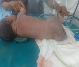
A 26 year old G2P1 lady delivered a term appropriate for gestational age, 3.38kg female baby via normal vaginal delivery who cried immediately after birth. APGAR score was noted to be 7/10 and 8/10 at the 1st and 5th minute respectively. Relevant antenatal scans showed a large cystic lesion 10x15cm in size with thin septations distal to the coccyx. Colour doppler was noted to be avascular. On examination the baby was found to be hemodynamically stable, with a sacral mass of 10x15cm with seropurulent discharge. In view of tachypnea the baby was shifted to NICU
on facemask oxygen at 5L/minute. In NICU, High flow nasal cannula was placed in view of tachypnea. A chest x-ray was done showing bilateral diffuse patchy opacities suspected to be due to meconium stained amniotic fluid. The baby was started on IV meropenem 135mg every 8th hourly. The child was subsequently posted for surgical resection of the mass on day 2 of life. On pre-anaesthetic evaluation, no other congenital anomalies were noted. On examination the cardiac and respiratory evaluation was normal. Echo was done which was noted to be normal as well. Routine blood investigations were done including a complete blood count, serum electrolytes, renal function tests and coagulation profile which were found to be within normal limits. Blood products were arranged in the pre-operative period and standard NPO orders were given.
On the day of surgery, the baby was taken into the operating theatre in a portable incubator. Standard ASA monitors of ECG, non-invasive blood pressure cuff and pulse oximeter was attached. A nasopharyngeal temperature probe was also placed. Two 24G intravenous cannula were secured and IV fluids were started. The patient positioned supine with both legs turned to right side in view of the sacrococcygeal mass and then preoxygenated with 100% oxygen for 3 minutes. Premedication was done with IV Glycopyrrolate 0.02mg IV, IV Midazolam 0.15 mg, Fentanyl 7mcg IV and trachea was intubated with a 3.0 microcuffed endotracheal tube after inhalational induction with sevoflurane 2% and muscle relaxation with atracurium 2mg IV. Patient was maintained on sevoflurane with nitrous oxide and oxygen with top ups of atracurium when required. Hand ventilation was done with Jackson-Reese circuit. Central venous access was attempted but ultimately abandoned due to difficulty in securing the same. The baseline blood pressure was noted to be around 70/40mmHg. Our facility did not have provision for arterial line invasive blood pressure monitoring and therefore we had to make do with non-invasive blood pressure monitoring. The procedure was done in lithotomy position and exceeded 4 hours duration. One episode of intra operative hypotension during which blood pressure fell to 50/30mmHg was noted for which IV dopamine infusion was started at 10mcg/kg/minute. Through-out the procedure the temperature was maintained with body warmer and fluid warmers in place. Blood loss exceeded the maximum allowable blood loss of 100ml which was replaced by packed red blood cells administered at 15ml/kg to a total of 55ml. 50ml of intravenous crystalloids were given in total. Abg was not done in the intra-operative period.
The baby was shifted to the NICU with endotracheal tube in place for further management in the postoperative period. Fresh frozen plasma at 15ml/kg was administered. Post-operative potassium value was noted to be 5.8mmol/L. Correction of the same was started. The neonate was extubated on post-operative day 3 and started on high flow nasal cannula which was later stopped. Oxygen via facemask was started. Inotropes were tapered and stopped on the same day. The following day the baby was weaned off to room air. Histopathology report revealed the tumour to be a sacrococcygeal mature cystic teratoma. An ultrasound abdomen was also done which showed no significant abnormality. The baby was hemodynamically stable and maintaining saturation on room air on post-operative day 5 and was discharged from the hospital (Figure 1).
Discussion
Sacrococcygeal teratoma is a rare tumour which presents frequently in infancy [2]. Surgical resection of large sacrococcygeal teratomas (SCTs) in premature neonates has been associated with significant perinatal mortality [3]. Certain predictors of poor outcome include delivery before 30 weeks, low birth weight, diagnosis before 20 weeks gestation, Apgar score less than 7, polyhydramnios, development of hydrops and placentomegaly.3 The tumour may become malignant during infancy and with increasing age the risk of malignancy grows. Hence at the earliest opportunity, complete surgical resection along with excision of the coccyx should be done [1].
Intraoperative management of SCT resection can be complicated due to difficulty positioning the patient in supine position to allow for endotracheal intubation, hypothermia, cardiovascular instability, associated coagulopathy, massive blood loss and tumour lysis [4]. In our patient, the legs were positioned laterally while the torso and head was turned to supine position to facilitate intubation.
Hypothermia is common in view of disproportionately large exposed surgical field to the body surface area.
Complications of hypothermia include coagulopathy and also delay in recovery due to hypothermia [5]. In this case the patient was adequately covered with drapes, fluid warming was done while administering fluids and blood products and a body warmer was kept to ensure temperature was maintained between 36-37 degrees Celsius. This was important due to the fact that the surgical procedure time exceeded 4 hours which placed the neonate at great risk of hypothermia.
Hyperkalemia, was noted in the post-operative period which may have been associated with manipulation of the tumour during resection or transfusion of packed red blood cells containing high levels of potassium. Tumour lysis due to surgical manipulation of the tumour can occur especially if multiple necrotic foci are noted. A case report by Kim JW et al demonstrated two cases in which cardiac arrest occurred due to hyperkalemia in surgical resection of SCT [3]. Vigilant monitoring is needed during surgical manipulation of tumour and blood transfusion.
Neonates are unable to mount an adequate adrenergic response and thus they are at high risk for hypotension and shock. As bleeding increases during surgery, mortality and morbidity rates increases as well [6]. A thorough pre- operative evaluation including discussion with surgeons is paramount to determine how much of blood loss is expected during the procedure as well as adequate reservation and issuing of blood products when needed. In our case the maximum allowable blood loss was exceeded and a transfusion of packed red blood cells was initiated. An episode of intra-operative hypotension occurred which resolved upon starting IV dopamine infusion at 10mcg/kg/minute. An already challenging case was made even more difficult with the lack of certain facilities in our setup including arterial line monitoring and intra-operative arterial blood gas. Therefore, extra vigilance and care was needed to ensure the neonate was kept hemodynamically stable with careful attention being paid to fluid input and output and thermoregulation.
Conclusion
Resection of large Sacrococcygeal teratomas can be challenging and requires meticulous planning, vigilance, and a multidisciplinary team approach. A strong understanding of neonatal physiology is needed and particular attention must be paid to thermoregulation and hemodynamic stability in the intra-operative period.
Conflict of Interest: No potential conflict of interest relevant to this article was reported.
Funding: Not applicable.
Acknowledgment: Not applicable. Disclaimer: Written informed consent was obtained from the parents of the neonate prior to publication.
References
-
Choudhury S, Kaur M, Pandey M, Jain A (2016) Anaesthestic management of sacrococcygeal teratoma in infants. Indian J Anaesth 60(5): 374-375.
-
Robinson S, Laussen PC, Brown TC, Woodward AA (1992) Anaesthesia for sacrococcygeal teratoma--a case report and a review of 32 cases. Anaesth Intensive Care 20(3): 354-358.
-
Kim JW, Gwak M, Park JY, Kim HJ, Lee YM, et al. (2012) Cardiac arrest during excision of a huge sacrococcygeal teratoma-A report of two cases. Korean J Anesthesiol 63(1): 80-84.
-
Abraham E, Parray T, Ghafoor A (2010) Complications with massive sacrococcygeal tumor resection on a premature neonate. J Anesth 24(6): 951-954.
-
Jafra A, Dwivedi D, Jain D, Bala I (2017) Giant sacrococcygeal teratoma: Management concerns with reporting of a rare occurrence of venous air embolism. Saudi J Anaesth 11(1): 124-125.
-
Kremer ME, Wellens LM, DerikxJP, Heij HA, Wijnen MHWA, et al. (2016) Hemorrhage is the most common cause of neonatal mortality in patients with sacrococcygeal teratoma. J Pediatr Surg 51(11): 1826-1829.
- Editorial on Multimodal Analgesia
- Surgical Incision Site Local Anaesthetic Infiltration and Superior Hypogastric Plexus Block in Total Abdominal Hysterectomy Under General Anaesthesia- A Placebo-Controlled, Randomized Clinical Trial
- Supraglottic Airway Insertion in Semi Fowler Position Due to Severe Thoracic Hyperkyphosis: A Case Report
- Anaesthetic Management of Cardiac Myxoma Patient with Systemic Involvement: A Case Report
- Current Problems in Pulmonary Respiratory Distress Syndrome (Literature Review)
- Evolution of Perioperative Hemodynamic Monitoring from the Hand on Pulse to Hypotension Prediction Index