
Investigate the Anticonvulsant and Neuroprotective Effects of Commercially Available Humid Acid by Utilizing both In Silico and In Vitro Approaches
Epilepsy is a chronic disorder of the brain that affects people worldwide. As per WHO, epilepsy is characterized by recurrent seizures, which are brief episodes of involuntary movement that may involve a part of the body (partial) or the entire body (generalized), and are sometimes accompanied by loss of consciousness and control of bowel or bladder function.
Introduction
Epilepsy is a chronic disorder of the brain that affects people worldwide. As per WHO, epilepsy is characterized by recurrent seizures, which are brief episodes of involuntary movement that may involve a part of the body (partial) or the entire body (generalized), and are sometimes accompanied by loss of consciousness and control of bowel or bladder function [1].
Epilepsy was one of the first brain disorders to be described. It was mentioned in ancient Babylon more than 3,000 years ago. The strange behaviour caused by some seizures has contributed through the ages to many superstitions and prejudices. From greek word attack, the word epilepsy is derived. In earlier times, People once thought that those with epilepsy were being visited by demons or gods. However, in 400 B.C., the early physician Hippocrates suggested that epilepsy was a disorder of the brain, and we now know that he was right [2].
Epilepsy is a major neurological disorder and upto 5% of the world population develops epilepsy in their lifetime. The current therapy of epilepsy with modern antiepileptic drugs is associated with side effects, dose-related and chronic toxicity as well as teratogenic effects and approximately 30% of the patients continue to have seizures with current antiepileptic drug therapy.
Traditional systems of medicines are popular in developing countries and upto 80% of the population relies on traditional medicines/ folk remedies for their primary health care need. Hence, there is a need to discover an Thesis alternative agent from natural sources [3].
Statement of The Problem
More than 2 million people in the United States have experienced an unprovoked seizure or been diagnosed with epilepsy. For about 80 percent of those diagnosed with epilepsy, seizures can be controlled with modern medicines and surgical techniques. However, about 25 to 30 % of people with epilepsy will continue to suffer from seizures with the current available treatment. Doctors call this situation intractable epilepsy. Having a seizure does not necessarily mean that a person has epilepsy. Only when a person has had two or more seizures is he or she considered to have epilepsy [2].
Approximately 50 million people currently live with epilepsy worldwide. An estimate shows that people suffering from epilepsy (i.e. continuing seizures or with the need for treatment) at a given time is between 4 and 10 per 1000 people. However, some studies shows that the proportion is much higher in low- and middle-income countries, between 7 and 14 per 1000 people.
Globally, each year epilepsy was diagnosed on estimating 2.4 million people. In high-income countries, annual new cases are between 30 and 50 per 100 000 people in the general population. This figure can be up to two times higher in low- and middle-income countries.
Various factors such as higher incidence of road traffic injuries, birth-related injuries, variations in medical infrastructure, availability of preventative health programmes and awareness among people can be the reason for these. Close to 80% of people with epilepsy live in low- and middle-income countries. It is estimated that there are more than 10 million persons with epilepsy in India. Its prevalence is about 1% in our population. The prevalence is higher in the rural (1.9%) compared to urban population (0.6%). In the Bangalore Urban Rural Neuroepidemiological Survey (BURNS), estimated that a prevalence rate of 8.8/1000 population was observed, with the rate in rural communities (11.9) being twice that of urban areas (5.7).
Epilepsy accounts for 0.6% of the global burden of disease, a time-based measure that combines years of life lost due to premature mortality and time lived in less than full health. In terms of health care needs, premature death and lost work productivity, epilepsy has significant economic implications. An Indian study conducted in 1998 calculated that the cost per patient of epilepsy treatment was as high as 88.2% of the country’s per capita Gross National Product (GNP), and epilepsy-related costs, which included medical costs, travel, and lost work time, exceeded $2.6 billion/year (2013 USD) [1].
Estimates suggest that available medication controls the seizures in only 50% of patients or decreases the incidence in only 75% of patients. The search for agents with anticonvulsant activity with more selectivity and lower toxicity continues to be an area of investigation in future.
Definitions
Epilepsy These are a group of CNS disorders characterized by paroxysmal cerebral dysrhythmia, manifesting as brief episodes(seizures) of loss or disturbance of consciousness, with or without characteristic body movements(convulsions), sensory or psychiatric phenomena [4, 5].
Seizures A seizure is a sudden surge of electrical activity in the brain.
Convulsion A convulsion is a condition in which body muscles contract and relax rapidly and repeatedly, results in an uncontrolled shaking of the body [6]. Theoretical Basis Despite the successful development of various new antiepileptic drugs (AEDs) in recent decades, the search for new therapies with better efficacy and tolerability remains an important goal. The discovery and development of a new AED relies heavily on the preclinical use of animal models to establish efficacy and safety prior to first trials in humans. This approach has been very successful and crucially contributed to the development of numerous clinically effective AEDs. In the discovery and development of new AEDs, animal models of seizures or epilepsy serve a variety of purposes. First, they are used for identifying novel AEDs. Second, animal models are used to evaluate the possible specific efficacies of the compound against different types of seizures or epilepsy if the antiepileptic activity of a novel compound was detected. Third, specific models of AED-resistant seizures are used to investigate whether the novel drug has advantages towards clinically established AEDs for therapy of difficult-to-treat types of seizures or epilepsies. Fourth, animal models are used to characterize the preclinical efficacy of novel compounds during chronic administration. Such chronic studies can serve different objectives, for instance evaluation of whether drug efficacy changes during prolonged treatment, e.g. because of development of tolerance. Fifth, in view of the possibility that chronic brain dysfunctions, such as epilepsy, might lead to altered sensitivity to drug adverse effects, models with epileptic animals are useful to study whether epileptogenesis alters the adverse effect potential of a given drug. Sixth, animal models can be used to estimate effective plasma concentrations of new AEDs for first clinical trials. And finally, seventh, animal models are crucial in discovering therapies that may prevent or modify the development of epilepsy after brain insults.
Not all animal models of seizures and/or epilepsy can be used for all of the above-described purposes. Furthermore, the intention of the experiment is essential for selection of a suitable animal model. For instance, simple seizure models such as the maximal electroshock seizure (MES) test, allowing to test high numbers of compounds for anticonvulsant activity in relatively short time, will be preferred above more complex models in screening approaches of anticonvulsant drug development.
For AED discovery, which necessitates screening of large numbers of compounds, animal models should be easy-to- perform, time- and cost-efficient, and predictive of clinical activity. This explains that two simple seizure models in mice and rats, the MES and pentylenetetrazole (PTZ) tests, which have been developed >60 years ago, are still the most widely used animal seizure models employed in the search for new AEDs [7, 8]. Experimental models These models for testing antiepileptic drugs have also shed light on the etiopathogenesis of epilepsy. Maximal electroshock seizures: Brief high intensity shock is applied to the head of a rodent produces tonic flexion- tonic extension- clonic convulsions. The tonic phase (especially extensor) is selectively abolished by drugs effective in generalized tonic clonic seizure. Activity in this model represents action on spread of seizure discharge. Pentylenetetrazol clonic seizures (PTZ): Injection of PTZ in rats or mice produces clonic convulsions which are prevented by drugs effective in absence seizures. Activity in this model represents action on seizure focus itself.
Chronic focal seizures: Produced by application of alumina cream on the motor cortex of monkey. Kindled seizures: Brief bursts of weak electrical impulses are applied to the brain (especially amygdala) intermittently over days. After- discharges increase progressively and tonic-clonic seizures are produced after 10-15 shocks; with time spontaneous seizures have a self-perpetuating and reinforcing effect: more neuronal circuits are facilitated and recruited in the seizure process. Kindling is probably involved in the genesis of clinical epilepsy [5]. Purpose of The Study The current therapy of epilepsy with modern antiepileptic drugs (AEDs) is associated with side effects, dose-related and chronic toxicity, as well as teratogenic effects, and approximately 30% of the patients continue to have seizures with current antiepileptic drugs therapy. The discovery of novel antiepileptic drugs relies upon the preclinical employment of animal models to establish efficacy and safety prior to the introduction of the AEDs in human volunteers. Natural products from folk remedies have contributed significantly in the discovery of modern drugs and can be an alternative source for the discovery of AEDs with novel structures and better safety and efficacy profiles. For the detection of antiepileptic activity, several plants are used for the treatment of epilepsy in different systems of traditional medicine and these plants have shown activity when tested in modern bioassays and many such plants are yet to be scientifically investigated. Medicinal plants used for the therapy of epilepsy in traditional medicine have been shown to possess promising anticonvulsant activities in animal models of anticonvulsant screening.
Anxiety
Neuropsychopharmacology may be defined as the branch of interdisciplinary neuroscience devoted to the study of drugs that have an effect on the nervous tissue and alter the behavior. Neuropharmacology deals with the study of effects of the drug on nerve cells, their synapses and circuit whereas the study of effects of drugs on behaviors, including emotional and cognitive mental activities, is Psychopharmacology. It associates the frontiers of fundamental neuroscience to the management of psychiatric and neurological diseases. This branch of science seeks to comprehend how drugs selectively affect the CNS to induce sleep, relieve pain, reduce fever, suppress muddled movement, prevent seizures or enhance attention. Neuropsychopharmacology seeks to understand how drugs can treat mania, anxiety, schizophrenia or depression without disturbing the consciousness. This field seeks to uncover the biological basis for intricate mental states. The goal of this field is not only to understand the nature of the alterations in biology which direct to distorted emotions and thought processes, but also to develop therapeutically priceless specific molecules which regulate the specific biologic underpinnings- namely, the as yet vague sequences of multineuronal interactions by which the behaviors emerge.
Anxiety may be defined as an unpleasant state of mental uneasiness, apprehension, nervousness and obsession or concern about something uncertain. The major symptoms include; arousal, tenseness, increased autonomic activity like respiration, blood pressure and heart rate. Tightness in chest, palpitations, perspirations etc. it is a common symptom in a variety of distinct mental illnesses and is a predominant symptom in panic disorders, phobias and obsessive-compulsive disorder.
The major types of anxiety include: Panic Disorder: Psychiatric condition associated with multiple disabling panic attacks. In between the panic attacks, an excessive time is spent by the individual in thinking about future panic attacks. Generalized Anxiety Disorder: It is characterized by persistent and excessive worries. The patient worries about various life events such as job performances, marital status, money, social status etc. Posttraumatic Stress Disorder: Caused due to the exposure of individual to life-threatening or terrifying life events. The individual re-experiences the traumatic event as flashbacks or as intrusive recollections. Obsessive–Compulsive Disorders: The root cause of the disorder is intrusive, repetitive compulsions and / or thoughts. Marked distress is the hallmark of this disorder. There occur irrational thoughts and acts which impair normal functioning. Drug therapy
Antianxiety Drugs
Benzodiazepines: Diazepam, lorazepam, alprazolam Azapironees: Buspirone Sedative antihistaminic: Hydroxyzine Beta blocker: Propranolol
Review of Literature
Epilepsy
Epilepsy is a chronic CNS disorder characterized by brief episodes of seizures and excessive EEG discharge. It is usually associated with loss of consciousness, violent spasmodic contractions of skeletal muscles (convulsions) and autonomic hyperactivity [9].
Epilepsy is one of the most common neurological disorders. Worldwide, the prevalence is estimated to be 0.5- 1%, and there is a lifetime incidence of 1- 3%. It has important medical, social and psychological consequences. Epilepsy is a heterogeneous symptom complex, a chronic disorder characterized by recurrent seizures. Seizures resulting from abnormal discharge of cerebral neurons and are finite episodes of brain dysfunction. It is estimated that in India (with population more than 1 billion), there will be 6- 10 million people with epilepsy, accounting for nearly 1/5 of global burden. The current treatment of epilepsy with modern antiepileptic agents is associated with side effects, dose-related and chronic toxicity, as well as teratogenic effects, and approximately 30% of the patients continue to have seizures with current antiepileptic drugs therapy. Therefore, there is a great need for the development of cheap, effective and safe anticonvulsant agents from plants and other sources [10]. Nature of Epilepsy The term epilepsy is used to define a group of neurological disorders all of which exhibit periodic seizures. Not all seizures involve convulsions. Seizures are associated with episodic high-frequency discharge of impulses by a group of neurons (sometimes referred to as focus) in the brain. What starts as a local abnormal discharge may then spread to other areas of the brain. The site of the primary discharge and the extent of its spread determine the symptoms that are produced, which range from a brief lapse of attention to a full convulsive fit lasting for several minutes, as well as odd sensations or behaviours.
The particular symptoms produced depend on the function of the region of the brain that is affected. Thus, involvement of the motor cortex causes convulsions, involvement of the hypothalamus causes peripheral autonomic discharge, and involvement of the reticular formation in the upper brain stem lead to loss of consciousness [11]. Types Generalised Seizures 1. Generalized Tonic-Clonic Seizures (Major Epilepsy, Grand Mal): commonest, lasts 1-2 min. Prolonged sleep and depression of all CNS functions after the usual sequence that is aura-cry-unconsciousness-tonic spasm of all body muscles-clonic jerking followed by 2. Absence Seizures (Minor Epilepsy, Petit Mal): prevalent in children, lasts about ½ min. Sudden loss of consciousness, no muscular component or little bilateral jerking, patient apparently freezes and stares in one direction.
3. Atonic Seizures (Akinetic Epilepsy): unconsciousness with relaxation of all muscles due to excessive inhibitory discharges. Patient may fall.
4. Myoclonic Seizures: shock-like momentary contractions of muscles of a limb or the whole body.
5. Infantile Spasms (Hypsarrhythmia): These type of epilepsy seen in infants and probably not a form of epilepsy. Intermittent muscle spasm and progressive mental deterioration.
Partial seizures 1. Simple Partial Seizures (Cortical Focal Epilepsy): Lasts ½-1 min. Often secondary. Depending on the area of cortex involved, convulsions are confined to a group of muscles or localized sensory disturbance, without loss of consciousness.
2. Complex Partial Seizures (Temporal Lobe Epilepsy): attacks of bizarre and confused behaviour and purposeless movements, emotional changes lasting 1-2 min along with impairment of consciousness. An aura often precedes. The seizure focus is located in the temporal lobe.
3. Simple Partial or Complex Partial Seizures Secondarily Generalized: The partial seizure occurs first and evolves into generalized tonic clonic seizures with loss of consciousness [5].
Causes
All forms of epilepsy have their origin in the brain. The different types of epilepsies are not based on a single underlying mechanism but are multifactorial in origin. Epilepsy results when many neurons in union, under a high excited stage, deliver massive discharges abolishing a finely organized pattern of the integrative activity of the brain.
John Jackson proposed that these seizures are caused by occasional, sudden, excessive, rapid and local discharges of grey matter and once initiate by the abnormal focus, the seizures attack the neighboring normal brain resulting into generalized convulsions. This abnormal focus may originate as a result of local biochemical changes, ischemia or the loss of vulnerable cell inhibitory systems. However, certain physiological changes may trigger the focus and thus facilitate the spread of abnormal electrical activity to normal tissue. Such factors include.
Genetic Factors
Several types of epilepsy have now been linked to defective genes for ion channels, the “gates” that control the flow of ions in and out of cells and regulate neuron signaling. Another gene, which is missing in people with progressive myoclonus epilepsy, codes for a protein called cystatin B. This protein regulates enzymes that break down other proteins. Another gene, which is altered in a severe form of epilepsy called LaFora’s disease, has been linked to a gene that helps to break down carbohydrates.
Other Disorders
In some cases, epilepsy may develop as a result of brain damage from other diseases. For example, brain tumors, alcoholism, and Alzheimer’s disease frequently lead to epilepsy because they alter the normal workings of the brain. Strokes, heart attacks, and other conditions that diminishes the supply of oxygen towards brain, also can cause epilepsy in some cases. About 32 percent of all cases of newly developed epilepsy in elderly people appears to be due to cerebrovascular disease, which reduces the supply of oxygen to brain cells. Meningitis, AIDS, viral encephalitis, and other infectious diseases and also ahydrocephalus -- a condition in which excess fluid builds up in the brain can lead to epilepsy. Epilepsy also can result from intolerance to wheat gluten (also known as celiac disease), or from a parasitic infection of the brain known as neurocysticercosis.
Epilepsy is having connection with a variety of metabolic diseases such as cerebral palsy, pyruvate dependency, tuberous sclerosis, Landau-Kleffner syndrome, and autism. Epilepsy is just one of a set of symptoms commonly found in people with these disorders.
Head Injury, Prenatal Injury and Developmental Problems
The developing brain is susceptible to many kinds of injury. Some conditions like Maternal infections, poor nutrition, and oxygen deficiencies that may affect the brain of a developing baby. These conditions may lead to cerebral palsy, which often is associated with epilepsy, or they may cause epilepsy that is unrelated to any other disorders.
Poisoning
Exposure to lead, carbon monoxide, and many other poisons may cause seizures. They also can result from exposure to street drugs and from overdoses of antidepressants and other medications.
Seizures are often triggered by factors such as lack of sleep, alcohol consumption, stress, or hormonal changes associated with the menstrual cycle. For some people, a seizure can also be triggered by light flashing at a certain speed or the flicker of a computer monitor and this type of epilepsy is known as photosensitive epilepsy. Smoking cigarettes also can trigger seizures. The nicotine in cigarettes acts on receptors for the excitatory neurotransmitter acetylcholine in the brain, which increases neuronal firing. Seizures are not triggered by sexual activity except in very rare instances [2].
Aetiologically, the epilepsies are classified into four groups: idiopathic, symptomatic, cryptogenic and progressive. The idiopathic epilepsies are thought to be genetically determined and are usually associated with particular clinical characteristic and specific electroencephalography (EEG) findings. Structural abnormality of the brain can result in symptomatic epilepsies and are acquired condition. Epilepsy is classified as cryptogenic when no clear abnormality or putative risk factor is identified for what is presumed to be a symptomatic or acquired epileptic condition. The term progressive epilepsy is used when epilepsy is associated with an evolving neurological condition [12, 13]. Symptoms Repeated seizure is the major cause of epilepsy. The individual should see a doctor If one or more of the following symptoms are present, especially if the symptoms recur:
- A convulsion with no temperature (no fever)
- Confused memory or short spells of blackout
- Intermittent fainting spells, during which loss of bowel or bladder control, followed by extreme
- tiredness
- For a short period, the person is unresponsive to instructions or questions
- The person becomes stiff, suddenly, for no apparent reason
- The person suddenly falls
- The person shows sudden bouts of blinking without apparent stimuli
- Sudden bouts of chewing, without any apparent reason
- For a short time, the person seems dazed and unable to communicate
- Repetitive movements that seem inappropriate
- The person becomes fearful for no apparent reason; they may even panic or become angry
- Peculiar changes in senses, such as smell, touch, and sound
- The arms, legs, or body jerk, in babies these will appear as a cluster of rapid jerking movements The following conditions need to be eliminated as they may appear as similar symptoms and are sometimes misdiagnosed as epilepsy:
- High fever with epilepsy-like symptoms
- Fainting
- Narcolepsy - recurring episodes of sleep during the day
- Cataplexy - periods of extreme weakness
- sleep disorders
- nightmares [13] Syndromes Cases of epilepsy may be arranged into epilepsy syndromes on the basis of specific features that are present. These features include, the seizure types, EEG findings, the age that seizure begins. Identifying an epilepsy syndrome is useful as it helps determine the underlying causes as well as what anti-seizure medication should be tried.
Since the onset of seizures is commonly early, the ability to categorize a case of epilepsy into a specific syndrome occurs more often with children. Less serious examples are benign rolandic epilepsy (2.8 per 100,000), childhood absence epilepsy (0.8 per 100,000) and juvenile myoclonic epilepsy (0.7 per 100,000). Severe syndromes with diffuse brain dysfunction caused, at least partly, by some aspect of epilepsy, are also commonly known as epileptic encephalopathies. These are associated with frequent seizures that are resistant to treatment and severe cognitive dysfunction, for instance Lennox–Gastaut syndrome and West syndrome. Genetics is believed to play an important role in epilepsies by a number of mechanisms. Simple and complex modes of inheritance have been identified for some of them. However, extensive screening have failed to identify many single gene variants of large effect.
Syndromes in which causes are not clearly identified are difficult to match with categories of the current classification of epilepsy. Categorization for these cases was made somewhat arbitrarily. In case of 2011 classification (idiopathic category) includes syndromes in which the general clinical features and/or age specificity strongly point towards a genetic cause. Some childhood epilepsy syndromes are included in the unknown cause category in which the cause is presumed genetic, for instance benign rolandic epilepsy. Others are included in symptomatic in some cases despite a presumed genetic cause, for example Lennox- Gastaut syndrome. Clinical syndromes in which epilepsy is not the main feature (e.g. Angelman syndrome) were categorized symptomatic but it was argued to include these within the category idiopathic. Classification of epilepsies and particularly of epilepsy syndromes will change with advances in research [14].
Pathophysiology
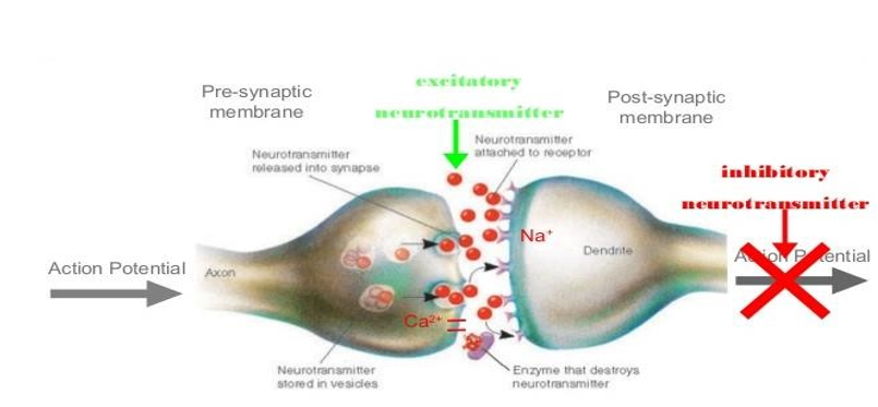
Action potential is the basic mechanism of neuronal excitability. Action potential is a hyperexcitable state can result from increased excitatory synaptic neurotransmission, decreased inhibitory neurotransmission, an alteration in voltage-gated ion channels, or an alteration of intra- or extra-cellular ion concentrations in favor of membrane depolarization. Membrane potential can varies with activation of ligand- gated channels, whose conductance is affected by binding to neurotransmitters; or with activation of voltage- gated channels, whose conductance is affected by changes in transmembrane potential; or with changes in intracellular ion compartmentalization. The major neurotransmitters in the brain are glutamate, gamma-amino-butyric acid (GABA), acetylcholine (ACh), norepinephrine, dopamine, serotonin, and histamine. The major excitatory neurotransmitter is the amino acid glutamate. All ionotropic glutamate receptors are permeable to Na+ and K+, and it is the influx of Na+ and outflow of K+ through these channels that contribute to membrane depolarization and generation of the action potential. The NMDA receptor also has a Calcium channel and in resting state, calcium channel is blocked by Magnesium ions, but under conditions of local membrane depolarization, Mg++ is displaced and the channel becomes permeable to Ca++. Influx of Ca++ tends to further depolarize the cell, and is thought also to contribute to Ca++ mediated neuronal injury under conditions of excessive neuronal activation (such as status epilepticus and ischemia), potentially leading to cell death, a process termed excitotoxicity.
The major inhibitory neurotransmitter, GABA, interacts with 2 major subtypes of receptor:
GABAA and GABAB receptors. GABAA receptors are found postsynaptically, while GABAB receptors are found presynaptically, and can thereby modulate synaptic release. GABAA receptors are permeable to Cl- ions in adult brain and action potential is inhibited by upon activation Cl- influx hyperpolarizes the membrane. Therefore, substances which are GABAA receptor agonists, such as barbiturates and benzodiazepines, are well known to suppress seizure activity. Rather than Cl- channels, GABAB receptors are associated with second messenger systems, and due to their presynaptic location, attenuation of transmitter release occurs. The second messenger systems often result in opening of K+ channels, leading to a hyperpolarizing current. Certain GABAB agonists, such as baclofen, have been reported to exacerbate hyperexcitability and seizures [15].
Diagnosis
Abnormal electrical activity during and following a seizure can be detected by electroencephalography (EEG) recording from electrodes distributed over the surface of the scalp. Various types of seizure can be recognized on the basis of the nature and distribution of the abnormal discharge. Modern brain imaging techniques, such as magnetic resonance imaging and positron emission tomography, are now routinely used in the diagnosis of epilepsy to identify structural abnormalities (eg. Lesions, tumors) that cause certain epilepsies [11]. Treatment Once epilepsy is diagnosed, it is important to begin treatment as soon as possible. Once seizures and their consequences become established, research suggests that current available medication and other treatments may be less successful in treating epilepsy.
Medications
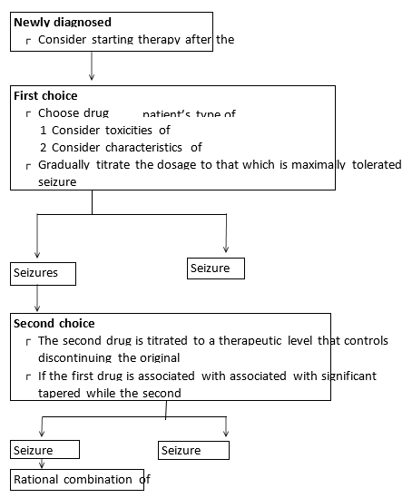
By far the most common approach to treating epilepsy is to prescribe antiepileptic drugs. Doctors diagnosing a patient with newly developed epilepsy often prescribe antiepileptic agents like carbamazepine, valproate, lamotrigine, oxcarbazepine, or phenytoin first, unless the developed epilepsy is a type that is known to require a different kind of treatment. For absence seizures, ethosuximide is often the primary treatment. Other commonly prescribed drugs include clonazepam, phenobarbital, and primidone. Some relatively new epilepsy drugs include tiagabine, gabapentin, topiramate, levetiracetam, and felbamate [2].
Classification
- Barbiturate: Phenobarbitone
- Deoxybarbiturate: Primidone
- Hydantoin: Phenytoin, Fosphenytoin
- Iminostilbene: Carbamazepine, Oxcarbazepine
- Succinimide: Ethosuximide
- Aliphatic carboxylic acid: Valproic acid, Divalproex
- Benzodiazepines: Clonazepam, Diazepam, Lorazepam, Clobazam
- Phenyltriazine: Lamotrigine
- Cyclic GABA analogues: Gabapentin, Pregabalin
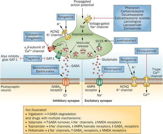
- Newer drugs: Topiramate, Zonisamide, Tiagabine [5]. Mechanism of Action • The currently available anticonvulsant agents are thought to act by three main mechanisms:
Surgery to Treat Underlying Conditions
When seizures are caused by a brain tumor, hydrocephalus, or other conditions that can be treated with surgery, doctors may operate to treat these underlying conditions. In many cases, once the underlying condition is successfully treated, a person’s seizures will disappear as well. Surgery to Remove a Seizure Focus Removal of a seizure focus, or small area of the brain where seizures originate is the most common type of surgery for epilepsy. This type of surgery, which doctors may refer to as a lobectomy or lesionectomy, is appropriate only for focal seizures that originate in just one area of the brain.
Indications of Surgery
- Medically intractable seizures
- Seizures significantly affect the quality of life
- Localized seizure focus
- Presence of signs predictable of seizure persistence Contraindications of Surgery
- Benign, self-limited epilepsy syndrome
- Neurodegenerative and metabolic disorders
- Non-compliance with drugs
- Severe family disfunctions
5. Associated psychosis
Multiple Subpial Transection
When seizures originate in part of the brain that cannot be removed, surgeons may perform a procedure called a multiple subpial transection Corpus Callosotomy In children with severe seizures that start in one half of the brain and spread to the other side, Corpus callosotomy, or severing the network of neural connections between the right and left halves, or hemispheres, of the brain, is done. Hemispherectomy and Hemispherotomy These procedures remove half of the brain’s cortex, or outer layer. These are used mainly in children who are suffering from seizures that do not show response to medication because of damage that involves only half the brain, as occurs with conditions such as Rasmussen’s encephalitis, Sturge- Weber syndrome, and hemimegencephaly. Devices The vagus nerve stimulator was approved by the U.S. Food and Drug Administration (FDA) in 1997 for use in people with seizures that are not well-controlled by medication. The vagus nerve stimulator is a battery-powered device that is surgically implanted under the skin of the chest, much like a pacemaker, and is attached to the vagus nerve in the lower neck. This device delivers short bursts of electrical energy to the brain via the vagus nerve. Researchers are studying whether transcranial magnetic stimulation (TMS), a procedure which uses a strong magnet held outside the head to influence brain activity, may reduce seizures. They also hope to develop implantable devices that can deliver drugs to specific parts of the brain [2].
Diet
Ketogenic diet is one of the oldest methods of treating childhood epilepsy. In children with refractory seizures who have failed drug therapy and are not candidate for epilepsy surgery, this therapy is as or more effective than the addition of new anti-epileptic drug. For example, it remains a reasonable alternative for children with Lennox-Gastaut syndrome refractory to standard drug therapy.
The ketogenic diet consists of a higher proportion of fats and small amounts of carbohydrates and protein. The basis of the therapeutic effectiveness of the ketogenic diet is because of the ketosis that develops when the brain is relatively deprived of glucose as an energy source and must shift to utilization of ketone bodies as the primary fuel [16].
Antiepilepsy Activity
Avanthi E et al., (2016)
Evaluated the antiepileptic activity of clove oil by MES model in mice. A total of thirty mice were taken, they were given an electroconvulsive shock. Thirty mice were divided into five groups of six animals each, the control group received distilled water 5ml/kg i.p, standard received Inj. Sodium valproate 200 mg/kg i.p. another group received Sesame oil – 10ml/kg i.p(control), test groups received Clove oil- 0.075 ml/kg i.p., Clove oil-0.1ml/kg i.p respectively. All the injections were given 30 minutes before the test. The results showed Clove oil produced significant antiepileptic effect at all the doses [17]. K Sandeep Kumar et al., (2015) Evaluated the antiepileptic activity of ethanolic extract of Biophytum sensitivum in animal models. The anticonvulsant activity was assessed using MES and PTZ using albino mice. The extract reduced the duration of tonic hind limb extension and delayed the onset of tonic clonic convulsion. The result showed that the ethanolic extract of the plant beneficial in both tonic clonic and absence seizures [10]. Nirmala D (2015)
Performed a study which involves in detecting anticonvulsant activity from Annacyclus pyrethrum roots by using maximal electroshock seizure (MES) in a dose- dependent way. MES-induced tonic seizures can be prevented either by drugs that inhibit voltage dependent Na+ channels, such as phenytoin, valproate and lamotrigine or by drugs that block glutamatergic excitation mediated by the NMDA receptor such as felbamate. The study showed that ethanolic extract from roots of A. pyrethrum can inhibit voltage dependent Na+ channels as phenytoin in MES induced tonic seizures [18]. Gummalla Pitchaiahet al., (2015) Evaluated the anticonvulsant activity of methanolic extract of Allium cepa (Onion) bulbs in Swiss albino mice. The anticonvulsant effect was assessed using maximal electroshock (MES) and Isoniazid (INH) induced seizure models. Methanolic extract (200 and 400 mg/kg) showed significant reduction in the duration of hind limb extensor phase in electroshock convulsions; protected the mice against the Isoniazid induced convulsions. The results showed significant improvement in brain GABA levels after Allium cepa treatment [19]. Santilna K S et al., (2014) Studied the anticonvulsant activity study of Artemisia nilagirica. The leaves part of the plant was dried, powdered and subjected to maceration using diethyl ether, chloroform and ethanol. The results showed that the alkaloids, flavonoids and terpenoids were identified to be present in all three solvents extracts. The result obtained suggests that the ethanolic and chloroform extracts of Artemisia nilagirica may be beneficial in the treatment of epilepsy [20]. Ravindra C Sutar et al., (2014) Evaluated the anticonvulsant activity of leaf extract of Holoptelea integrifolia. The petroleum ether and methanolic extract of the leaves was evaluated using Pentylenetetrazole (PTZ) induced convulsions in mice and maximal electroshock (MES) induced Convulsions and lithiumpilocarpine induced status epilepticus in rats. The petroleum ether extract and methanolic extract delayed onset of PTZ- induced convulsions and also prolonged the onset of tonic convulsions in mice. Both the extracts failed to protect the rats from MES induced convulsions. The extracts also protected rats against seizures induced by lithium-pilocarpine. The results indicate that petroleum ether and methanol extracts contained such phytochemial compounds which are active in case of Pentylenetetrazole (PTZ) and lithium pilocarpine induced status epilepticus, which support the ethnomedicinal application of the plant as an anticonvulsant agent [21]. Dilnawaz Pathan et al., (2014) Evaluated the anticonvulsant effect of ethanolic extract of roots of Picrorhiza kurroa on electrically and chemically induced seizures. The extract was studied for its anticonvulsant effect on maximal electroshock-induced seizures and pentylenetetrazole, picrotoxin induced seizures in mice. It has been observed in the present study that extract (100 mg/kg) showed significant increase in latency to clonic convulsions and reduced mortality. The results shows that Picrorhizakurroa possess anticonvulsant activity against Pentylenetetrazole, Maximal electroshock and Picrotoxin induced convulsions in mice [22]. Ganapathi G. Varmaet al., (2014) Performed the evaluation of antiepileptic activity of methanolic leaves extract of Tragia involucrata in mice. In vivo screening models like maximal electroshock-induced convulsion (MES), pentylenetetrazole (PTZ) and picrotoxin (PTX) induced models are used to evaluate the antiepileptic effects of the extracts. In the MES induced convulsion, methanolic extract (800 mg/kg), showed high significant inhibition on tonic hind limb extension and decrease in duration of stupor period. In PTZ and PTX induced model extract (400 mg/kg and 800 mg/kg) showed delay on the onset of convulsions, decreased duration of convulsion and reduced mortality significantly. The results showed that Tragiainvolucrata possesses dose dependent antiepileptic activity [23]. Mehrdad Modaresi et al., (2014) Studied the antiepileptic activity of hydroalcoholic extract of Ocimum basilicum in mice. The experimental groups comprised control, sham, and four treatment groups receiving the extract at 100, 250, 300, and 350 mg/kg doses 65 minutes before PTZ injection. The obtained results of using different doses of the extract indicated that the mice receiving the extract at 100 and 250 mg/kg doses exhibited the highest and lowest frequency of myoclonic twitches, respectively [24]. Chinchawade A B et al., (2013) Observed the anticonvulsant activity of chloroform extract of bark & root of Erythrina variegate. The pentyleneterazole (PTZ) and the maximal electroshock seizure (MES) models were used for assessing the anticonvulsant effects of the chloroform extract in mice and rats. The extract produced significant protection against PTZ-induced and MES induced convulsions in rat. The results obtained from this study indicate that the chloroform root and bark extract of Erythrinavariegata may be beneficial in both absence and tonic clonic seizures [25]. Abubakar K et al., (2013) Evaluated the anticonvulsant effect of methanolic extract of Evolvulus alsinoides in mice using pentyleneterazole (PTZ) and the maximal electroshock seizure (MES) model. The extract significantly increased the latency of PTZ induced seizure. In the MES test a dose dependent decrease in the duration of seizure was also observed. These findings suggest that the methanol extract of the plant contains bioactive principles that may be beneficial in the treatment of epilepsy [26].
Ashish P Anovadiya et al., (2013)
Performed the antiepileptic and memory retention activity of Curcumin perse and in combination with antiepileptic drugs. In this study, antiepileptic activity of curcumin and its combination with phenytoin and sodium valproate were studied in chronic model (14 days) of Maximal Electroshock Seizure (MES) and Pentylenetetrazole (PTZ) induced seizure respectively. Curcumin (100 mg/kg) reduced clonic phase and significantly inhibited PTZ induced seizure. Addition of curcumin to sub therapeutic dose of sodium valproate showed synergistic effect..Curcumin found to be effective in absence seizure alone and as add on with sodium valproate [27]. Prabhat Singh et al., (2012) Studied antiepileptic activity of aqueous extract of fruits of Tricosanthes dioica. The antiepileptic efficacy of aqueous extract was evaluated by hand limb extension induced by MES and PTZ induced seizures in mice models. The aqueous extract was showed significant antiepileptic activity in both models and it was found to be due to activity against generalized tonic- clonic and cortical focal seizures [28]. Vikas Saroch et al., (2012) Evaluated the anticonvulsant Activity of Apasmarari rasa. Apasmarari rasa was subjected to assess the LD 50 and anti- convulsant activity on Male Albino rats was by means of MES (Maximal Electro convulsing Shock) Method. A supramaximal strength was 150mA in rats for 0.2 seconds and stimulus was applied via ear clip electrodes. The animal dose of Phenytoin (7.2mg/kg), Smritisagar rasa (18mg/kg) and Apasmarari rasa (5.4mg/ kg) was given orally to different groups. The animals were observed for a period of 180 minutes after being subjected to electro convulsions. Both standard drugs also shown good results when it comes to HLE (hind limb extension), butother factors such as time duration of flexion, tonus, clonus, recovery time amongst others in test drug group (Apasmarari rasa) showed significantly better results [29]. Vipin K Garget al., (2011) Evaluated the anticonvulsant activity of ethanolic extract of Cynodon dactylon. The anticonvulsant activity was studied using maximal electroshock (MES) and Pentylenetetrazol (PTZ) induced convulsions in mice. The extract suppressed hind limb tonic extensions (HLTE) induced by MES and also exhibited protector effect in PTZ-induced seizures. The results showed that the ethanolic extract of Cynodon dactylon has anticonvulsant effect in the both models suggesting their possible depressant action in the central nervous system [30]. Shyamjith Manikkoth et al., (2011) Performed Phyllanthus amarus on maximal electroshock-induced seizures (MES) and pentylenetetrazole (PTZ) induced seizures. The aqueous and ethanolic extracts of the leaves and stems of P. amarus significantly abolished the hind limb extension induced by MES. The same dose also significantly protected the animals from PTZ induced tonic convulsions [31]. Harish Babu B et al., (2010) Performed anticonvulsant activity of the methanolic extract of Martynia annua on Maximal Electroshock(MES) and Pentylenetetrazole (PTZ) induced seizures models in albino wistar rats. These studies showed, the mean duration of extensor phase of test group reduced to significant level as compared to control group. In Pentylenetetrazol induced seizure test, onset of myoclonic spasm and clonic convulsion was delayed in the test group. The study concluded Martynia annua possesses an anticonvulsant effect which results from the potentiation of the activity of GABA [32].
N. S. Vyawahareet al., (2009)
Evaluated the anticonvulsant activity of roots of Argyreia speciosa in mice. The mice were pretreated with different doses of Argyreia speciosa extract for 10 days and then, they were subjected to either pentylenetetrazole or maximal electroshock seizures treatment. The hydroalcoholic extract of Argyreia speciosa at the dose of 200 and 400 mg/kg significantly delayed the latency to the onset of first clonus and significantly reduced the duration of hind limb extension. This study shows that the hydroalcoholic extract possesses anticonvulsant activity against pentylenetetrazole and maximal electroshock seizures [33]. KarunakarHegde et al., (2009)
Studied the anticonvulsant activity of Carissa carandas root extract in experimental mice. The ethanolic extract was studied for its anticonvulsant effect on maximal electroshock- induced seizures and pentylenetetrazole-, picrotoxin-, bicuculline- and N-methyl-dl-aspartic acid induced seizures in mice. The data suggest that the ethanolic root extract reduced the duration of seizures produced by maximal electroshock as well as delayed the latency of seizures produced by pentylenetetrazole and picrotoxin [34].
Scope and Plan of Work
According to network pharmacology, medicines affect complex biological networks as opposed to single targets or pathways. Accordingly, rather than focusing on a single target, a drug’s effectiveness is determined by its ability to affect a network of targets or pathways. It includes two major approaches Network Biology and Polypharmacology. Network Pharmacology is an integrative and systems approach to understand complex relationship between molecules in cells whereas, polypharmacology is the ability of the molecule or ligand to interact with multiple proteins. Therefore, here various databases were used to identify various targets of disease and phytoconstituents and the common targets identified between them were used for further analysis i.e., In-silico studies. As targeting a single protein can increase the chances of failure therefore, multiple proteins were targeted by the ligand selected to see the actions of ligand on those receptors, acting as a potential molecule.
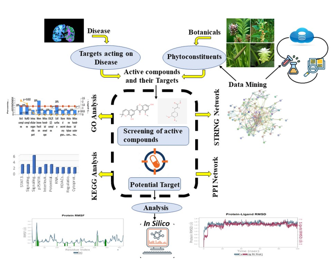
This pictorial explains the work that has been carried out. It explains how using various databases the top proteins and ligands were identified and how in-silico studies helped in understanding the effect of ligand on top proteins. This explains how network pharmacology and molecular modelling approach can help treating Alzheimer’s Disease.
Aim
To evaluate the antiepileptic and anti-anxiety activities of Phyllanthus reticulatus using network pharmacology approach.
Objectives
- To identify the phytoconstituents present in Phyllanthus reticulatus using literature survey, IMPAAT and Dr Dukes databases.
- To perform pharmacokinetic screening of the selected phytoconstituents using quikprop tool of Schrodinger.
- To predict the phyto-targets of Phyllanthus reticulatus using Swiss Target Prediction and the BindingDB databases.
- To predict the therapeutic targets of epilepsy and anxiety using Disgenet database.
- To identify the common targets between phyto-targets and disease targets using venny tool.
- To build a protein-protein interaction network using string database
- To perform topological analysis using Cytoscape software to identify the key proteins in the network.
- To perform gene ontology and pathway analysis using Funrich software and reactome database.
- To perform molecular docking and molecular dynamics to assess the binding affinity and binding stability of the protein ligand complex.
Plan of Work
Phase I
- Literature survey
- Identification of phytoconstituents
- ADMET Screening
- Identification of phyto and disease targets.
Phase II
- Identification of common targets
- Construction of protein-protein interaction network.
- Topological Analysis Phase III
- Gene ontology study
- Pathway Analysis Phase IV
- Molecular docking
- Molecular dynamics
Materials & Methods
Plant Profile
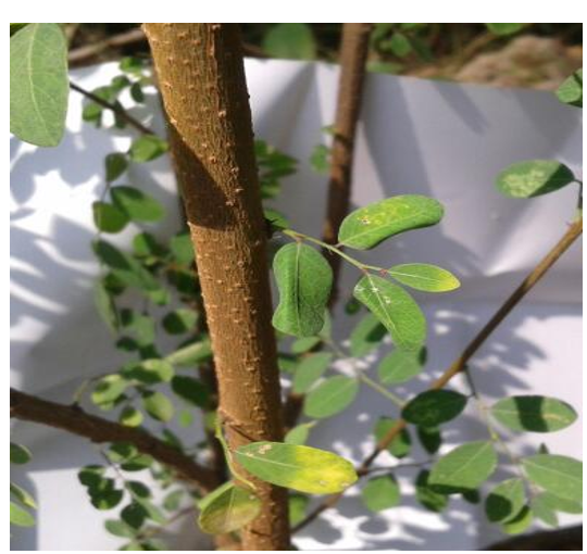
Phyllanthus reticulatus Poir. is a shrub with smooth or lenticellate branches reaching a maximum height of 10 feet that belongs to the family Euphorbiaceae. The leaves of the plants are traditionally used as diabetic and have also been reported to possess diuretic, astringent, and astringent properties. The leaf extract of P. reticulatus Poir. has been reported to possess antimicrobial properties against Gram- negative bacteria and the whole plant extract has been reported to have antioxidant property.
| Kingdom | Plantae |
|---|---|
| Order | Malpighiales |
| Family | Euphorbiaceae |
| Genus | Phyllanthus |
| Species | P. Reticulatus |
Table 1: Scientific classification of Phyllanthus reticulatus Poir.
Collection of Botanicals and Their Phytochemicals
A careful review of the literature utilizing databases like PubMed and ScienceDirect led to the identification of a list of phytoconstituents present in Phyllanthus reticulatus poir. The Indian Medicinal Plants Phytochemistry and Therapeutics Database (IMPPAT) (https://cb.imsc.res.in/ imppat/) an Dr. Duke’s Phytochemical and Ethnobotanical Databases (https://phytochem.nal.usda.gov/phytochem/ search) were used to identify the active phytoconstituents found in Phyllanthus reticulatus poir. From the PubChem Database, the 2D structures of the active phytoconstituents were downloaded in Structure Data File format. (https:// pubchem.ncbi.nlm.nih.gov/) (https://www.sciencedirect. com/) [35, 36, 37].
ADME Screening of Identified Phytochemicals
QikProp tool in Schrodinger was used to predict the ADME properties of the obtained active phytoconstituents. Schrodinger’s QikProp was used to forecast how possible drug compounds would be absorbed, distributed, metabolized, and excreted [38].
Screening of Target and Disease Proteins
Swiss Target Prediction and the BindingDB databases were used to predict the targets proteins of the active phytoconstituents. The foundation of BindingDB is the idea that substances with comparable structural characteristics may bind to the same target proteins. The targets of the chosen phytoconstituents were predicted using the specialised function “Find my compounds target” in BindingDB. The target prediction probability was set at 0.85. Swiss Target Prediction (http://www.swisstargetprediction. ch/), which compares a library of 370000 known actives on more than 3000 proteins in 2D and 3D space, suggests the most likely macromolecular targets for selected compounds. The DisGeNet database (https://www.disgenet.org/), was used to find the treatment targets related to epilepsy and anxiety. DisGeNet is a platform for discovering human genes and the diseases they are linked to [36, 37].
Identification of Common Targets
The targets that are both implicated in the aetiology of the selected diseases (epilepsy and anxiety) and serve as targets for the phytoconstituents present in Phyllanthus reticulatus poir were determined by an analysis using a Venn diagram. The Venny tool was used to conduct this analysis. (https://bioinfogp.cnb.csic.es/tools/venny/) [36, 39].
Analysis of Protein-Protein Interaction
The STRING database (https://string-db.org/) was used to create the association between the discovered common targets. The STRING database’s “multiple proteins” option was picked, the common targets were typed into the “list of names” box, and “Homo sapiens” was selected as the organism. The “SEARCH” button was then used in the subsequent step to create a protein-protein interaction network of the shared target proteins [40, 41].
Topological Analysis
The protein-protein interaction network of 76 common targets created in STRING database was exported to the Cytoscape software by clicking on the “Export” option > “Send network to Cytoscape”.
The network of proteins appeared on the Cytoscape terminal.
Go to Apps> CytoNCA > Open. A window will open showing the centralities.
Click on the checkbox of degree centrality (DC), betweenness centrality (BC) and closeness centrality (CC) and click on Analyze. The topological parameters of the protein-protein interaction network were analyzed on the basis of these centralities.
Click on the “Node Table” to access the analyzed proteins on the basis of the centralities. The proteins are arranged in descending order to obtain the list of proteins from highest degree centrality to the lowest.
The degree centrality is given reference over other centralities [42].
Gene Ontology (Go) Enrichment Analysis
The Gene Ontology (GO) enrichment analysis was done with the help of FunRich software, which is a useful tool for visualising and analysing functional enrichment in genes and proteins. It is an effective tool for examining the functional importance of the genomic and proteomic data. The enrichment analysis was carried out for biological process (BP), molecular function (MF) and cellular process (CP).
Molecular Docking & Molecular Simulation Dynamics Studies
Virtual Screening and molecular interaction analysis was performed using Schrodinger software to identify the most suitable lead molecule acting on the desirable target [43, 44, 45, 46, 47, 48].
Protein Preparation The 3D structure of protein was created using the protein preparation wizard in Schrodinger Maestro. Bond orders were assigned, the CCD database was used, missing hydrogen atoms were added, missing side chains were filled in, hydrogen bonds were optimized to prevent steric clashes, zero-order bonds to metals and disulfide bonds were formed, selenomethionines were converted to methionines, water molecules were removed from het groups that were further than 5Å, and Epik was used to keep the het state at its default pH. Restrained minimization, which converges heavy atoms to an RMSD of 0.30Å, was carried out using the OPLS3 force field [49, 50].
Ligand Preparation Using Schrodinger maestro’s LigPrep wizard, the 2D structures of all the chosen phytoconstituents were converted into a simple 3D structure. The potential ionization state was produced using Epik at the desired pH of 7.0 2.0 for accurate tautomer enumeration and to evaluate the protonation state in biological status. A maximum of 32 stereoisomers can be produced per ligand by retaining certain chiralities. The processes that followed used the lowest penalty state and the OPLS3 force field settings [51, 52].
Receptor Grid Generation The Schrodinger maestro’s Receptor Grid generation function’s default settings were used to create the receptor grid files. The ligand required for docking was chosen so that a grid would be formed around it. The grid size was set to “Dock ligands similar in size to the Workspace ligand” and the grid’s centroid served as the box’s center. The partial charge cut-off was set at 0.25 and the van der Waals radius scaling factor was set to 1.0 when defining the receptor [51, 52].
Molecular docking The top five selected target proteins (3EMl, 6N4B, 1S8C, 2BKS, and 6NJS) were docked with the predetermined phytoconstituents using Schrodinger Maestro’s extra precision (XP) glide. It takes into consideration the penalties imposed on non-cis/trans amide bonds. In this case, the partial charge cut-off was set to 0.15 and the van der Waals scaling factor was set to 0.80. The final score was computed using an energy-minimized posture and a glide score. The best-bound ligands to the active site were determined based on the glide score with the highest negative value [38, 43].
Steps to be followed to perform molecular docking are listed below:
Open Schrodinger Maestro > Click on File > Go to “Change working directory” > Go to the file location where you want to save all the data & select the file.
Go to File > Click on “Import Structures” > Select the Protein > Import. Delete all the chains of the protein except the one required for docking. Also, remove all the water molecules and all the ligands. Keep only one ligand or co- crystal which is required for docking with the protein.
Protein preparation- Go to taskbar > type “Protein preparation” and click on Protein preparation wizard > type the protein name in the jobname dialog box > Click on Preprocess. Processed protein will be obtained.
- Go to File > Click on “Import Structures” > Select all the ligands > Import.
- Ligand preparation – Go to taskbar > type “ligprep” > Select all the ligands from the project table > type the file name in the jobname dialog box > Run. Prepared ligands will be obtained.
- Receptor grid generation- Select the prepared protein> Go to taskbar > type “Receptor Grid Generation” > Click on the ligand/ co-crystal that is required for docking with the protein. A grid will be generated around it. Once a grid is generated, type the file name in the jobname dialog box and Click on Run.
- Molecular Docking – Go to taskbar > type “Ligand Docking” > Receptor grid- Select “From file” > Browse the File name and select the zip file from the receptor grid files generated in the Receptor grid generation process. Then, go to “Use ligands from” > Select Project table > Go to the project table > Select all the prepared ligands from the project table. Click on “use input partial charges”. Go to Settings> Select XP (extra precision) and select Flexible for ligand sampling> type the file name in the jobname dialog box> Run.
- Once, docking is completed, Go to Table > Select the docking results> Click on Data > Export > Spreadsheet> type the filename> Save.
MMGBSA
The Prime MM-GBSA method was used to calculate the binding free energy (ΔGbind) of the selected phytoconstituents and the top 5 pharmacological targets. The most optimal poses of protein-ligand complexes generated using Glide XP docking were selected in order to calculate the binding free energy (ΔGbind), as shown in the equation below.
ΔGbind =Gcomplex−(Gprotein+Gligand)
where Gcomplex, Gprotein, and Gligand are the free energies of the complex, protein, and ligand, respectively.
It has been demonstrated that, despite questionable approximations in conformational entropy and the quantity of water molecule free energy, the docking data from MM- GBSA can be employed as an additional scoring function. The outcomes also showed that MM-GBSA can distinguish between a ligand’s real and decay states in addition to rescoring datasets [38, 43].
Steps to be followed to perform MMGBSA are listed below:
- Open Schrodinger Maestro > Click on File > Go to “Change working directory” > Go to the file location where you want to save all the data & select the file.
- Click on File> Import Structures> Select the pv. maegnz file from the docking files of the required docked ligand and protein for which MMGBSA needs to be performed.
- Go to Taskbar > type MM-GBSA and click on it. A Prime MM-GBSA dialog box will appear. Select “Take complexes from separated protein and ligand structures”. Take ligands from: > Click on Selected entries from the project table> Select all the prepared ligands from the project table.
- Take receptor from: > Click on Workspace entry> Select the prepared protein from the workspace.
- Click on input ligand partial charges and click on “All” in protein flexibility.
- Type the file name in the jobname dialog box and click on Run.
- Once, MMGBSA is completed, Go to Table > Select the MMGBSA results> Click on Data > Export > Spreadsheet> type the filename> Save.
Molecular Dynamics
Molecular dynamics simulation (MDS) provides proof that docked complexes are stable and that the binding posture identified by molecular docking is correct. To evaluate the confirmational changes of the protein-ligand complex in the solvated system, the MD simulation was carried out using the Desmond module of Schrodinger 2020-1, LLC, New York, NY.
Under orthorhombic periodic boundary conditions and with a 10 A buffer between the protein atoms and box edges, the protein-ligand complex was solvated using the TIP3P water model. The solvated system was neutralised by adding 0.15M NaCl counter ions. The system was then minimised using the OPLS3e force field default values. The smooth particle mesh Ewald approach was used to predict the long-range electrostatic interactions with a tolerance of 1e09. At a cut-off radius of 9.0 A, the short-range Van der Waals and Coulomb interactions were calculated. The MD simulation was run in an isothermal-isobaric ensemble (NPT) for a total of 40 ns with a time interval of 2 fs at 300 K and 1 bar of pressure. The Nose-Hoover chain thermostat method and Martyna- Tobias-Klein method were employed together at 100 and 200 ps, respectively. For bonded, short-range non-bonded, and long-range electrostatic forces, 2, 2, and 6 fs of the RESPA (Reference System Propagator Algorithm) multiple time- step techniques were used, respectively. Every 100ps, data was collected, and the generated trajectories were examined. The behaviour of the trajectories was assessed using the Simulation Interaction Diagram tool in the Desmond MD package based on root mean square deviation (RMSD) and root mean square fluctuation (RMSF). MD results in protein- ligand interactions, RMSD, and RMSF [38, 44, 47, 48].
Steps to be followed to perform MD are listed below:
- Open Schrodinger Maestro > Click on File > Go to “Change working directory” > Go to the file location where you want to save all the data & select the file.
- Click on File> Import Structures> Select the out file from the MMGBSA files of the required protein for which MD needs to be performed. Also, select the prepared ligand for which MD needs to be run.
- Firstly, Click on System Builder > Solvation > Predefined- TIP3P Solvent Model, Box Shape- Orthorhombic > Click on Minimize volume. Then, Go to Ions > Click on Recalculate > Type the jobname > Run it with the default options.
- Then, click on the result obtained from System Builder> Energy Minimization> Load. The molecule will be loaded. Simulation time set to 100ps> Type the jobname >Run it with the default options.
- Finally, click on the result obtained from Energy Minimization> Click on Molecular Dynamics > Load. The molecule will be loaded. Simulation time set to 100 ns > Type the file name in the jobname dialog box > Run.
- Once, MD is completed, Simulation Interaction Diagram is run to generate the results. The results are obtained in the form of desmond pdf, raw data and images.
Results
Identification of Botanicals and Retrieval of Their Active Constituents
A total of 25 phytoconstituents present in Phyllanthus reticulatus were identified through a comprehensive literature review and with the help of databases like IMPPAT and Dr. Duke’s Phytochemical and Ethnobotanical Database. The list of botanicals is presented in table 2 and the SMILES of the selected phytoconstituents is given in table 3. The major active constituents were identified and downloaded from the PubChem database in SDF (Structured Data File) format.
| S.no | Part | Phytoconstituent |
|---|---|---|
| 1 | Stem | Hovetrichoside C |
| 2 | Root | Methyl gallate |
| 3 | Leaf | Ellagic acid |
| 4 | Bark | Hovetrichoside A |
| 5 | Root | Carthamoside A1 |
| 6 | Leaf | Humic acid |
| 7 | Root | Fulvic acid |
| 8 | Leaf | Betulinic acid |
| 9 | Root | Betulin |
| 10 | Stem | Pinoresinol |
| 11 | Root | Corilagin |
| 12 | Leaf | Syringaresinol |
| 13 | Stem | Epifriedelanol |
| 14 | Root | Beta-Sitosterol |
| 15 | Bark | Macranthoside A |
| 16 | Bark | Lyoniresinol |
| 17 | Leaf | Methylellagic acid |
| 18 | Root | Octacosanol |
| 19 | Root | Tricosanol |
| 20 | Leaf | Friedelin |
| 21 | Leaf | Glochidonol |
| 22 | Root | Pentacosane |
| 23 | Bark | Sorghumol |
| 24 | Root | Taraxerol |
| 25 | Root | Hydroxyfriedelanone |
| Part | Smiles | |
| Hovetrichoside C | C1=CC(=CC=C1CC2(C(=O)C3=C(O2)C=C(C=C3OC4C(C(C(C(O4)CO)O)O)O)O)O)O | |
| Methyl gallate | COC(=O)C1=CC(=C(C(=C1)O)O)O | |
| Ellagic acid | C1=C2C3=C(C(=C1O)O)OC(=O)C4=CC(=C(C(=C43)OC2=O)O)O | |
| Hovetrichoside A | COC1=C(C=CC(=C1)C(CO)C(C2=CC(=C(C=C2)O)OC)OC3C(C(C(C(O3)CO)O)O)O)O | |
| Carthamoside A1 | CC=CC#CC#CCCCOC1C(C(C2C(O1)COC(O2)(C)C)O)O | |
| Humic acid | C1C2C=CC1C(C2C(=O)O)(C(=O)O)[N+](=O)[O-] | |
| Fulvic acid | CC1(CC2=C(CO1)C(=O)C3=C(O2)C=C(C(=C3C(=O)O)O)O)O | |
| Betulinic acid | CC(=C)C1CCC2(C1C3CCC4C5(CCC(C(C5CCC4(C3(CC2)C)C)(C)C)O)C)C(=O)O | |
| Betulin | CC(=C)C1CCC2(C1C3CCC4C5(CCC(C(C5CCC4(C3(CC2)C)C)(C)C)O)C)CO | |
| Pinoresinol | COC1=C(C=CC(=C1)C2C3COC(C3CO2)C4=CC(=C(C=C4)O)OC)O | |
| Corilagin | C1C2C(C(C(C(O2)OC(=O)C3=CC(=C(C(=C3)O)O)O)O)OC(=O)C4=CC(=C(C(=C4C5=C(C(=C(C=C5C(=O) O1)O)O)O)O)O)O)O | |
| Syringaresinol | COC1=CC(=CC(=C1O)OC)C2C3COC(C3CO2)C4=CC(=C(C(=C4)OC)O)OC | |
| Epifriedelanol | CC1C(CCC2C1(CCC3C2(CCC4(C3(CCC5(C4CC(CC5)(C)C)C)C)C)C)C)O | |
| Beta-Sitosterol | CCC(CCC(C)C1CCC2C1(CCC3C2CC=C4C3(CCC(C4)O)C)C)C(C)C | |
| Macranthoside A; | CC1C(C(C(C(O1)OC2C(C(COC2OC3CCC4(C(C3(C)CO)CCC5(C4CC=C6C5(CCC7(C6CC(CC7)(C)C)C(=O) O)C)C)C)O)O)O)OC8C(C(C(C(O8)CO)O)O)O)O | |
| Lyoniresinol | COC1=CC(=CC(=C1O)OC)C2C(C(CC3=CC(=C(C(=C23)OC)O)OC)CO)CO | |
| Methylellagic | COC1=C(C=C2C3=C1OC(=O)C4=CC(=C(C(=C43)OC2=O)O)O)O | |
| Octacosanol | CCCCCCCCCCCCCCCCCCCCCCCCCCCCO | |
| Tricosanol | CCCCCCCCCCCCCCCCCCCCCCCO | |
| Friedelin | CC1C(=O)CCC2C1(CCC3C2(CCC4(C3(CCC5(C4CC(CC5)(C)C)C)C)C)C)C | |
| Glochidonol | CC(=C)C1CCC2(C1C3CCC4C(C3(CC2)C)(CCC5C4(C(CC(=O)C5(C)C)O)C)C)C | |
| Pentacosane | CCCCCCCCCCCCCCCCCCCCCCCCC | |
| Sorghumol | CC(C)C1CCC2C1(CCC3(C2(CC=C4C3CCC5C4(CCC(C5(C)C)O)C)C)C)C | |
| Taraxerol | CC1(CCC2(CC=C3C4(CCC5C(C(CCC5(C4CCC3(C2C1)C)C)O)(C)C)C)C)C | |
| Hydroxyfriedelan | CC1C(=O)CCC2C1(CCC3C2(CCC4(C3(CCC5(C4CC(C(C5)O)(C)C)C)C)C)C)C | |
Table 2: List of Phytoconstituents present in Phyllanthus reticulatus.
ADME Properties Using Qikprop
The ADME properties of the selected phytoconstituents were studied with the help of QikProp tool of Schrodinger software version 13.1. The properties like CNS activity, molecular weight, predicted octanol/water partition coefficient, predicted brain/ blood partition coefficient and Lipinski’s rule of five were obtained. Havetrichoside A crossed blood brain barrier within the optimal range of (-3.0-1.2) with the values -1.97 and -1.16 respectively. The results are presented in the table 4.
| Ligands | Properties or Descriptors | ||||
| CNS | mol MW | QPlogPo/w | QPlogBB | Rule of Five | |
| Humic Acid | -2 | 227.173 | 0.496 | -1.371 | 0 |
| Fulvic Acid | -2 | 308.244 | 0.572 | -1.872 | 0 |
| Methyl Gallate | -2 | 184.148 | -0.19 | -1.444 | 0 |
| Betulinic acid | -1 | 456.707 | 6.242 | -0.536 | 1 |
| Betulin | 0 | 442.724 | 5.981 | -0.389 | 1 |
| Pinoresinol | 0 | 358.39 | 2.922 | -0.691 | 0 |
| Corilagin | -2 | 634.46 | -3.001 | -4.893 | 1 |
| Syringaresinol | -1 | 418.443 | 3.149 | -0.862 | 0 |
| Epifriedelanol | 1 | 428.74 | 7.075 | 0.222 | 1 |
| β-Sitosterol | 0 | 414.713 | 7.622 | -0.354 | 1 |
| Ellagic Acid | -2 | 302.197 | -1.295 | -2.395 | 0 |
| Macranthoside A | -2 | 913.107 | 0.463 | -4.703 | 1 |
| Lyoniresinol | -2 | 420.458 | 2.364 | -1.665 | 0 |
| Methylellagic Acid | -2 | 316.223 | -0.536 | -1.908 | 0 |
| Havetrichoside A | -2 | 482.483 | -0.455 | -1.197 | 0 |
| Havetrichoside C | -2 | 450.398 | -1.162 | -2.346 | 0 |
| Carthomoside A1 | -1 | 350.411 | 2.854 | -0.935 | 0 |
| Octacosanol | 1 | 410.75 | 7.075 | -1.665 | 2 |
| Tricosanol | 0 | 340.6 | 7.622 | -1.908 | 2 |
| Friedelin | -2 | 426.7 | -1.295 | -1.197 | 2 |
| Glochidonol | -2 | 440.7 | 0.463 | -2.346 | 2 |
| Pentacosane | -2 | 352.7 | 2.364 | -0.935 | 2 |
| Sorghumol | 0 | 426.7 | -0.536 | -1.665 | 2 |
| Taraxerol | -2 | 426.72 | -0.455 | -1.908 | 2 |
| Hydroxyfriedelanone | 0 | 442.72 | -1.295 | -1.197 | 2 |
Table 3: Pharmacokinetic Properties of the selected Phytoconstituents.
Identification of Phyto-Targets and Disease Targets
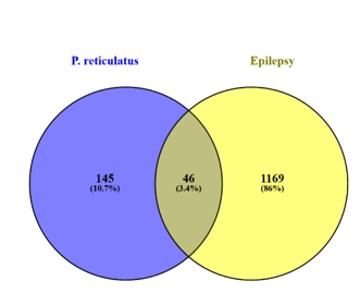
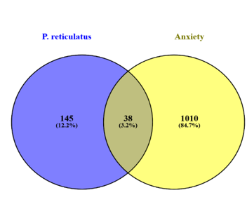
A total of 145 targets of the phytochemicals were identified by entering the canonical SMILES by giving input in the databases like SwissTargetPrediction and BindingDB. A total of 1010 targets of anxiety and 1169 targets of epilepsy were identified with the help of the DisGeNET database.
Identification of Common Targets
A total of 38 common targets for anxiety were identified between the total targets of the disease and the total targets identified for all the phytoconstituents. A total of 46 common targets for anxiety were identified between the total targets of the disease and the total targets identified for all the phytoconstituents. These common targets were identified with the help of Venny 2.1.0 online tool which helps in analysing Venn diagrams for analysing the connections between various datasets. The results are given in figure 6 and 7. and the list of common targets is given in the tables 5 and 6.
| SERPINE1 | DAO | ADORA2A | HDAC2 | TLR9 |
| GABRA1 | PTGS2 | SHBG | MMP12 | EDNRA |
| ESR2 | ESR1 | GABRA4 | P2RX7 | CSF1R |
| SRC | MAOA | OPRM1 | DYRK1A | ACHE |
| CNR2 | CDK5 | PIK3CA | CAMKK2 | GABRA5 |
| FLT4 | CES2 | SLC6A2 | INSRR | MAOB |
| GSK3B | GABRA2 | SLC6A4 | GABRA6 | |
| GSR | GABRA3 | MAPK3 | SCN9A |
Table 4: List of common targets for anxiety.
| ESR1 | HIF1A | PRKCD | ERN1 | |
| CNR2 | CDK5 | GABRA4 | PIK3CD | MAPK8 |
| INSR | GABRA2 | PIK3CG | PIK3CB | MAPK10 |
| EGFR | GABRA3 | PIK3CA | CDK1 | HSP90AA1 |
| GSK3B | ADORA1 | SLC6A4 | DYRK1A | SCN9A |
| AKT1 | ADORA2A | MTOR | CAMKK2 | DPP4 |
| BACE1 | SLC5A2 | RELA | CXCR2 | CASP3 |
| BRAF | ADK | HDAC2 | NTRK1 | CSF1R |
| PTGS2 | SHBG | GABRA5 | HSP90AB1 | GABRA6 |
| ACHE |
Table 5: List of common targets for epilepsy.
Building of the Target Network
The 97 targets identified with the help of Venny 2.1.0 tool were added to the STRING Database to obtain the PPI network. The results are given in the figures 8 and 9. The PPI network helps in understanding interactions between different proteins and their functional relationships.
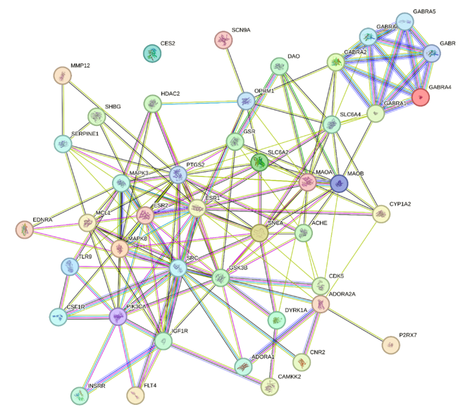
Proteins are represented by circular nodes in the PPI diagram and their interactions are represented by lines called edges. The purple edges represent interactions determined by using curated databases and pink edges represent experimentally determined protein-protein interactions. The green, red and blue edges represent predicted determined protein-protein interactions based on gene neighbourhood, gene fusions and gene co-occurrence respectively. The yellow and black edges represent protein- protein interactions based on text mining and co-expression respectively.
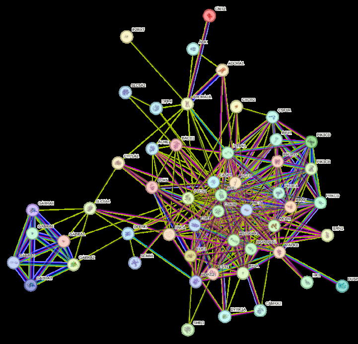
Proteins are represented by circular nodes in the PPI diagram and their interactions are represented by lines called edges. The purple edges represent interactions determined by using curated databases and pink edges represent experimentally determined protein-protein interactions. The green, red and blue edges represent predicted determined protein-protein interactions based on gene neighbourhood, gene fusions and gene co-occurrence respectively. The yellow and black edges represent protein- protein interactions based on text mining and co-expression respectively.
Topological Analysis
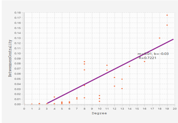
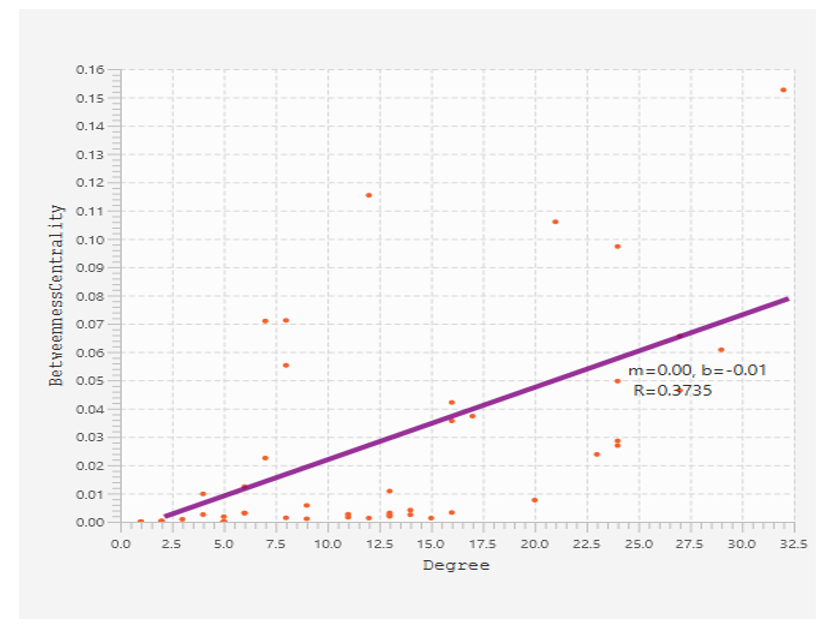
Top 10 proteins were identified from the 38 and 46 common proteins for anxiety and epilepsy through topological analysis with the help of Cytoscape software. The degree and betweenness centrality of top 10 proteins were assessed using CytoNCA plugin of cytoscape software. GABAR1 was found to have highest degree (26) and betweenness centrality (2269.54), followed by GABAR6 with degree value of 18 and betweenness centrality value of 897.89 respectively for anxiety. GABAR1 was found to have highest degree (32) and betweenness centrality (338.04), followed by PRKCD with degree value of 29 and betweenness centrality value of 258.19 respectively for epilepsy. The results are given in the table 7 and table 8 and in figures 10 and 11.
| Protein | Degree | Betweenness Centrality |
| GABRA1 | 26 | 2269.54 |
| GABRA6 | 18 | 897.89 |
| INSRR | 15 | 400.56 |
| SLC6A4 | 13 | 374.6 |
| SNCA | 13 | 265.12 |
| GABRA1 | 12 | 201.79 |
| ESR2 | 11 | 159.32 |
| MCL1 | 11 | 417.25 |
| SHBG | 8 | 598.32 |
| CDK5 | 8 | 89.12 |
Table 6: Topological analysis of common targets of anxiety using Cytoscape Software.
| Protein | Degree | Betweenness Centrality |
| GABRA1 | 32 | 338.04 |
| PRKCD | 29 | 258.19 |
| SHBG | 27 | 162.09 |
| CNR2 | 27 | 142.74 |
| SLC5A2 | 24 | 131.16 |
| ACHE | 24 | 118.07 |
| GABRA5 | 24 | 109.82 |
| HK1 | 24 | 95.04 |
| ERN1 | 23 | 94.67 |
| HDAC2 | 20 | 64.28 |
Table 7: Topological analysis of common targets of epilepsy using Cytoscape Software.
Gene Ontology (GO) Enrichment Analysis
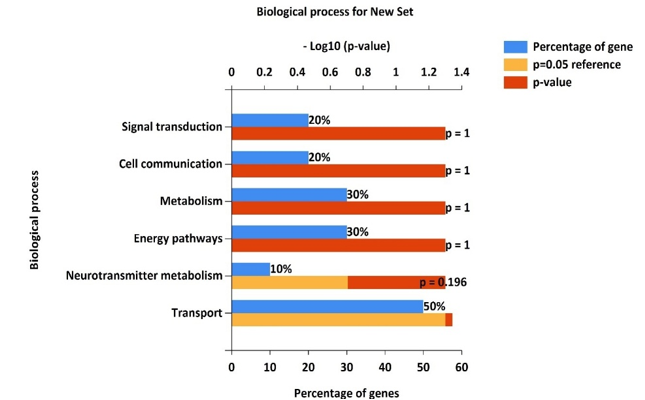
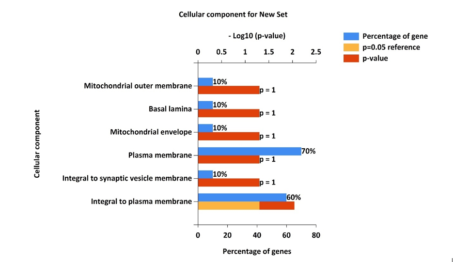
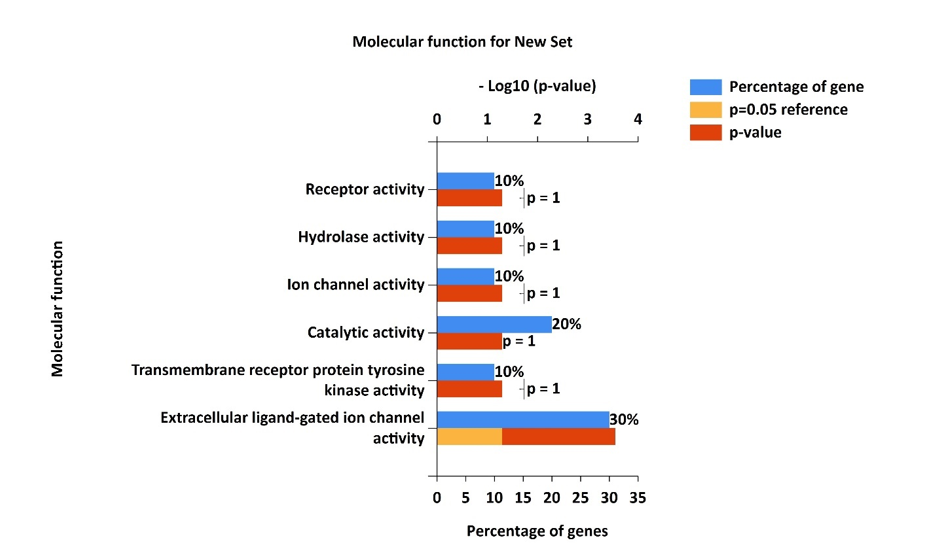
The Gene Ontology (GO) enrichment analysis was done with the help of FunRich software. The top 10 proteins that were identified were involved in a variety of cellular components (CC), biological processes (BP) and molecular functions (MF). In the case of biological processes (BP), the top proteins were involved in signal transduction, cell communication, metabolism energy pathways neurotransmitter metabolism and transport. For cellular components, the top 10 targets were found to be enriched in mitochondrial outer membrane, mitochondrial envelope, basal lamina, plasma membrane and synaptic vesicle membrane. In the case of molecular functions (MF), the targets were found to be associated with receptor activity, hydrolase activity, ion channel activity and extracellular ligand gated ion channel activity. The results are given in figures 12-14.
Pathway Analysis
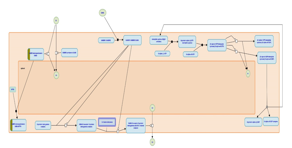
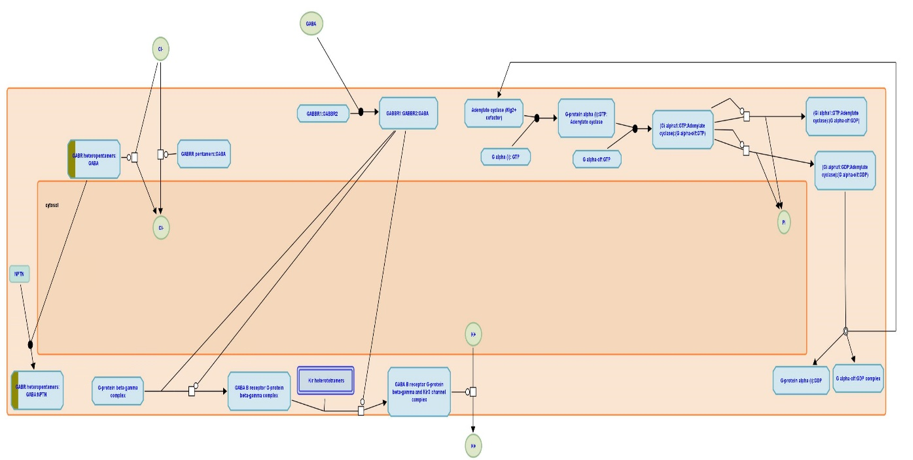
The pathway enrichment analysis was done with the help of Reactome software. The pathway analysis revealed that the top proteins were involved in activation of GABA A receptors. Gamma aminobutyric acid (GABA) receptors are the major inhibitory receptors in human synapses. They are of two types. GABA A receptors are fast-acting ligand gated chloride ion channels that mediate membrane depolarization and thus inhibit neurotransmitter release. GABA B receptors are slow acting metabotropic Gprotein coupled receptors that act via the inhibitory action of their Galpha/Go subunits on adenylate cyclase to attenuate the actions of PKA. In addition, their Gbeta/gamma subunits interact directly with N and P/Q Ca2+ channels to decrease the release of Ca2+. GABA B receptors also interact with Kir3K+ channels and increase the influx of K+, leading to cell membrane hyperpolarization and inhibition of channels such as NMDA receptors. The pathway is given in figure 15 and 16.
Molecular Docking
Molecular Docking studies were done for the top 17 phytoconstituents present in Phyllanthus reticulatus: humic acid, fulvic acid, methyl gallate, betulinic acid, botulin, pinoresinol, corilagin, syringaresinol, epifriedelanol, β-sitosterol, ellagic acid, macranthoside A, lyoniresinol, methylellagic acid, havetrichoside A, havetrichoside C and carthomoside A. The phytoconstituents that showed more than 2 violations for Lipinski’s rule were not used for further studies. The above ligands were docked against 6X3X protein Schrodinger Suite software version 13.1. The dock score for the ligand, havetrichoside C was found to better than the other ligands. The results of molecular docking are presented in the table 9 and the 2D interactions are represented in the figure 17.
| Phytoconstituent | g-score |
|---|---|
| (Kcal/mol) | |
| Havetrichoside C | -8.54772 |
| Havetrichoside A | -7.06173 |
| Methyl gallate | -5.21619 |
| Carthomisde A1 | -4.95656 |
| Ellagic acid | -4.69665 |
| Fulvic acid | -4.55037 |
| Pinoresinol | -4.50028 |
| Methyl ellagic acid | -4.27449 |
| Syringaresinol | -3.54985 |
| Betullinic acid | -2.93162 |
| Betulin | -2.43139 |
| Humic acid | -2.12076 |
| Corilagin | -1.60855 |
| Epifriedelanol | -1.19869 |
| B-Sitosterol | -0.77514 |
| Macronthoside A | -0.68591 |
| Lyoniresinol | -0.54896 |
| Diazepam | -7.57729 |
Table 8: Dock score of the selected phytoconstituents.
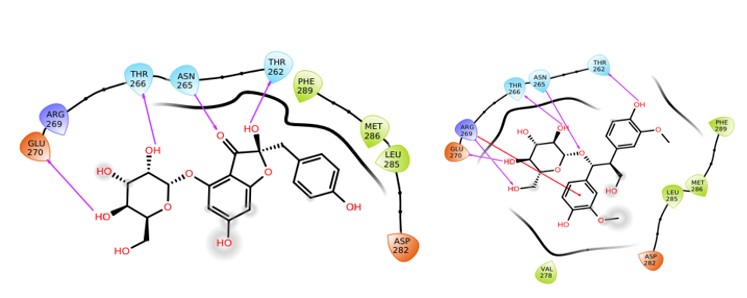
Havetrichoside C Havetrichoside A
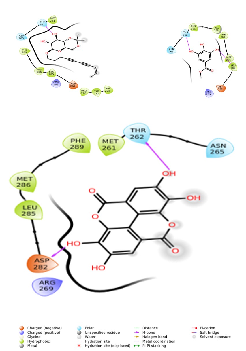
| Carthomoside A | Methyl Gallate |
Ellagic Acid
MM/GBSA
The MM/GBSA studies were performed for all the protein- ligand complexes using the Schrodinger software version 13.1. The results showed havetrichoside C to be better than other ligands. The MM/GBSA result of havetrichoside C against proteins 6X3X was found to be –56.19 kcal/mol compared to the standard (diazepam) value of -48.52 kcal/ mol. The MM/GBSA results are presented in the table 10.
| Phytoconstituent | g-score | |
|---|---|---|
| (Kcal/mol) | ||
| Havetrichoside C | -56.19244526 | |
| Havetrichoside A | -51.02232839 | |
| Methyl gallate | -37.24929666 | |
| Carthomisde A1 | -48.33516714 | |
| Ellagic acid | -54.5902698 | |
| Fulvic acid | -35.2281953 | |
| Pinoresinol | -41.84076116 | |
| Methyl ellagic acid | -54.82350247 | |
| Syringaresinol | -36.16180423 | |
| Betullinic acid | -42.52459809 | |
| Betulin | -36.01424758 | |
| Humic acid | -33.41592773 | |
| Corilagin | -19.97457074 | |
| Epifriedelanol | -40.78280735 | |
| Β-Sitosterol | -9.755844956 | |
| Macronthoside A | -28.45980336 | |
| Lyoniresinol | -35.03162511 | |
| Diazepam | -48.52823893 | |
Table 10: Free energy score of the selected phytoconstituents.
Molecular Dynamics
Ligand- Protein complex of havetrichoside C with 6X3X were studied using Desmond Schrodinger Suite software version 13.1. A constant-temperature, constant-pressure ensemble (NPT) with an atmospheric pressure was set to last for 100 ns at 310 K and 1.013 bar to run the MD simulation. OPLS4 force field was used to develop the complexes. The RMSD of the havetrichoside C- 6X3X complex had converged approximately around 15ns and thereafter there is no shift in the RMSD value. The RMSD results are represented in figure 18.
The RMSF values fluctuate along the protein sequence, indicating variations in the flexibility or mobility of different residues. Regions with higher RMSF values suggest greater flexibility, while regions with lower RMSF values indicate more rigid or stable conformations. Peaks in the RMSF curve represent residues with significant fluctuations, likely indicating regions of increased flexibility or structural dynamics. Valleys in the RMSF curve correspond to residues with relatively lower fluctuations, suggesting regions of stability or constrained motion. The amino acids from 250 to 300 are involved in formation of stable interactions between the protein and the ligands. The RMSF results are represented in figure19.
Different types of interactions are shown between the protein and ligand. The important interactions are: VAL-258: Shows a significant fraction of Hydrophobic interactions. MET-261: Exhibits a high proportion of Hydrophobic and H-bond interactions.THR-262: Predominantly involved in Hydrophobic interactions with a moderate fraction of H-bonds. ASN-265: Primarily engaged in Hydrophobic and Ionic interactions. THR-266: Appears to participate in Hydrophobic, Ionic, and Water bridge interactions. ARG-269: Demonstrates a considerable fraction of Ionic interactions. GLU-270: Involved in Ionic and Hydrophobic interactions. ASP-282: Shows Hydrophobic and Ionic interactions. LEU- 285: Engaged mainly in Hydrophobic interactions. MET- 286: Indicates Hydrophobic interactions. PHE-289: Shows a fraction of Hydrophobic interactions. The protein-ligand contacts are represented in figure 20.
The ligand molecule has various functional groups, such as hydroxyl (OH) and carbonyl (C=O) groups, which can form interactions with specific amino acid residues of the protein. Aminoacid residue, ASN 265 maintains a stable interaction with the ligand for 66 % of the simulation time. Thr 266 and Thr 262 maintain stable interactions for 42 % and 35 % of the simulation time. The results are represented in figure 21.
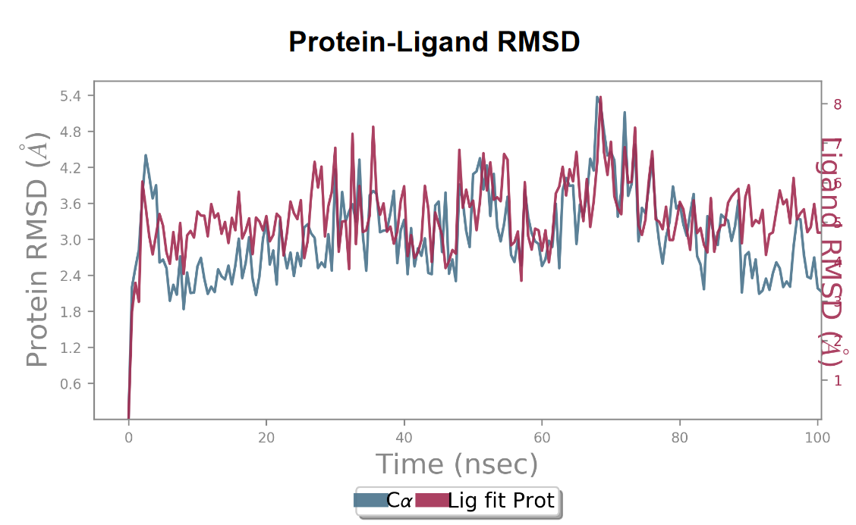
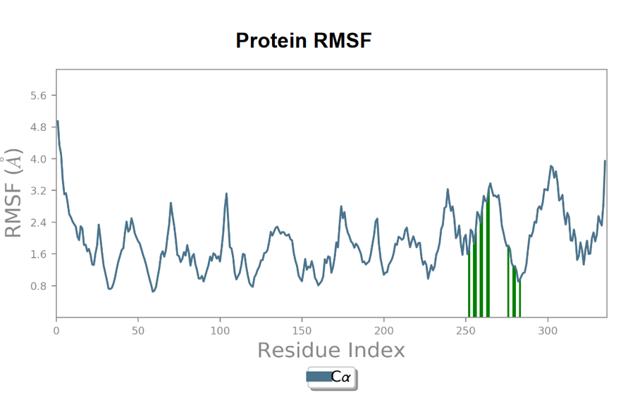
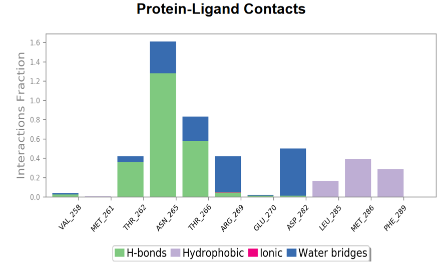
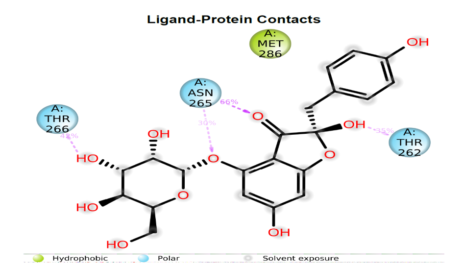
Discussion
The aim of the current study is to evaluate the anti- anxiety and anti-epileptic activity of Phyllanthus reticulatus through network pharmacology and molecular modelling approach.
Network pharmacology is a distinctive in-silico approach that targets multiple proteins involved in the pathogenesis of a disease instead of a single protein. It has enabled a paradigm shift from the classical existing ‘one disease–one target–one drug’ dogma to the current ‘multicomponent, multi-target’ approach [53, 54]. It aims to comprehend the ways in which biological networks are affected by pharmaceuticals and how these networks react to these perturbations. It has become a vital technique in the search for new drugs and can be applied to discover novel therapeutic targets, forecast drug effectiveness and toxicity, and enhance drug combinations. Network pharmacology, as opposed to classical pharmacology, focuses on the network level interactions between drugs and biological systems to provide an understanding of the mechanism of action of drug.
Phyllanthus reticulatus, a medicinal plant, exhibits a plethora of pharmacological activities. Its petroleum ether and ethanolic leaf extracts demonstrated significant antidiabetic effects in alloxan-treated mice, particularly at 1000 mg/kg dose [55]. Moreover, the leaves showed potent in vitro antiplasmodial activity against chloroquine-sensitive and chloroquine-resistant strains of Plasmodium falciparum [56]. Additionally, the aqueous extract of plant’s aerial parts exhibited hypocholesterolemic activity by reducing cholesterol levels and improving HDL cholesterol [57]. Its methanolic stem bark extract displayed antimicrobial and cytotoxic properties [58], while the ethanolic extract of aerial parts showed hepatoprotective activity against CCl4- induced liver damage in rats [59]. Furthermore, the leaf extracts exhibited significant antibacterial [60], antinociceptive, anti-hyperglycemic [61], analgesic, and anti-inflammatory activities [62]. The entire plant’s methanolic and ethanolic extracts also showed potent antioxidant activity [63]. Finally, two fractions of the ethanolic extract displayed anti-hepatitis B viral activity, suggesting the plant’s potential in treating various diseases [64].
The literature survey and data mining revealed the presence of 25 active phytoconstituents in Phyllanthus reticulatus. The 25 phytoconstituents are: humic acid, fulvic acid, methyl gallate, betulinic acid, botulin, pinoresinol, corilagin, syringaresinol, epifriedelanol, β-sitosterol, ellagic acid, macranthoside A, lyoniresinol, methylellagic acid, havetrichoside A, havetrichoside C, carthomoside A, Octacosanol, Tricosanol, Friedelin, Glochidonol, Pentacosane, Sorghumol, Taraxerol and Hydroxyfriedelanone [65].
The phytoconstituents of Phyllanthus reticulatus were subjected to prediction of pharmacokinetic properties like drug-likeliness, blood-brain barrier penetration, partition coefficient etc. Among the 25 phytoconstituents of Phyllanthus reticulatus, 17 compounds exhibited favorable pharmacokinetic properties. These 17 phytoconstituents were used for further molecular docking, MM GBSA and molecular dynamics study to identify the best phytoconstituents that could be effective in the management of epilepsy and anxiety. The 17 phytoconstituents that exhibited favorable pharmacokinetic properties are: humic acid, fulvic acid, methyl gallate, betulinic acid, botulin, pinoresinol, corilagin, syringaresinol, epifriedelanol, β-sitosterol, ellagic acid, macranthoside A, lyoniresinol, methylellagic acid, havetrichoside A, havetrichoside C and carthomoside A.
A total of 145 targets of the phytochemicals were identified by entering the canonical SMILES by giving input in the databases like SwissTargetPrediction and BindingDB. A total of 1010 targets of anxiety and 1169 targets of epilepsy were identified with the help of the DisGeNET database. A total of 38 common targets for anxiety were identified between the total targets of the disease and the total targets identified for all the phytoconstituents. A total of 46 common targets for anxiety were identified between the total targets of the disease and the total targets identified for all the phytoconstituents. The most important therapeutic targets in epilepsy and anxiety were identified with the help of topological parameters like degree centrality and betweenness centrality. Degree centrality measures the number of connections a node (protein/target) makes with other nodes in a network. Betweenness centrality determines the influence of a node (protein/target) in controlling the interaction between a pair of nodes (protein/target) passing through this node in the network. Closeness centrality measures the inverse of average distance of a node from all other nodes in a network. It is widely accepted that nodes with higher degree, betweenness and closeness centrality values may represent important targets and play a crucial role in a biological network [66, 67]. Topological analysis revealed that GABAA receptor to be the most important target involved in the pathogenesis of anxiety and epilepsy.
The pathway enrichment analysis was done with the help of Reactome software. The pathway analysis revealed that the top proteins were involved in activation of GABA A receptors. Gamma aminobutyric acid (GABA) receptors are the major inhibitory receptors in human synapses [68, 69]. They are of two types. GABA A receptors are fast-acting ligand gated chloride ion channels that mediate membrane depolarization and thus inhibit neurotransmitter release. GABA B receptors are slow acting metabotropic Gprotein coupled receptors that act via the inhibitory action of their Galpha/Go subunits on adenylate cyclase to attenuate the actions of PKA. In addition, their Gbeta/gamma subunits interact directly with N and P/Q Ca2+ channels to decrease the release of Ca2+ [70, 71]. GABA B receptors also interact with Kir3K+ channels and increase the influx of K+, leading to cell membrane hyperpolarization and inhibition of channels such as NMDA receptors [72, 73]. The molecular docking, MMGBSA and molecular dynamics studies showed that havetrichoside c exhibits better binding affinity and stability with GABA receptor compared to other phytoconstituents present in Phyllanthus reticulatus. Havetrichoside C, a naturally occurring compound that exhibits a range of bioactive properties, making it a subject of interest in drug discovery and development. Studies have shown that havetrichoside C possesses potent antioxidant properties, which can help combat oxidative stress-induced damage in cells and tissues. Additionally, it has been found to have anti-inflammatory effects, potentially offering therapeutic benefits in various inflammatory conditions. Furthermore, havetrichoside C has demonstrated promising anticancer activity in preclinical studies. Research suggests that it may inhibit the growth of cancer cells and induce apoptosis, making it a potential candidate for cancer treatment. Moreover, this compound has been investigated for its neuroprotective effects, showing potential in protecting neurons from damage and degeneration. Such properties could have implications in the treatment of neurodegenerative diseases like Alzheimer’s and Parkinson’s.
Xian-wen Ye, et al. conducted a study to analyze the pharmacologic mechanisms of Coptidis rhizoma for the treatment of Alzheimer’s disease using network pharmacology approach. They found STAT3 to be a major target of Coptidis rhizoma and concluded that inhibition of STAT3 would be a promising treatment for Alzheimer’s disease. A study conducted by Yangwen Luo et al. looked into the therapeutic potential of Sceletium tortuosum for the treatment of neurodegenerative disorders, including Alzheimer’s disease. The findings revealed that MAOB might be a target of Sceletium tortuosum and that its inhibition might raise the levels of dopamine and other monoamines in the brain, potentially enhancing cognitive function in Alzheimer’s disease. Jarrel J, et al. employed network pharmacology approach to look at the molecular causes of Alzheimer’s disease and to find prospective therapeutic targets in TCM. The findings revealed that HDAC2 could be a possible target and that blocking it could halt neuronal degeneration and cognitive impairment [73].
In diseases with multifactorial origins like epilepsy and anxiety, combination therapy has been proven to be more efficacious than monotherapy. The current study has identified that havetrichoside c could be effective in prevention and treatment of epilepsy and anxiety through activation of GABA receptors involved in the pathogenesis of epilepsy and anxiety.
Conclusion
The results of the current study reveal that phytoconstituents can serve as excellent candidates in the management of epilepsy and anxiety by modifying the functions of GABA receptors. Further in vivo studies using animal models are required to confirm the therapeutic potential of the havetrichoside c in the prevention and treatment of epilepsy and anxiety.
References
-
WHO (2014) Epilepsy.
-
MedicineNet, Seizures: Symptoms and Types
-
Neeru, Kashyap S, Sharma N (2016) Study of antiepileptic activity of Roylea elegans wall (aerial parts). World Journal of Pharmacy and Pharmaceutical Sciences 5(7): 439-453.
-
Shodh Ganga: a reservoir of Indian theses
-
Tripathi KD, Essentials of medical pharmacology, Jaypee brothers medical publishers(p) ltd, 6th (edn), 401-402
-
Epilepsy Foundation
-
Loscher W (2011) Critical review of current animal models of seizures and epilepsy used in the discovery and development of new antiepileptic drugs. EJOE 20(5): 359-368
-
Gupta B (2012) Medicinal plants as agent of anticonvulsant activity. Int J Res Dev Pharm L Sci 1(3): 126-134
-
Goyal RK (2007) Dessari and Gandhi’s elements of pharmacology. 17th(Edn.), BS Shah Prakashan, pp: 288- 289.
-
Kumar KS, Kota K, Jarinabanu T, Tahashildar J, Ragavendhra P, et al. (2015) Antiepileptic activity of ethanolic extract of Biophytum sensitivum (L.) DC. In Animal models. Int J Curr Res Aca Rev 3(7): 23-30 .
-
Rang HP, Dale MM, Ritter JM, Flower RJ, Rang and Dale’s pharmacology, Elsevier publications, 7th (edn) 540-543
-
Epilepsy in the WHO Eastern Mediterranean Region
-
Medical News Today
-
https://www.med.unc.edu/neurology/files/child.pdf/ Epilepsy%20BASICORE.pdf
-
Harvey RA, Champe P (2009) Lipincott’s illustrated review: Pharmacology. 4th (Edn.), Lippincott Williams, Philadelphia, USA, pp: 564.
-
Avanthi E, Kumar P, Lokesh BN, Guruprasad Y. (2016); The study of antiepileptic activity of clove oil by MES model in mice. Indian Journal of Pharmacy and Pharmacology 3(3): 103-107
-
Nirmala D (2015) Studies on anticonvulsant activity of Annacyclus pyrethrum in albino mice. Asian J Pharm Clin Res 8(4): 178-187
-
Pitchaiah G, Anusha VL, Hemalatha CH, Anil Kumar Y, Sravani K, et al. (2015) Anxiolytic and anticonvulsant activity of methanolic extract of Allium cepa Linn (Onion) bulbs in Swiss albino mice. Journal of Pharmacognosy and Phytochemistry 4(2): 131-134
-
Santilna KS, Mahesh NM, Suresh J (2014) Anticonvulsant Activity Study of Artemisia nilagirica. IJPPR 6(4): 826- 830
-
Sutar RC, Kasture SB, Kalaichelvan VK (2014) Evaluation of anticonvulsant activity of leaf extracts of Holoptelea integrifolia (roxb) planch in experimental animals. Int J Pharm Pharm Sci 6(4): 308-311.
-
Pathan D, Ambavade S (2014) Anticonvulsant activity ethanolic extract of Picrorhiza kurroa. Pharmacophore 5(1): 141-146
-
Varma GG, Mathai BK, Das K, Gowda G, Rammohan S, et al (2014) Evaluation of antiepileptic activity of methanolic leaves extract of Tragia involucrata Linn. in mice. International Letters of Natural Sciences 17(1): 167-179.
-
Modaresi M, Pouriyanzadeh A, Asadi-Saman M (2014) Antiepileptic activity of hydroalcoholic extract of basil in mice. J HerbMed Pharmacol 3(1): 57-60
-
Chinchawade AB, Deshmukh DB, Gaikwad DD, Grampurohit ND (2013). Anticonvulsant Activity of Chloroform Extract of Bark & Root of Erythrina variegata L. IJPCR 5(1): 23-25.
-
Abubakar K, Ugwah-Oguejiofor CJ, Usman MN, Abubakar SB, Abdulkadir R et al. (2013). Evaluation of the Anticonvulsant Effect of the Methanol Extract of Evolvulus alsinoides in Mice. Sch. Acad. J. Pharm 2(6): 436-441.
-
Anovadiya AP, Sanmukhan JJ, Vadgama VK, Tripath CB (2013) Evaluation of antiepileptic and memory retention activity of Curcumin perse and in combination with antiepileptic drugs. Asian J Pharm Clin Res 6(2): 145-148.
-
Singh P, Garg VK, Sharma PK, Gupta s (2012) Antiepileptic activity of aqueous extract of Tricosanthes dioica Roxb. Asian J Plant Sci Res 2 (1): 45-47.
-
Saroch V, Hiremath RS, Patil PA (2012) Anticonvulsant Activity of Apasmarari rasa – An Experimental Study, International Journal of Ayurvedic Medicine 3(1): 48- 52.
-
Garg VK, Paliwal SK (2011) Anticonvulsant activity of ethanolic extract of Cynodon dactylon. Der Pharmacia Sinica 2 (2): 86-90.
-
Manikkoth S, Deepa B, Joy AE, Rao SN (2011) Anticonvulsant activity of Phyllanthus amarus in experimental animal models. International Journal of Applied Biology and Pharmaceutical Technology 2(4): 144-147.
-
Babu HB, Mohana LS, Saravana KA (2010) Studies on phytochemical and anticonvulsant property of Martyniya annua linn. International Journal of Phytopharmacology 1(2): 82-86.
-
Vyawahare NS, Bodhankar SL (2009) Anticonvulsant Activity of Argyreia speciosa in Mice. Indian Journal of Pharmaceutical Sciences 71(2): 131-134.
-
Hegde K, Thakker SP, Joshi AB, Shastry CS, Chandrashekhar KS (2009) Anticonvulsant activity of Carissa carandas Linn. Root Extract in Experimental Mice. Trop J Pharm Res 8 (2): 117-125.
-
Buddhadev SG, Buddhadev SS (2017) A complete review on ativisha –Aconitum heterophyllum. An International Journal of Pharmaceutical Sciences 8(1): 111-114.
-
Pahal S, Gupta A, Choudhary P, Chaudhary A, Singh S et al. (2021) Network pharmacological evaluation of Withania somnifera bioactive phytochemicals for identifying novel potential inhibitors against neurodegenerative disorder. J Biomol Struct Dyn. 40(21): 10887-10898.
-
Thirumal Kumar D, Shree Devi MS, Udhaya Kumar S, Sherlin A, Mathew A, et al. (2022) Understanding the activating mechanism of the immune system against COVID-19 by Traditional Indian Medicine: Network pharmacology approach. In: Advances in Protein Chemistry and Structural Biology. Academic Press Inc. 275-379.
-
Fatriansyah JF, Rizqillah RK, Yandi MY, Fadilah, Sahlan M (2022) Molecular docking and dynamics studies on propolis sulabiroin-A as a potential inhibitor of SARS- CoV-2. J King Saud Univ Sci 1: 34(1).
-
Yang Y, He Y, Wei X, Wan H, Ding Z, et al. (2022) Network Pharmacology and Molecular Docking-Based Mechanism Study to Reveal the Protective Effect of Salvianolic Acid C in a Rat Model of Ischemic Stroke. Front Pharmacol 27: 12
-
Wang K, Wang Y, Yan J, Hou C, Zhong X, et al. (2022) Network Pharmacology and Molecular Docking Integrated Strategy to the Screening of Active Components and Mechanisms of Stephaniae Tetrandrae Radix on Breast Cancer. Processes 10(11).
-
Zhang M, Yang J, Zhao X, Zhao Y, Zhu S, et al. (2021) Network pharmacology and molecular docking study on the active ingredients of qidengmingmu capsule for the treatment of diabetic retinopathy. Sci Rep 11(1).
-
Li H, Guo L, Ding X, An Q, Wang L et al. (2022) Molecular Networking, Network Pharmacology, and Molecular Docking Approaches Employed to Investigate the Changes in Ephedrae Herba before and after Honey- Processing. Molecules 27(13).
-
Hasan MM, Khan Z, Chowdhury MS, Khan MA, Moni MA, et al. (2022) In silico molecular docking and ADME/T analysis of Quercetin compound with its evaluation of broad-spectrum therapeutic potential against particular diseases. Inform Med Unlocked 1: 29.
-
Rajagopal K, Varakumar P, Aparna B, Byran G, Jupudi S, et al. (2021) Identification of some novel oxazine substituted 9-anilinoacridines as SARS-CoV-2 inhibitors for COVID-19 by molecular docking, free energy calculation and molecular dynamics studies. J Biomol Struct Dyn 39(15): 5551-5562.
-
Bakal RL, Jawarkar RD, Manwar JV, Jaiswal MS, Ghosh A, et al. (`2022) Identification of potent aldose reductase inhibitors as antidiabetic (Anti-hyperglycemic) agents using QSAR based virtual Screening, molecular Docking, MD simulation and MMGBSA approaches: Identification of potent aldose reductase inhibitors as antidiabetic (Anti-hyperglycemic) agents. Saudi Pharmaceutical Journal 30(6): 693-710.
-
Thangavel N, Albratty M (2023) Benchmarked molecular docking integrated molecular dynamics stability analysis for prediction of SARS-CoV-2 papain- like protease inhibition by olive secoiridoids. J King Saud Univ Sci 35(1).
-
Jupudi S, Rajagopal K, Murugesan S, Kumar BK, Raman K, et al. (2022) Identification of Papain-Like Protease inhibitors of SARS CoV-2 through HTVS, Molecular docking, MMGBSA and Molecular dynamics approach. South African Journal of Botany 151: 82-91.
-
He F, Wang X, Wu Q, Liu S, Cao Y, et al. (2023) Identification of potential ATP-competitive cyclin-dependent kinase 1 inhibitors: De novo drug generation, molecular docking, and molecular dynamics simulation. Comput Biol Med 155
-
Fatriansyah JF, Rizqillah RK, Yandi MY, Fadilah, Sahlan M (2022) Molecular docking and dynamics studies on propolis sulabiroin-A as a potential inhibitor of SARS- CoV-2. J King Saud Univ Sci 34(1).
-
Hasan MM, Khan Z, Chowdhury MS, Khan MA, Moni MA, et al. (2022) In silico molecular docking and ADME/T analysis of Quercetin compound with its evaluation of broad-spectrum therapeutic potential against particular diseases. Inform Med Unlocked. 29.
-
He F, Wang X, Wu Q, Liu S, Cao Y, et al. (2023) Identification of potential ATP-competitive cyclin-dependent kinase 1 inhibitors: De novo drug generation, molecular docking, and molecular dynamics simulation. Comput Biol Med 155.
-
Obaidullah AJ, Alanazi MM, Alsaif NA, Alanazi AS, Albassam H, et al. (2022) Network Pharmacology- and Molecular Docking-Based Identification of Potential Phytocompounds from Argyreia capitiformis in the Treatment of Inflammation. Evidence-based Complementary and Alternative Medicine 2022.
-
Jupudi S, Rajagopal K, Murugesan S, Kumar BK, Raman K, et al. (2022) Identification of Papain-Like Protease inhibitors of SARS CoV-2 through HTVS, Molecular docking, MMGBSA and Molecular dynamics approach. South African Journal of Botany 151: 82-91.
-
Chandran U, Mehendale N, Patil S, Chaguturu R, Patwardhan B et al. (2017) Network Pharmacology. In: Innovative Approaches in Drug Discovery: Ethnopharmacology, Systems Biology and Holistic Targeting. Elsevier Inc. p. 127-64.
-
Kumar S, Kumar D, Deshmukh RR, Lokhande PD, More SN, et al. (2008) Antidiabetic potential of Phyllanthus reticulatus in alloxan-induced diabetic mice. Fitoterapia 79 (1): 21-23.
-
Omulokoli E, Khan B, Chhabra SC (1997) Antiplasmodial activity of four Kenyan medicinal plants. Journal of Ethnopharmacology 56 (2): 133-7.
-
Maruthappan V, Sakthi KS (2010) Effects of Phyllanthus reticulatus on lipid profile and oxidative stress in hypercholesterolemic albino rats. Indian Journal of Pharmacology 42 (6): 388-391.
-
Begum T, Rahman SM, Rashid AM (2006) Phytochemical and Biological Investigations of Phyllanthus reticulatus. Dhaka University Journal of Pharmaceutical Sciences 5 (1-2): 21-23.
-
Das KB, Bepary S, Datta KB, Chowdhury Azad AAK, Ali, et al. (2008) Hepatoprotective activity of Phyllanthus reticulatus. Pakistan Journal of Pharmaceutical Sciences 21(4): 333-337.
-
Shruthi SD, Ramachandra YL, Padmalatha RS, Shettty VA (2010) Antibacterial potential of leaf extracts from Kirganelia reticulate Baill. International Journal of Pharmaceutical Research and Development 2 (6): 0974-9446.
-
Rahmatullah M, Ghosh KC, Almamun A, Hossain MT, Ahmed SA, et al. (2010) pharmacological study on Antinociceptive and Anti-hyperglycemic effects of methanol extract of leaves of Phyllanthus reticulates Poir. In swiss albino mice. Advances in Natural and Applied Sciences 4 (3): 229-232.
-
Saha A, Masud AM, Bachar CS, Kundu KJ, Datta KB, et al. (2007) The analgesic and anti-inflammatory activities of extracts of Phyllanthus reticulatus in mice model. Pharmaceutical Biology 45 (5): 335-359.
-
Maruthappan V, Shree KS (2010) A report on the antioxidant activity of the powder of the entire plant of Phyllanthus reticulatus Poir. Indian Journal of Pharmacology 4(4): 265-269.
-
Das BP, Mohammad S, Pavel AM, Bhattacharjee R, Das B, et al. (2011) Anti-hepatitis B viral activity of Phyllanthus reticulatus. Bangladesh Pharmaceutical Journal 14 (1): 11-14.
-
Saleh-E-In MM, Kar P, Roy A, Kowalski R (2022) Phytochemical account: Phyllanthus reticulatus Poir. J Phytomol Pharmacol 1(1): 19-29.
-
Hopkins AL (2008) Network pharmacology: The next paradigm in drug discovery. Nature Chemical Biology. Nature Publishing Group 4: 682-690.
-
Chandran U, Mehendale N, Patil S, Chaguturu R, Patwardhan B et al. (2017) Network Pharmacology. In: Innovative Approaches in Drug Discovery: Ethnopharmacology, Systems Biology and Holistic Targeting. Elsevier Inc 127-164.
-
Olsen RW, Tobin AJ (1990) Molecular biology of GABAA receptors. The FASEB Journal 4(5): 1469-1480.
-
Mehta AK, Ticku MK (1999) An update on GABAA receptors. Brain research reviews 29(2-3): 196-217.
-
Macdonald RL, Olsen RW (1994) GABAA receptor channels. Annual review of neuroscience 17(1): 569- 602.
-
Olsen RW, Sieghart W (2009) GABAA receptors: subtypes provide diversity of function and pharmacology. Neuropharmacology 56(1): 141-148.
-
Mody I, Pearce RA (2004) Diversity of inhibitory neurotransmission through GABAA receptors. Trends in neurosciences 27(9): 569-575.
-
Lobo IA, Harris RA (2008) GABAA receptors and alcohol. Pharmacology Biochemistry and Behavior 90(1): 91-94
- Gallic and Citric Acid Present in the Peels of Tropical Fruits as an Alternative in the Fight against Cancer
- Treating the Forehead Lines with Combination of Forehead and Glabellar Botulinum Toxin Among Japanese Patients
- Clinical Evaluation of Patients Suffering from Breast Cancer & Determination of Treatment Therapies and Better Strategies Related to Breast Cancer
- Medieval Recipes by Al-Zahrāwī for Heart Palpitations Treatment
- Etiology and Prescription Errors of Myocardial Infarction in Different Health Care Systems of Azad Kashmir
- Early Diagnosis and Multidisciplinary Management of Turner Syndrome: A Paediatric Case Study