Effects of Antipronation Taping on Single-Limb Stance Static Balance in Subjects with Pronated Foot-an Experimental Study
Aim of the Study: To investigate the effect of antipronation taping on subjects with pronated foot during single limb stance. Background: Abnormal foot pronation causes passive instability of the joints of foot. The pronated foot, therefore, is unstable during weight bearing. The effect of antipronation taping on pronated foot during single limb stance lacks evidence. Study Design: Single-group repeated measures design. Methodology: Ten subjects (3 men, 7 women) with pronated foot (navicular drop = 13.0 ± 3.7mm) participated in this study conducted at the Mary Varghese Institute of Rehabilitation, Christian Medical College, Vellore. The subjects were asked to stand in unilateral stance on the dominant leg on the force platform of the balance master for 10 seconds. The degree of sway in the anteroposterior (AP) axis, the transverse axis and the velocity of moment (mm2/s) were calculated before and after application of antipronation taping. A mean of three recordings was taken for analysis. Results: There was a significant reduction in the scores of velocity of moment, anteroposterior sway and mediolateral sway after taping of the pronated foot. (P-value = 0.005) Conclusion: The study shows that antipronation taping improves single limb stance balance in subjects with pronated foot.
Introduction
The foot is the most distal segment in the lower extremity and represents a relatively small base of Ann Physiother Occup Ther
support upon which the body maintains balance (particularly in single-leg stance).Excessively supinated or pronated foot postures influence peripheral (somatosensory) input via changes in joint mobility or surface contact area or, secondarily; through changes in muscular strategies to maintain a stable base of support [1, 2].
Although three distinct arches function to support the foot, the medial longitudinal arch (MLA) has been found to be the arch of clinical significance [3, 4, 5, 6]. The pronated (flat) foot is associated with excessive subtalar joint pronation, which stretches the spring ligament and the tendon of the Tibialis posterior muscle resulting in the loss of the MLA [7].
Many people with pes planus demonstrate a gait with no toe-off, often associated with a large plantar weight- bearing surface [8, 9]. Symptoms of pronated foot include, shortening of the everter muscles of the foot (the Peroneal muscles), tenderness of the plantar fascia and laxity of the supporting structures of the medial side of the foot (the medial ligament or deltoid ligament and the tibialis posterior tendon) [10]. Over time, this functional deformity will develop into a chronic structural deformity, and abnormal stresses will be transferred to the more proximal areas, affecting the knees, hips, and low back [11, 12, 13, 14, 15].
Treatment for pes planus revolves around reducing the stresses that caused the problem and muscle strengthening program to strengthen the anterior and posterior tibialis and intrinsic foot muscles. Other treatments include arch taping or support, ultrasound to heal damaged tissues, stretching of tight muscle groups, and orthotic devices [16, 17]. Several studies have shown that anti-pronation taping improves arch height and controls pronation during both static and dynamic activity [18, 19, 20, 21, 22, 23, 24, 25].
However its effect on single limb stance has not been established. Therefore this study aims to find out the effect of antipronation taping on single limb stance of subjects with pronated foot.
Subjects and Method
Participants
Ten subjects (3 men, 7 women), with normal Body Mass Index (18.5-24.9 kg/m2), of age group between 18 and 40 years, who had pronated foot (navicular drop =
13.0 ± 3.7mm), were included in the study after obtaining a written consent. This Single-group repeated measures experimental study was conducted at the Mary Verghese Institute of Rehabilitation, Christian Medical College, Vellore. Each subject who was recruited was asked to come for a single study visit and the total duration for the study was 6 months. Subjects with any pre-existing balance disorders, musculoskeletal problems (previous history of ankle and knee injuries, or previous lower limb fractures), neurological problems, history of dizziness and history of alcohol and drug abuse were excluded from the study.
Procedure
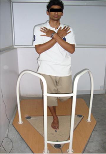
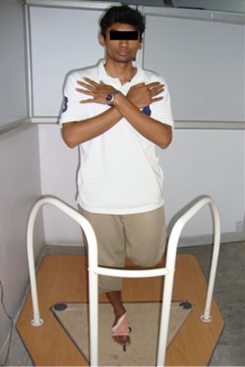
The postural sway was determined by the Balance Measuring and Training Equipment (BMTE) (Metitur Oy, Jyvaskyla, Finland). The subjects were made to stand bare foot on the force platform of the balance master with arms crossed across their chest and eyes open (Figure 1). The centre of pressure was displayed on the screen. The test limb was maintained in full extension, with toes towards the anterior direction of the force plates and the non-test limb positioned to 90o of knee flexion. Subject was asked to perform 10 second single limb stance as motionless as possible. A practice trial was done to make the subject comfortable with the procedure. Three test trials of 10 seconds each were done with a gap of 5 to 10 seconds between each test trial. The antipronation taping was then applied and the same procedure repeated again (Figure 2). The velocity of moment, anteroposterior sway and mediolateral sway were measured before and after taping and the mean of the three test trials were taken for analysis.
A change in the distribution of pressure reflects the amount of sway from front to back, and from side to side. The dispersion index is a measure of the variation of pressure about the mean centre of pressure over the duration of the session. A large dispersion index indicates a great amount of sway, whereas a small value is indicative of a limited sway.
The Application of Antipronation Taping
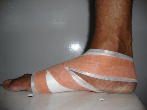
The subject was placed in long sitting with the lower leg supported on a table and the foot extending past the table. Using 5cm hypoallergenic tape, two anchors were applied around the metatarsal heads, overlapping by two- thirds.
The 3.8cm rigid tape was then applied beginning at the dorsal aspect of the forefoot encircling the posterior aspect of the calcaneum. The tape was then applied obliquely across the lateral aspect of the calcaneum and the plantar aspect of the foot, proceeding towards the medial longitudinal arch, gently lifting it up before attaching it again to the distal and dorsal aspect of the first ray. Another strip was applied overlapping the previous one by two-thirds. Finally another anchor was applied over the distal half of the first metatarsal head using a hypoallergenic tape followed by a rigid tape Figure 3 [29, 30, 31, 32, 33, 34]. Time duration taken for this taping technique was approximately 5-10 minutes.
Results
The study was a single - group repeated measures design, to investigate the effect of antipronation taping on subjects with pronated foot during single limb stance. Ten subjects, with pronated foot participated in the study after written consent. The degree of sway in the AP axis, the transverse axis and the velocity of moment (mm2/s) were measured before and after taping during the single limb stance on the balance master. A mean of three recordings was taken for analysis.
| Variables | Participants | ||||
|---|---|---|---|---|---|
| Gender | |||||
| Male | 3 (30) | ||||
| Female | 7(70) | ||||
| Navicular drop | 13.0 ± 3.7 mm |
Table 1: Descriptive statistics of Participant Characteristics. (Proportion of gender, Mean ± S.D. of Navicular drop).
The data was analysed using the Wilcoxon Signed Rank Test, also known as the Wilcoxon Matched Pairs Test, which is a non-parametric test used to test the median difference in paired data. This test is the non- parametric equivalent of the paired t-test. The pre and post-test for velocity of moment, anterposterior sway and mediolateral sway are significant at 0.05 level.
| N | Minimum | Maximum | Mean | Std. Deviation | P value | |||||||||||||||
|---|---|---|---|---|---|---|---|---|---|---|---|---|---|---|---|---|---|---|---|---|
| Velocity of moment_pre | 10 | 49.5 | 74.9 | 60.32 | 9.0391 | |||||||||||||||
| 0.005 | ||||||||||||||||||||
| Velocity of moment_ post | 10 | 23.1 | 59.9 | 34.84 | 12.9721 | |||||||||||||||
| anteroposteriorsway_pre | 10 | 11.43 | 25.63 | 16.91 | 3.65948 | |||||||||||||||
| 0.005 | ||||||||||||||||||||
| anteroposteriorsway_post | 10 | 9.1 | 14.5 | 11.91 | 1.9828 | |||||||||||||||
| medio-lateral sway_pre | 10 | 13.2 | 24.9 | 17.043 | 4.2396 | |||||||||||||||
| 0.005 | ||||||||||||||||||||
| medio-lateral sway_post | 10 | 11.0 | 20.3 | 14.154 | 2.8944 | |||||||||||||||
| Valid N (list wise) | 10 |
Table 2: Pre- post comparison of velocity of moment, anterposterior sway and mediolateral sway using Wilcoxon Signed Rank Test.
The Table 2 shows descriptive statistics of the velocity of moment, anteroposterior sway and mediolateral sway before and after the application of antipronation taping. It shows that there is a significant difference between the pre and post mean scores of the three components.
Discussion
This study was designed to find the efficacy of antipronation taping on pronated foot during single limb stance. Ten subjects (3 men, 7 women), with normal Body Mass Index (18.5-24.9 kg/m2), of age group between 18 and 40 years, who had pronated foot (navicular drop = 13.0 ± 3.7mm), were included in the study after obtaining a written consent Table 1. Each subject was tested for velocity of moment, anteroposterior sway and mediolateral sway before (pre-taping) and immediately after the application of antipronation tape (post- taping).The mean and standard deviation of pre and post taping application were compared. The analysis of data was done using Wilcoxon Signed Rank Test (Table 2).
The findings from this study show that the balance in single limb stance is decreased in pronated foot (pre taping) which correlates with Cobb SC, Tis LL, et al, which have shown that people with a pronated foot have poorer standing postural control. It also supports the existing evidence demonstrated by Tsai LC, Yu B, et al. who have found that individuals with pronated feet are at a greater risk for loss of balance and falls when they are required to stand in unilateral stance for functional activities [30].
The results from this study show a significant improvement in single limb stance balance after the application of antipronation taping (P-value = 0.005). Studies done by Vicenzino B, Holmes CF, Lange B and Del Rossi G [21, 31, 32, 33], shows that the augmented Low Dye tape is an effective tool for placement of the subtalar joint into the neutral position.
Antipronation taping are meant to provide temporary external support for the medial longitudinal arch [35, 36, 37]. As the foot bears weight, the tape helps maintain the shape and height of the arch, preventing it from falling medially. The strapping also reduces motion at the midtarsal joints (talonavicular and calcaneocuboid joints), altering how the forefoot adapts to the ground and reducing the amount of pressure placed on that region [32, 37].
This is in agreement with the literature, in which researchers have investigated the effect of antipronation taping techniques on static foot posture and reported such techniques to be effective in controlling vertical navicular height [18, 19, 20, 37].
Many studies have shown that subjects with pronated feet have impaired balance in single limb stance, and that antipronation taping helps in maintaining subtalar joint in neutral position [9, 21, 30, 31, 32, 33]. The findings from this study suggest that antipronation taping is effective in improving the balance during single limb stance in subjects with pronated foot as shown by changes in velocity of moment, anteroposterior sway and mediolateral sway immediately after taping.
Traditionally researchers have focused on improvement in the navicular height to be the clinical implication of antipronation taping. The vertical navicular height which is a measure of the medial longitudinal arch of the foot, decreases with pronation of the foot [18, 19, 20, 37]. Taping also reduces pressures in the forefoot and shifts midfoot pressures laterally to help prevent or reduce over pronation. This may explain the reason for improved balance after the antipronation taping [37].
In unilateral limb stance position the intrinsic muscles of the foot and ankle that help support the arch are more active to support the foot and aid in balance [37]. This may have contributed to improved balance in single limb stance position.
It has been suggested that abnormal biomechanics in the foot, such as low arch height and pronation, may increase the risk of soft tissue injuries on the medial side of the lower extremity and at the knee [37, 38]. The findings from this study suggest that anti-pronation taping improves single limb stance balance. This study is the first of its kind to demonstrate that antipronation taping improves balance during single limb stance in subjects with pronated foot. The single limb stance may replicate the single-legged–stance phase of walking and may therefore reduce the incidence of soft tissue injuries. However, further work is warranted to evaluate such a possibility.
Conclusion
This study shows that antipronation taping improves the balance significantly during single limb stance in subjects with pronated foot. Hence therapists and athletic trainers can use antipronation taping for balance training in rehabilitation and sports training programs for subjects with excessive pronation of foot. What we already know?
- Subjects with pronated feet have impaired balance in single limb stance.
- Antipronation taping helps in maintaining subtalar joint in neutral position. What we learn from this article?
• Antipronation taping improves balance during single limb stance in subjects with pronated foot.
Acknowledgement
We are thankful to Dr. George Tharion, Professor, Department of Physical Medicine and Rehabilitation, Christian Medical College, Vellore, who has always been a source of inspiration in education and research. Conflicts of Interest: The authors stated that they had no interests which might be perceived as posing a conflict or bias.
References
-
Hertel J, Gay MR, Denegar CR (2002) Differences in postural control during single-leg stance among healthy individuals with different foot types. J Athl Train 37(2): 129-132.
-
Franco AH (1987) Pes cavus and pes planus: analyses and treatment. Phys Ther 67(5): 688-694.
-
Coplan JA (1989) Rotational motion of the knee: A comparison of normal and pronating subjects.J Orthop Sports PhysTher 10(9): 366-369.
-
Hunt CC (1980) Subtalar joint pronation: influence of balanced insoles. Unpublished thesis. University of Maryland, College Park.
-
McPoil TC, Brocato RS (1985) The foot and ankle: biomechanical evaluation and treatment. In: Davies GI (Eds.), Orthopedic and Sports Physical Therapy, pp: 313-341.
-
Root ML, Orien WP (1977) Normal and Abnormal Function of the Foot: Clinical Biomechanics, Los Angeles, CA: Clinical Biomechanics Corp.
-
Hoppenfeld S (1976) Physical Examination of the Spine and Extremities. Appleton-Century-Crofts, New York, pp: 197-235.
-
Kessler RM, Hertling D (1983) Management of Common Musculoskeletal Disorders: Physical Therapy Principles and Methods. PA Harper, Row Publishers Inc., Philadelphia, pp: 448-503.
-
Cobb SC, Tis LL, Johnson BF, Higbie EJ (2004) The effect of forefoot varus on postural stability. J Orthop Sports Phys Ther 34(2): 79-85.
-
Cooper D, Fair J (1979) Managing the pronated foot. The Physician and Sports medicine 7(5): 131.
-
Schulties SS, Draper DO (1995) A modified low-Dye taping technique to support the medial longitudinal arch and reduce excessive pronation. J Athl Train 30(3): 266-268.
-
McPoil TG, Knecht HG, Schuit D (1988) A survey of foot types in normal females between the ages of 18 and 30 years. J Orthop Sports Phys Ther 9(12): 406- 409.
-
Lattanza L, Gray GW, Kantner RM (1988) Closed versus open kinematic chain measurements of subtalar joint eversion: implications for clinical practice. J Orthop Sports Phys Ther 9(9): 310-314.
-
Simkin A, Leichter I, Giladi M, Stein M, Milgrom C, et al. (1989) combined effect of foot arch structure and an orthotic device on stress fractures. Foot ankle 10(1): 25-29.
-
DahleL LK, Mueller MJ, Delitto A, Diamond JE (1991) Visual assessment of foot type and relationship of foot type to lower extremity injury. J Orthop Sports Phys Ther 14(2): 70-74.
-
Franko AH (1987) Pescavus and pesplanus, Analyses and treatment. Phys Ther 67(5): 688-694.
-
SL Higgs (1937) Flat feet. Post grad Med J 13(144): 354-357.
-
Vicenzino B, Feilding J, Howard R (1997) An investigation of the anti-pronation effect of two taping methods after application and exercise. Gait Posture 5(1): 1-5.
-
Vicenzino B, Griffiths SR, Griffiths LA (2000) Effect of anti-pronation tape and temporary orthotic on vertical navicular height before and after exercise. J Orthop Sports Phys Ther 30(6): 333-339.
-
Ator R, Gunn K, McPoil TG (1991) The effect of adhesive strapping on medial longitudinal arch support before and after exercise. J Orthop Sports Phys Ther 14(1): 18-23.
-
Vicenzino B, M Franettovich, T McPoil, T Russell, G Skardoon, et al. ( 2005) Initial effects of anti- pronation tape on the medial longitudinal arch during walking and running. Br J Sports Med 39(12): 939- 943.
-
Kaufman KR, Brodine SK, Shaffer RA, Johnson CW, Cullison TR, et al. (1999) The effect of foot structure and range of motion on musculoskeletal overuse injuries. Am J Sports Med 2(5): 585-593.
-
Tomaro JE, Burdett RG, Chadran AM (1996) Subtalar joint motion and the relationship to lower extremity overuse injuries. J Am Podiatr Med Assoc 86(9): 427- 432.
-
Williams DS, McClay IS, Hamill J (2001) Arch structure and injury patterns in runners. Clin Biomech (Bristol, Avon) 16(4): 341-347.
-
Vicenzino B (2004) Foot orthotics in the treatment of lower limb conditions: a musculoskeletal physiotherapy perspective. Man Ther 9(4): 185-196.
-
Brody DM (1982) Techniques in the evaluation and treatment of the injured runner. Orthop Clin North Am 13(3): 541-558.
-
Cote KP, Brunet ME, Gansneder BM, Shultz SJ (2005) Effects of Pronated and Supinated Foot Postures on Static and Dynamic Postural Stability. J Athl Train 40(1): 41-46.
-
Donatelli RA (1990) Abnormal biomechanics. In: Donatelli RA, (Ed.), The Biomechanics of the Foot and Ankle. PA: FA Davis Co, Philadelphia, 15-17: 32-65.
-
Russo SJ, Chipchase L (2001) The effect of low dye taping on peak plantar pressures of normal feet during gait. Australian Journal of Physiotherapy 47(4): 239-244.
-
Tsai LC, Yu B, Mercer VS, Gross MT (2006) Comparison of Different Structural Foot Types for Measures of Standing Postural Control. J Orthop Sports Phys Ther 36(12): 942-953.
-
Holmes CF, Wilcox D, Fletcher JP (2002) Effect of a Modified, Low-Dye Medial Longitudinal Arch Taping Procedure on the Subtalar Joint Neutral Position Before and After Light Exercise. J Orthop Sports Phys Ther 32(5): 194-201.
-
Lange B, Chipchase L, Evans A (2004) The effect of Low-Dye taping on plantar pressures, during gait, in subjects with navicular drops exceeding 10 mm. J Orthop Sports Phys Ther 34(4): 201-209.
-
Del Rossi G, Fiolkowski P, Morodyski MB, Bishop M, Rimble M, et al. (2004) Forhow long do temporary techniques maintain the height of the longitudinal arch? Phys Ther Sport 5(2): 84-89.
-
O Sullivan K, Kennedy N, ONeill E, Ni Mhainin U (2008) The effect of low-dye taping on rear foot motion and plantar pressure during the stance phase of gait. BMC Musculoskelet Disord 9: 111.
-
Beam JW (2006) Orthopedic Taping, Wrapping, Bracing & Padding. PA: FA Davis Company, Philadelphia, pp: 44-46.
-
Perrin DH (2005) Athletic Taping and Bracing. 2nd (Edn.), Champaign, IL Human Kinetics, pp: 40-43.
-
Newell T, Simon J, Docherty CL (2015) Arch-Taping Techniques for Altering Navicular Height and Plantar Pressures During Activity. J Athl Train 50(8): 825- 832.
-
Williams DS, McClay IS, Hamill J (2001) Arch structure and injury patterns in runners. Clin Biomech (Bristol, Avon) 16(4): 341-347.
- Electrolyte Considerations for Athletes
- Comprehensive Rehabilitation in Adults with Diabetic Peripheral Neuropathy: A Literature Review on Frequency, Intensity, and Duration Parameters
- Exercise Duration and Its Association with ADHD Symptom Severity in Children and Adolescents: A Parent-Reported Survey Study
- Adaptation of the Adult Neurophysiology of Pain Questionnaire for Use in Pediatrics
- A Non-Pharmacological Multidisciplinary Pain Program within a Hospital Wellness Program: A Mixed Methods Study
- The Effect of Frenkel's Exercise with PNF on Functional Reach in Stroke Survivors: A Randomized Control Trial