Effects of Indigo and Red Clover Extracts on MCF-7 and MCF-12A Cells
<p>Extracts from different types of plants can be utilized for their medicinal properties. Indigo and red clover contain anticancer compounds due to their specific bioactive molecules such as trisindoline, polyphenols, flavonoids, flavanols, and tannins in indigo, and the isoflavones biochanin A, daidzein, genistein, and formononetin in red clover. It is important to consider the cytotoxic effects of these natural compounds on normal human cells as well as cancer cells to determine if these extracts could be implemented therapeutically in the future. For this research, an MTS assay was utilized to test the effects of red clover and indigo extracts on the metabolism of both MCF-7 breast cancer cells and MCF-12A non-cancerous breast cells to determine if there is a significant difference in the cytotoxic effects of these compounds on cancer cells versus non-cancer cells. While indigo, red clover, and a mixture of indigo and red clover all appeared to exhibit inhibition on the MCF-7 cells at 1000 μg/mL (p= 1.16 x 10^-5 to 0.000286), the 1:1 mixture of indigo and red clover yielded a mitogenic effect on the MCF-12A cells (p = 0.000288 to 3.65 x 10^-5), thus, demonstrating a synergistic relationship.</p>
Introduction
Anticancer Activity of Indigo
There are specific compounds in indigo and red clover extracts that exhibit an anticancer effect, such as the metabolites of indigo, including indirubin and its derivatives, which have demonstrated anticarcinogenic activity [1, 2, 3]. Extracts of indigoid compounds were harvested from Escherichia coli cells that were transformed with an oxygenase gene called ipoA and tested on cultured cancer cells. The specific indigo metabolite that was classified as trisindoline, purified from the mix of indigo metabolites, resulted in cytotoxic effects on HCT15 colorectal adenocarcinoma cells, MES- SA uterine sarcoma cells, and two variations of these types of cancer cells that were multi-drug resistant. Trisindoline had cytotoxic effects that were not only comparable to etoposide, which is a common chemotherapeutic drug, but it had a significant cytotoxic effect on multi-drug resistant cell lines that demonstrated resistance to etoposide. This is because trisindoline does not appear to be a substrate for the p-gylcoprotein that is often found in multi-drug resistant cells that works to pump out chemotherapeutic drugs from cells [1].
The bioactive substances in indigo that exhibit anticancer activity include polyphenols, flavonoids, flavanols, and tannins [2]. Performed experiments to determine the effect that indigo extracts could have on colon cancer, cervical cancer, liver cancer, breast cancer, gastric cancer, and laryngeal cancer cell lines, as well as normal human renal cells. Water, methanol, and ethanol extracts were made from the flowers, leaves, stems, and a seed of the indigo plants, and an MTT assay was performed to determine the effect of these extracts on the different types of cancer cells. The results were varied depending on the part of the plant that the extracts were taken from as well as the type of solution the extracts were in, the type of cancer cell that the assay was performed on, and the concentration of the extracts; however, there were cytotoxic effects observed on those cancer cell lines [2]. In another study by Daugherty, et al. lyophilized root extractions from Amorpha fruticosa, which is indigo bush or false indigo, demonstrated cytotoxic effects on PC-12 cells, which are carcinogenic adrenal neural cells [4].
Discovered that the compound tryptanthrin in indigo extracts is especially bioactive and demonstrates inhibitory effects on murine myelomonocytic leukemia WEHI-3B JCS cells and also can prevent the growth of intestinal tumors [5]. Indirubin is another bioactive compound in indigo that was shown to have anti- leukemic effects [5]. Lastly, the polyphenol kaempferol was discovered to be bioactive and possesses anticancer activity as well [6].
Anticancer Activity of Red Clover
The reason why red clover has anticancer effects is because its extracts have been proven to have both antiangiogenic and anti-inflammatory responses [3]. Red clover contains isoflavones, which are a group of phytoestrogens displaying anticancer activity. Red clover contains the isoflavones formononetin and biochanin A, which upon absorption into the human body are converted to daidzein and genistein respectively. Daidzein and genistein were tested via chorioallantoic membrane assays, yielding antiangiogenic effects. This was also supported because when daidzein and genistein were methylated, a much lesser antiangiogenic effect was observed. The anti-inflammatory effects of red clover may be due to the presence of daidzein, genistein, and biochanin A. The production of specific proteins associated with the promotion of angiogenesis, such as IL- 8, matrix metalloproteinase 13, inhibin [beta] A, follistatin, and fibronectin, is down-regulated due to isoflavones like daidzein and genistein. In addition, these types of isoflavones help to upregulate angiogenesis inhibitors, such as plasminogen activator inhibitor-1, endostatin, angiostatin and thrombospondin-1 [3].
Supporting the idea that red clover may have anticancer activity, a horizontal migration assay performed by Mannella et al., showed that genistein and isoflavones from red clover stopped the pro-migration of breast cancer cells that is normally induced by 17 beta- estradiol (E2) [7]. While 17 beta-estradiol resulted in increased horizontal cell migration of T47-D breast cancer cells, genistein and isoflavones from red clover inhibited the E2 dependent capacity of these breast cancer cells to migrate. In estrogen receptor positive T47- D breast cancer cells, 17 beta-estradiol induced actin cytoskeleton rearrangements, which lead to the development of pseudopodia. This rearrangement is what causes cancer cells to have increased motility. Given 17 beta-estradiol in conjunction with genistein or isoflavones from red clover, this remodeling of the cytoskeleton was inhibited. In addition, the cell invasion of the T47-D breast cancer cells was prevented as well when these cells were administered 17 beta-estradiol along with genistein and red clover isoflavones (Table 1).
- Plant
- Compound
- Effect
- Authors Year
- Antiinflammatory/growth
- Indigo
- Trytanthrin
- Heo
- 2013 inhibiting
- Indigo
- Indirubin
- Anticarcinogenic
- Yoo
- 2008
- Indigo
- Trisindoline
- Cytotoxix/ anticarcinogenic
- Yoo
- 2008
- Indigo
- Kaempferol
- Anticancer
- Kimura 2014
- Red Clover
- Biochanin A
- Anti-inflammatory
- Krenn 2009
- Red Clover
- Daidzein
- Antiangiogenic/antiinflammatory
- Krenn 2009
- Red Clover Formononetin
- Anticancer
- Krenn 2009
- Red Clover
- Genistein
- Antiangiogenic/antiinflammatory
- Krenn 2009
Table 1: Summarizes the compounds present in indigo
Through all previous studies, it was clear that indigo and red clover extracts have the ability to inhibit cancer cell proliferation, migration, and induce apoptosis in cancer cells. The goal of this study was to expand on the previous research by focusing solely on breast cancer cells and non-cancerous breast cells. A novel approach was used by mixing the two extracts of indigo and red clover together to test for a synergistic effect on cells. This study examines the effects of red clover and indigo extracts against MCF-7 breast cancer cells and extends on past research by testing the effects of these compounds on MCF-12A non-cancerous breast cells as well.
Materials and Methods
Extracts
In order to test the effects of indigo and red clover on MCF-7 and MCF-12A cells, extracts were made in order for stock solutions to be created. Baptisia australis is the plant that the indigo extracts were made from, and Trifolium pratense is the plant that was used to make the red clover extracts. Indigo extracts were made from the roots, stems, and leaves of the indigo plant. Red clover extracts were made from the blossoms and leaves of the red clover plant. These extracts were made in a methanol solution using a Soxhlet extractor, followed by rotary evaporation in order to remove the methanol. Lyophilization was used to ensure that residuals were removed from the extract in order to obtain the dried product. The amount of indigo extract produced was 10.186 grams, and the amount of red clover extract produced was 7 grams. Stock solutions of 10 mg/mL of the indigo and red clover extracts were made by re- suspending the extracts in fresh growth medium.
Cell Culturing
It was necessary to culture the MCF-7 and MCF-12A cells first in order to have viable cells that could be tested with red clover and indigo extracts. Human carcinoma cell lines were obtained from the American Type Culture Collection (ATCC Manassas, VA, USA) and cultured according to standard mammalian tissue culture protocols and using sterile techniques. MCF-7 (ATCC® HTB-22, Manassas, VA) cells were cultured in Dulbecco’s Modification of Eagle’s Medium (DMEM, 1x) (Corning™ 15017CM, Manassas, VA) with 4.5 g/L glucose without L- glutamine and sodium pyruvate. All media was supplemented with 10% FBS/10,000 IU/mL (S11150, Atlanta Biologicals, Flowery Branch, GA) and 1% Penicillin/10,000 µg/mL Streptomycin (ATCC® 30- 2300™, Manassas, VA). MCF-12A (ATCC® CRL-10782, Manassas, VA) cells were cultured in Dulbecco’s Modification of Eagle’s Medium: F-12 (ATCC® 30-2006™, Manassas, VA), which contains 2.5 mM L-glutamine, 15 mM HEPES, 0.5 mM sodium pyruvate, and 1200 mg/L sodium bicarbonate. In addition, this medium was additionally supplemented with 20 ng/mL human epidermal growth factor (E9644-5X.2MG, Sigma Aldrich, St. Louis, MO), 100 ng/mL Cholera toxin (C8052-1mg, Sigma Aldrich, St. Louis, MO), 0.01 mg/mL insulin, (Gibco 12585-014, Fisher Scientific, Pittsburgh, PA), 500 ng/mL hydrocortisone, (H0135-1MG, Sigma Aldrich, St. Louis, MO), 5% Horse Serum (ATCC® 30-2040™, Manassas, VA), and 1% Penicillin/ Streptomycin, (ATCC® 30-2300™, Manassas, VA). MCF-7 and MCF-12A cells were incubated at 37ºC under a 5% flowing CO2 incubator. MCF-7 and MCF-12A cells were cultured following the ATCC protocol in a T-75 flask.
MTS Assay
An MTS assay was necessary in order to quantify the cytotoxic and/or mitogenic effects that the extracts had on the cells. MCF-7 cells and MCF-12A cells were inoculated at 1*106 cells/mL in three 96 well plates and incubated for 48 hours at 37oC and 5% CO2. Only columns 3-10 on the 96 well plates were actually plated with cells. The following dilutions were made from the 10 mg/mL stock solutions: 1:10, 1:20, 1:100, 1:1000, 1:2000, and 1:10,000. This gave us solutions of the following concentrations respectively: 1000 µg/mL, 500 µg/mL, 100 µg/mL, 10 µg/mL, 5 µg/mL, and 1 µg/mL. These dilutions were plated on columns 4-9 in order of decreasing concentration. One plate was treated with indigo, one plate with red clover, and one plate with a 1:1 mixture of indigo and red clover. Each plate contained three negative controls and one positive control: column 1 contained medium, column 2 contained medium and extract (1000 µg/mL), column 10 contained medium and cells, and column 3 was the positive control because it contained medium, MCF-7 cells or MCF-12A cells, and hydrogen peroxide (1.5%). The treated plates were incubated for 48 hours at 37ºC and 5% CO2, and then the medium and extracts from the wells were removed and the cells were washed using sterile filtered phosphate buffered saline (PBS). After 100 µL of new medium was added to the wells, 20 µL of MTS assay was also administered to them. All of the plates were incubated for a minimum of one hour at 37ºC and 5% CO2 before data was collected by reading each plate in the enzyme-linked immunosorbent assay (ELISA) plate reader. The ELISA plate reader was preset for 450 nm because that is the closest filter setting to the maximum absorbance of converted MTS at 490nm, and it gave the absorbance values for all of the different solutions in the wells [4].
extract were not significantly different with p values ranging from 0.0169 to 0.756; however, test treatments of 500 and 100 μg/mL indigo extract were significantly different with p values of 0.0015 to 6.7*10^-5. Comparing the MCF-7 to the MCF-12A cells as displayed in Figure 2, all treatments were not significantly different from each other (p = 0.007 to 0.897) except MCF-7 and MCF-12A cells at 500 μg/mL (p = 0.00013) of red clover. As displayed in Figure 3, the controls of medium, medium with 1000 μg/mL of 1:1 mixed extracts, medium with hydrogen peroxide and cells, and medium with cells were not significantly different with p values ranging from 0.032 to 0.97. All test treatments comparing MCF-7 to MCF-12A cells were significantly different from each other with p values ranging from 0.00012 to 6.19*10^-7.
Results
For the data that was obtained from each plate, alpha = 0.05. The Bonferoni adjustment was used to compensate for a type 1 error, which is the increase of the possibility of incorrectly rejecting the null hypothesis; therefore, alpha = 0.0025. As displayed in Figure 1, controls of medium, medium with 1000 μg/mL of indigo extract, and medium with cells and hydrogen peroxide were significantly different with p values ranging from 0.00011 to 0.0018. Comparing MCF-7 and MCF-12A cells, test treatments of 1000, 10, 5, 1, and 0 μg/mL of indigo Figure 1: Effects of indigo against MCF-7 and MCF-12A cells, respectively, 48-hours.
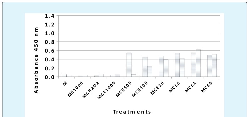
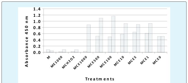
Figure 3: Effects of a (1:1) mixture of indigo and red clover against MCF-7 and MCF-12A Comparing the effects of indigo extract on MCF-7 and MCF-12A cells for 48 hours of exposure using the alpha of 0.0025 resulted in a significant inhibitory effect on the MCF-7 cancer cells for the 1000, 500 and 100 μg/mL concentrations (p= 0.00017 to 0.000578). In contrast, the effects of indigo extract at the 500, 100, 10, and 5 μg/mL concentrations resulted in a mitogenic effect on the MCF- 12A non-cancerous cells. But at the 1000 μg/mL concentration, indigo extract significantly inhibited both the MCF-7 and MCF-12A cells (p = 000251 and 4.72 x 1-^- 5, respectively.) These results indicate that at the 500 and 100 μg/mL concentrations of indigo extract, MCF-7 cancer cells are inhibited and the MCF-12A cells are mitogenically increased in numbers.
Comparing the effects of red clover extract to MCF-7 and MCF-12A cells for 48 hours of exposure using the alpha of 0.0025 resulted in a non-significant inhibitory effect on the MCF-7 cancer cells for the 500, 100, 10, 5, and 1 μg/mL concentrations compared to medium and cells alone (p= 0.073 to 0.722). In contrast, the effects of red clover extract at the 1000, 500, 100 μg/mL concentrations resulted in a significant inhibitory effect on the MCF-12A non-cancerous cells. But at the 1000 μg/mL concentration, red clover extract significantly inhibited both the MCF-7 and MCF-12A cells (p = 0.000286 and 0.00011, respectively.) These results indicate that at the 1000 μg/mL concentration of red clover extract against MCF-7 cancer cells and the MCF-12 A non-cancerous cells, they are both significantly inhibited. However, at the 500 to 0 μg/mL red clover concentrations, the MCF-7 cancer cells grow freely but inhibited the MCF-12A non-cancerous cells from 1000 to 5 μg/mL, therefore, yielding a non-desired result.
Comparing the effects of a 1:1 mixture of indigo and red clover extracts on MCF-7 and MCF-12A cells for 48 hours of exposure using the alpha of 0.0025 resulted in a significant inhibitory effect on the MCF-7 cancer cells for the 1000 μg/mL concentration (p= 1.16 x 10^-5). However, the 500 to 1 μg/mL concentrations were not significantly different to medium and cells alone. In contrast, the effects of 1:1 mixture of indigo and red clover extract at the 1000, 500, 100, 10, 5, and 1 μg/mL concentrations resulted in a mitogenic effect on the MCF- 12A non-cancerous cells, (range of p = 0.000288 to 3.65 x 10^-5). These results indicate that the 1000 μg/mL concentrations of the 1:1 mix of indigo and red clover extract has the desired outcome.
Polyphenols, flavonoids, flavanols, and tannins are found in indigo. Some of the specific compounds in indigo that have been shown to demonstrate anticancer activity include the following: tryptanthrin, the polyphenol kaempferol, indirubin and its derivatives, and trisindoline [1, 5, 6]. For red clover, isoflavones are the bioactive compounds that compose it. The specific isoflavones in red clover include the following: formononetin, biochanin A, genistein, and daidzein [3]. It is possible that the polyphenols and flavonoids in indigo contribute the most to its observed anticancer activity, and specifically that the genistein and daidzein in red clover contribute the most to its observed anti-proliferative response on cancer cells [3, 5]. Therefore, the mixture of polyphenols, flavonoids, and the isoflavones genistein and daidzein could have a synergistic anticancer effect on cells.
Conclusion
The red clover extract alone did not exhibit the desired results but gave the reverse of the desired result; whereas the indigo extract alone yielded desirable results with the 500 and 100 μg/mL concentrations due to an increased growth of the MCF-12A non-cancerous cells and lowered growth of the MCF-7 cancer cells; however, neither the 500 and 100 μg/mL concentrations were significantly different to cells and medium alone for the MCF-7 cells. At the 1000 μg/mL concentration of indigo extract, both the cancer and non-cancerous cells were significantly inhibited. The effects of the 1:1 mixture of indigo and red clover extracts on MCF-7 and MCF-12A cells for 48 hours of exposure yielded the most promising results. At the 1000 μg/mL concentration, the MCF-7 cancer cells were significantly decreased compared to non-cancerous cells and were similar in value to the positive kill control of cells with hydrogen peroxide. By contrast, the MCF-12A non-cancerous cells were mitogenically increased at a concentration of 1000 μg/mL. This suggests that the 1:1 mixture of indigo and red clover extracts at the 1000 μg/mL concentration may exhibit a synergistic effect by inhibiting breast cancer cells and not harming non-cancerous breast cells, and therefore could provide a safe breast cancer treatment.
References
-
Yoo M, Choi SU, Choi KY, Yon GH, Chae JC, et al. (2008) Trisindoline synthesis and anticancer activity. Biochemical and Biophysical Research Communications 376(1): 96-99.
-
Heo BG, Park YJ, Park YS, Bae JH, Cho JY, et al. (2014) Anticancer and antioxidant effects of extracts from different parts of indigo plant. Industrial Crops & Products 56: 9-16.
-
Krenn L, Paper DH (2009) Inhibition of angiogenesis and inflammation by an extract of red clover (Trifolium pratense L.). Phytomedicine 16(12): 1083- 1088.
-
Daugherty MK, Abramovitch D, Ivankovic D, Weinbrenner DR (2017) Cytotoxic Effects of Amorpha fruticosa Leaf, Stem, and Root Extractions on PC-12 Adrenal Neural Cells from Male Rattus Norvegicus. Advances in Cancer Prevention 2(1):120.
-
Heo BG, Jang HG, Cho JY, Namiesnik J, Jastrzebski Z, et al. (2013) Partial characterization of indigo (Polygonum tinctorium Ait.) plant seeds and leaves. Industrial Crops and Products 42: 429-439.
-
Kimura H, Ishihara T, Michida M, Ogawa S, Akihiro T, et al. (2014) Identification and quantitative analysis of polyphenolic compounds from the indigo plant (Polygonum tinctorium Lour). Nat Prod Res 28(7): 492-495.
-
Mannella P, Tosi V, Russo E, Zullino S, Pancetti F, et al. (2012) Effects of red clover extracts on breast cancer cell migration and invasion. Gynecological Endocrinology: Gynecol Endocrinol 28(1): 29-33.
- The Muculent Bleb-Mucinous Cystic Neoplasm-Hepatobiliary Region
- Insulin Sensitizers as Anti-Aging Agents: Unveiling Synergies with Albumin, GLP-1RA, Klotho Protein, and Metformin in the Quest to Combat Aging
- Reprogramming of GLP-1 Response at Prediabetes for the Prevention of Type 2 Diabetes: The Role of Albumin and GLP-1 Receptor Agonists
- The Mingled Allies-Combined Hepatocellular Carcinoma and Cholangiocarcinoma
- Compilation and Embodiment-Leydig Cell Tumour Testis
- Glucolipotoxicity: A Novel Different Perspective on the Causes of Cancer