Addenda and Appurtenant: Extra-Nodal NK/T Cell Lymphoma
An exceptional Non-Hodgkin’s lymphoma in Asia or Mexico may be denominated as the extra-nodal natural killer (NK)/T cell lymphoma. As an aggressive tumour, it may gradually eradicate the anatomical structures of the head and neck, decimate the nasal cavity and emerge within the para-nasal sinuses. Nasal or extra-nasal natural killer (NK)/T cell lymphoma may be situated within the nasal cavity, nasopharynx, and upper gastrointestinal or aero-digestive tract. Nasal blockage or nasal discharge, systemic symptoms of rhinitis or sinusitis and an acute invasive fungal rhino-sinusitis in the immune compromised may be demonstrated. Extra-nodal NK/T cell lymphoma arises within the skin, gastrointestinal tract, testis, lung, eye, soft tissue and spleen. A diagnostic nasal endoscopy or a computerized tomography (CT) scan of the para-nasal sinuses may be mandated. Cellular morphology of atypical, enlarged lymphoid cells with abundant or clear cytoplasm, irregular nuclear borders, vesicular nuclei, granular chromatin and inconspicuous nucleoli admixed with an inflammatory infiltrate of histiocytes, neutrophils, immunoblasts and esoinophils may be enunciated. Immune markers of T cell differentiation such as a CD2+, CD3+, CD4+, CD5+, CD7+, CD8+, CD43+,CD45RO+ or CD45+, cy (cytoplasmic) CD3+ and a nonreactive surface CD3- may be delineated. An immune reactive Linker for Activation of T cells (LAT) with cytotoxic granule-associated proteins Granzyme B, Perforin and TIA 1 may be expounding.
Preface
Extra-nodal natural killer (NK)/T cell lymphoma as an infrequent tumour comprises of an approximate 6% of the Non-Hodgkin’s lymphoma in the in East, Central and South Asia or Mexico [1, 2]. A mature natural killer (NK)/T cell lymphoma such as an extra-nodal NK/T cell lymphoma may constitute 22.4 % instances of Non- Hodgkin’s lymphoma in Asia, in contrast to a 4.3% to 5.1 % prevalence detected in United States and Europe. The nasal type of natural killer (NK)/ T cell lymphoma may depict an elevated incidence of an estimated 2% to 8% of the Non-Hodgkin’s lymphoma arising in the Asian subcontinent [1, 2]. Within the developed world (Europe and United States), the nasal type of NK/T cell lymphoma expounds a mere 2% or lower proportion of Non- Hodgkin’s lymphoma [2, 3]. Extra -nodal natural killer (NK)/T cell lymphoma may be designated as an aggressive tumour of the head and neck. Characterized by a gradual, inexorable eradication of the anatomical structures of the head and neck, it may particularly decimate the nasal cavity with an uncommon emergence within the paranasal sinuses. The mean age of representation of the lymphoma may be at 45 years and ranges from 19-80 years [2, 3].
Clinical Symptoms
The asymptomatic preliminary lesions of extra -nodal natural killer (NK)/T cell lymphoma may engender a deferred detection and consequent initiation of therapy. A history of nasal blockage or nasal discharge may be demonstrated with systemic symptoms of rhinitis or sinusitis. Immune compromised persons commonly display evidence of an acute invasive fungal rhino- sinusitis. A gradual destruction of the nasal cavity may be enunciated, accompanied by weight loss and fever [3, 4].
Disease Characteristics
Extra-nodal NK/T cell lymphomas may exhibit a heterogeneous coterie of lesions. The aggressive, predominant T cell lymphomas may be subcategorized contingent to the clinical features. The nasal or extra- nasal natural killer (NK)/T cell lymphoma may be situated within the nasal cavity, nasopharynx, and upper gastrointestinal or aero-digestive tract. The lymphoma frequently disseminates within the abutting anatomic structures such as the para-nasal sinuses, orbit or oral cavity. An extra-nodal NK/T cell lymphoma may be infrequent, in contrast to the nasal subtype of NK/T lymphoma [4, 5]. Extra-nodal NK/T cell lymphoma commonly arises within the skin, gastrointestinal tract, testis, lung, eye, soft tissue and spleen. The areas of dissemination may simulate the nasal NK/T cell lymphoma. The typical histology, immune phenotype, concomitant Epstein Barr viral (EBV) infection localized within the tumour and appropriate clinical elucidation may reinforce the diagnosis of extra nasal NK/T cell lymphoma [4, 5].
Investigative Assays
A diagnostic nasal endoscopy may depict a comprehensive eradication of the cartilaginous and bulk of the bony fraction of the nasal septum. The inferior or the middle nasal turbinate may occasionally be destroyed [5, 6, 7, 8]. A computerized tomography (CT) scan of the para- Anubha Bajaj. Addenda and Appurtenant: Extra-Nodal NK/T Cell Lymphoma. Cell Cellular Lif Sci J 2018, 3(2): 000133.
nasal sinuses demonstrates an elimination of the mid nasal structures with the nasal turbinate. Maxillary sinuses may infrequently be incriminated with an augmented mucosal lining. The hard palate may be fissured. An elevated density of the nasal/ para-nasal sinuses/ nasopharynx may be discerned by the computerized tomography (CT) scan. Infrequently the radiographic findings of an extra-nodal nasal NK/T lymphoma may be ambiguous [5]. Confirmation of diagnosis of a natural killer (NK)/T cell lymphoma may be achieved with the employment of a positron emission tomography (PET CT) and/or indiscrete punch biopsies of the nasal mucosa or nasopharynx. An “extra nasal” variant of the lymphoma may be categorized where on visual inspection, the nasopharynx may be devoid of the tumour or a histological validation of the lymphoma may be lacking.
Histological Elucidation
Nasal and extra-nasal NK / T cell lymphoma demonstrate an identical histology with a characteristic mucosal ulceration, glandular disruption with architectural distortion and a clear cell change of the mucosal glands. An angio-centric and angio invasive pattern of disease evolution along with extensive coagulation necrosis and an admixture of apoptotic bodies may be delineated [3, 5]. A diverse cellular morphology of atypical, enlarged lymphoid cells with abundant or clear cytoplasm, irregular nuclear borders, vesicular nuclei, granular chromatin and inconspicuous nucleoli may be enunciated. A prominent inflammatory infiltrate comprising of histiocytes, neutrophils, immunoblasts and esoinophils may be discerned. Peri- vascular and intravascular inflammatory infiltrates with fibrinoid necrosis of the blood vessels may be depicted. Pseudo-epitheliomatous hyperplasia of the superficial epithelium may occur infrequently. The morphological attributes may be limited and concurrent with site of origin of the tumour [3, 5].
Immune Phenotype
The categorical investigation of an extra-nodal NK/T cell lymphoma may constitute of the immune markers of T cell differentiation such as a CD2+, CD3+, CD4+, CD5+, CD7+, CD8+, CD43+,CD45RO+ or CD45+, cy (cytoplasmic) CD3+ and a non-reactive surface CD3-. Adjunct T cell associated antigens such as a T cell receptor (TCR) ᾳ β and TCR gamma delta may be non-reactive [5, 8]. The B cell markers such as CD10- or CD20- may be lacking and Copyright© Anubha Bajaj.
the association with vimentin may favour a diagnosis of a T cell lymphoma. The natural killer (NK) cell specific immune marker CD56+ may be elucidated though adjunctive immune markers of NK cells such as CD16- or CD57- or antigens of a T cell lineage may be non-reactive. Majority of the nasal subtypes of NK/T cell lymphoma may delineate the Epstein Barr virus (EBV+) though a few extra nasal NK/T cell lymphoma (non-nasal type) may lack the viral (EBV) concurrence [8, 9]. However, a profusion of T cell receptor or immunoglobulin gene rearrangements may be absent with the tumours of natural killer (NK) cells. The Epstein Barr virus (EBV) may be incriminated in the evolution of a Non-Hodgkin’s lymphoma such as an extra-nodal natural killer (NK)/T cell lymphoma. The Epstein Barr encoded RNA (EBER) may be reactive in an estimated half (50%) of the individuals afflicted with NK/T cell lymphoma. A human immune deficiency virus (HIV) infection may coexist with the Epstein Barr virus (EBV) infection in extra-nodal NK /T cell lymphoma [8]. The Epstein Barr viral ribonucleic acid (EBV- RNA) may be discerned by in situ hybridization (ISH) techniques. However, a natural killer (NK) cell neoplasm with the absence of a T cell receptor (TCR) genetic rearrangement may aberrantly exhibit CD5+ immune reactivity. A dual immune reaction of CD3+ with CD5+ may not adequately demonstrate a T cell versus an NK cell lineage [9, 10]. An angio destructive category of the NK/T cell lymphoma appearing emerging at dermal sites may depict an immune reactive CD54+ concomitantly with an immune reactive CD29+ ( β1 integrin) which permits cellular homing to the sites of inflammation. However, a CD54+ immune reactivity may be lacking with variants such as intravascular large B cell lymphoma. Immune reactivity to CD62L+ or L selectin, a homing molecule concurrent to high endothelial venules or capillaries, may also be elucidated [1, 2]. The T cell chemokine receptor CD183 or CXCR3, described as a G protein coupled serpentine receptor, may be exemplified with type I T helper cells, natural killer (NK) cells, macrophages and dendritic reticulum cells. The CD183 immune molecule may adhere to and interact with one of the CXC chemokines in order to initiate a chemotaxis of the immune cells towards the inflamed locales [4, 5].
Adjunctive Molecular Markers
The characteristic immune marker- Linker for Activation of T cells (LAT) may be defined as a trans- membrane protein necessary for the elucidation of thymocytes and T cell signalling. An immune reactive LAT may normally be delineated with variable categories of T cells, NK cells, mast cells and megakaryocytes [10, 11].
Anubha Bajaj. Addenda and Appurtenant: Extra-Nodal NK/T Cell Lymphoma. Cell Cellular Lif Sci J 2018, 3(2): 000133.
Additionally, cytotoxic granule-associated proteins such as Granzyme B, Perforin and TIA 1 may be expounded. The proteins ensure target cell death in conjunction with cytotoxic lymphocytes. The cytotoxic proteins may usually be exemplified within the NK cells and cytotoxic T lymphocytes. Granzyme B may be considered as a serine protease generally available within the secretory lysozymes. The protein may be engulfed by the target cells in the course of conjugation with cytotoxic lymphocytes [1, 2]. The conjugated molecule may activate the caspase dependent cell apoptosis. Perforin may emerge as a pore forming protein demonstrated in the secretory lysosomes of the cytotoxic T cells (CTL). It may permit the ingression of granzyme protein into the cytoplasm of the target cells. As a cytotoxic protein, it constitutes as a mandatory component of the target cell death. TIA 1 may a depicted as a protein of an RNA binding domain which augments cellular apoptosis via obscure mechanisms. Concomitant cell adhesion proteins and homing receptors appear within the neoplasm. The specific proteins regulate the mobilization of leukocytes and define the methodology of tissue specific dissemination of the lymphoma subtypes [1, 2]. Molecules such as cutaneous lymphocyte-associated antigen (CLA) may intercept the receptors homing to skin by concurring with its ligand elucidated on dermal and alternative sites. Cutaneous lymphocyte-associated antigen (CLA) may be discerned within the cutaneous instead of a non- cutaneous T cell lymphoma. As designated in the nasal type NK/T cell lymphoma, the emergence of CLA may be associated with an inferior prognosis [11, 12]. CD54 may be denominated as an intercellular adhesion molecule (associated with lymphocyte migration) appearing within the augmented venular and capillary endothelium. Proliferative activity as quantified by Ki 67 marker may range from 10% to 50% with various extra-nodal lymphomas.
Prognostic Determinants
Majority of the individuals with a nasal type of natural killer (NK)/ T cell lymphoma depict an early stage disease. An estimated three fourths (70%) of individuals may be in stage I or II during the detection of the disorder and thus elucidate a superior outcome [12, 13]. Extra nasal NK/T cell lymphoma may frequently demonstrate an advanced stage of disease with a majority (80%) appearing in stage III or IV. Concomitant emergence of B symptoms such as fever, malaise and weight loss greater than 10% of the body weight in preceding six months , an elevated serum lactate dehydrogenase (LDH), inferior gradation with the Eastern Cooperative Oncology Group Copyright© Anubha Bajaj.
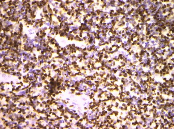
(ECOG) and an extra nasal representation of the disease may impact the disease free survival (DFS) [14, 15]. Individuals with extra-nasal disease may elucidate a worse overall survival (OS), in contrast to a nasal disease. Extra nodal NK/ T cell lymphoma may depict a median survival of 0.28 years or 3.36 months [16]. Prognostic models may be constructed in order to stratify the risk of tumour dissemination and mortality in patients with NK/T cells lymphoma. The models may be configured with the International Prognostic Index (IPI) and the Italian Prognostic Index for peripheral T cell lymphoma unspecified (PTCL –U). A prognostic index for a T cell lymphoma (PIT) may thus be articulated [7]. The International Peripheral T cell Lymphoma Project (I PTCL P) may be known to deliver suboptimal outcomes. The Korean Prognostic Index, essentially evolved for the Asian population, may connote superior prognostic evaluation of the neoplasm. The particular prognostic index incorporates parameters such as serum lactate dehydogenase (LDH) levels, regional lymph node enlargement and the presence of B symptoms along with Ann Arbor staging for appropriate stratification of the patients [7]. The contemporary Prognostic Index of Natural Killer cells (PINK) and the Prognostic Index of Natural Killer cells with Epstein Barr viral DNA data (PINK E) employs four distinct criterions to categorize the patients: age beyond 60 years, stage III/IV disease, distant lymph node involvement and a non-nasal variant of the disease. The specific indices (PINK and PINK E) may provide a superior concordance with probability of disease progression. The emergence of Epstein Barr virus (EBV+) infection may be cogitated as an independent prognostic factor for determining the overall survival [7] (Figures 1-12).
![Figure 1: Extra-nodal NK/T cell lymphoma: small atypical lymphoid cells [17].](/fulltextimages/3152/fig_1.jpeg)
Anubha Bajaj. Addenda and Appurtenant: Extra-Nodal NK/T Cell Lymphoma. Cell Cellular Lif Sci J 2018, 3(2): 000133.
![Figure 2: Extra nodal NK/T cell lymphoma: dermal ingress of atypical lymphoid cells [18].](/fulltextimages/3152/fig_2.jpeg)
![Figure 3: Extra nodal NK/T cell lymphoma: Angio- invasive configuration of aberrant lymphoid cells [17].](/fulltextimages/3152/fig_3.jpeg)
![Figure 4: Extra nodal NK/T cell lymphoma: angio- centric articulation of anomalous lymphoid cells [18].](/fulltextimages/3152/fig_4.jpeg)
Copyright© Anubha Bajaj.
![Figure 5: Extra nodal NK/T cell lymphoma – vascular incrimination with coagulative necrosis [19].](/fulltextimages/3152/fig_5.jpeg)
![Figure 6: Extra nodal NK/T cell lymphoma large foci of tumour necrosis [19].](/fulltextimages/3152/fig_6.jpeg)
![Figure 7: Extra nodal NK/T cell lymphoma: enlarged, aberrant lymphoid cells with mucosal ulceration [20].](/fulltextimages/3152/fig_7.jpeg)
![Figure 8: Extra nodal NK/T cell lymphoma: zonal coagulative necrosis with disseminated atypical lymphoid cells [21].](/fulltextimages/3152/fig_8.jpeg)
![Figure 9: Extra nodal NK/T cell lymphoma: clear cytoplasm, vesicular nuclei [22].](/fulltextimages/3152/fig_9.jpeg)
Anubha Bajaj. Addenda and Appurtenant: Extra-Nodal NK/T Cell Lymphoma. Cell Cellular Lif Sci J 2018, 3(2): 000133.
![Figure 10: Extra nodal NK/T cell lymphoma: intense aggregates of enlarged, anomalous lymphoid cells [23].](/fulltextimages/3152/fig_10.jpeg)
![Figure 11: Extra nodal NK/T cell lymphoma- immune reactive for CD56+ [24].](/fulltextimages/3152/fig_11.jpeg)
| Natural Killer (NK)/T cell Lymphomatoid | |
|---|---|
| Gastropathy |
The pseudo-malignant condition may depict a proliferation of natural killer (NK) cells .The self-limiting gastropathy implicates the gastric region or intestinal tract. Females may be commonly affected, in contrast to the males. Adults between 27 to 75 years may frequently delineate the lymphoma. The disorder may be clinically asymptomatic or individuals may depict ambiguous gastro-intestinal symptoms. Endoscopy demonstrates miniature, superficial lesions of one centimetre magnitude along with mucosal erosion, mucosal ulcer or elevated mucosa [4]. The mucosal aberrations may be accompanied by haemorrhage and superficial oedema. The morphological alterations manifest at solitary or multitudinous sites within the gastrointestinal tract. Radiographic imaging may not be able to decipher Anubha Bajaj. Addenda and Appurtenant: Extra-Nodal NK/T Cell Lymphoma. Cell Cellular Lif Sci J 2018, 3(2): 000133.
alternative locales of disease emergence. Individuals may depict a spontaneous resolution with a persistence or reappearance of lesions on follow up, particularly with the absence of cytotoxic chemotherapy. Histological examination demonstrates an extensive infiltrate of atypical lymphoid cells confined to the gastrointestinal mucosa [4]. Foci of epithelial invasion by the aberrant lymphoid cells may be exemplified. The anomalous cellular component comprises of medium to enlarged cells with indented or irregular nuclei and visible to inconspicuous nucleoli. Several cells may exhibit brightly eosinophilic granules confined to the cytoplasm. Angio- invasion may be absent along with a lack of fibrinoid necrosis. The immune histochemical profile may collocate the presence of immune reactive cytoplasmic CD3+ with an absence of surface CD3-, immune reactive CD56+ and TIA 1+. A lack of immune reactive CD5- , EBER - and genetic rearrangements of the T cell receptor (TCR) may be elucidated [4].
Enteropathy associated T Cell Lymphoma
Enteropathy associated T cell lymphoma (EATL) may be described as a tumour of intestinal intraepithelial T lymphocytes. A contemporary classification of dual subtypes may be enunciated. Type I as a classical subcategory. Type I enteropathy associated T cell lymphoma (EATL) may be considered as an infrequent disorder , though commonly delineated in the Western World, in concordance with celiac disease. The condition may be rare in Asians [4]. Histology Type I EATL may depict enlarged lymphocytes with a frequent admixture of an inflammatory infiltrate comprised of histiocytes and eosinophils. The abutting mucosal surface may delineate alterations concordant with gastrointestinal enteropathy. The phenotype of the disorder may be expounded by an immune reactive CD3+ and CD103+ while the CD5-, CD4-, CD8-/+,CD30+/-, CD56- immune molecules may be non-reactive or dubiously expressed [4]. Type II enteropathy associated T cell lymphoma (EATL) may be considered as a distinct entity for further classification. It may be connoted as a ϫᵟ T cell lymphoma. Type II EATL may be defined as an infrequent condition, restricted primarily to the Asian population.
Histology
Type II EATL may be configured of monomorphic, medium sized aberrant lymphocytes commingled with minimal quantities of inflammatory cells. A lack of Copyright© Anubha Bajaj.
necrosis may be characteristic. The abutting gastrointestinal mucosa may demonstrate an intra- epithelial lymphocytosis [4]. The phenotype displayed by the tumour cells may be an immune reactive CD3+, CD8+, CD56+ with a lack of immune reaction to CD5-, CD4-, CD30-. The T cell receptor (TCR) may commonly demonstrate a ϫᵟ phenotype, in contrast to a ᾳ β TCR subtype. The gastrointestinal mucosa with an intra- epithelial lymphocytic ingress adjacent to the lymphoma exhibits an immune phenotype which may be concordant with the lymphoma in an estimated one third (35%) of the instances and may differ from the malignant infiltration in an estimated two thirds (65%) individuals. Immune reactivity for CD8+ or CD56+ may be a frequent phenomenon [4].
Therapeutic Protocols
Radiation therapy may prove to be an efficacious modality for the management of NK/T cell lymphoma confined to a solitary site. However, a long term disease control may not be adequately achieved with the employment of singular radiotherapy. The administered dosage and the extent of fields incorporated may be valuable factors in evaluating the overall efficacy of radiation therapy [7]. A comprehensive inclusion of nasal/paranasal sinuses along with the nasopharynx within the radio-therapeutic fields may provide a competent prognosis. Restriction of radiotherapy to the primary lesions may amplify the probability of localized disease relapse. Systemic therapies such as cyclophosphamide, hydroxydaunorubicin, oncovin and prednisone (CHOP) may not prove to be efficacious for treating a NK/T cell lymphoma. The current chemotherapeutic regimen of methotrexate, leucovarin, ifosfamide, mesna, dexamethasone, etoposide and L– asparaginase (SMILE) may be beneficial and is frequently recommended [7]. However, the contemporary chemotherapy may be accompanied by significant toxicity. The De VIC regimen (dexmethasone, etoposide, ifosfamide, carboplatin) may prove to be efficacious when administered in combination with radiation therapy. The combination may be less toxic than the contemporary SMILE regimen. The application of concurrent radiation may be accompanied with a poor oral intake and substantial mucositis for duration of several weeks following cessation of therapy. The GELOX regimen (gemcitabine, oxaliplatin and L–asparaginase) may delineate a superior response when employed in combination with or sequential to radiotherapy in individuals of a preliminary stage NK/T cell lymphoma [7]. A multimodal treatment methodology may be Anubha Bajaj. Addenda and Appurtenant: Extra-Nodal NK/T Cell Lymphoma. Cell Cellular Lif Sci J 2018, 3(2): 000133.
necessitated for managing the infrequent natural killer (NK)/T cell lymphomas. The contemporary treatment algorithm may be focussed on reducing the considerable toxicity and co-morbidities associated with concurrent or sequential radiotherapy and chemotherapy, while amplifying the therapeutic efficacy with long term disease control. The current treatment strategies may be applicable for the disease of upper aero-digestive tract. Radiotherapy and chemotherapy, singularly or in combination may be employed. The optimal therapy for treating a nasal/ non nasal extra-nodal natural killer (NK)/T cell lymphoma remains obscure. The untreated disorder may be fatal in a majority of the patients and an estimated half (50%) of the mortality occurs due to systemic dissemination of the disease [7]. The disseminated disease displays a median survival of 9 months and the individuals on appropriate therapy may depict a median survival of 1.1 years. Addition of chemo- radiation in the therapeutic protocol may augment the prognostic predilection of the disease [7].
Conclusion
A nasal natural killer (NK)/ T cell lymphoma generally depicts an early stage disease. Extra nasal NK/T cell lymphoma demonstrates an advanced stage of disease with a majority (80%) of patients in clinical stage III or IV with B symptoms such as fever, malaise and weight loss greater than 10% of the body weight in preceding six months, an elevated serum lactate dehydrogenase (LDH) and an inferior gradation with the Eastern Cooperative Oncology Group (ECOG) which impacts the disease free survival (DFS) and a poor overall survival (OS). Numerous indices for risk stratification of patients may be employed such as the International Prognostic Index (IPI), the Italian Prognostic Index for peripheral T cell lymphoma unspecified(PTCL –U), a prognostic index for a T cell lymphoma (PIT), the International Peripheral T cell Lymphoma Project ( I PTCL P) and the Korean Prognostic Index. The latter incorporates parameters such as serum lactate dehydrogenase (LDH) levels, regional lymph node enlargement and the presence of B symptoms along with Ann Arbor staging. Prognostic Index of Natural Killer cells with Epstein Barr viral DNA data (PINK E) employs four distinct criterion such as age beyond 60 years, stage III/IV disease, distant lymph node involvement and a non-nasal variant of the disease and provides a superior concordance with disease progression. Radiation therapy may be efficacious for managing a solitary NK/T cell lymphoma. Combinations of cyclophosphamide, hydroxydaunorubicin, oncovin and prednisone (CHOP) may not be efficacious in treating a NK/T cell lymphoma.
Copyright© Anubha Bajaj.
The contemporary regimen of methotrexate, leucovarin, ifosfamide, mesna, dexamethasone, etoposide and L – asparaginase (SMILE) may be beneficial. The De VIC regimen (dexmethasone, etoposide, ifosfamide, carboplatin) or the GELOX regimen (gemcitabine, oxaliplatin and L –asparaginase) may be superior when employed in combination with or sequential to radiotherapy (Tables 1-5).
T cell prolymphocytic leukaemia T cell large granular lymphocytic leukaemia Chronic lymphoproliferative disorder of NK cells Aggressive NK cell leukaemia Systemic EBV+ T cell lymphoma of childhood Hydroa –vacciniforme like lymphoproliferative disorder Adult T –cell leukaemia /lymphoma Extra-nodal NK/T cell lymphoma –nasal type Enteropathy associated T cell lymphoma Monomorphic epitheliotropic intestinal T cell lymphoma Indolent T cell lymphoproliferative disorder of the gastrointestinal tract Hepatopslenic T cell lymphoma Subcutaneous panniculitis like T cell lymphoma Mycosis fungoides Sezary syndrome Primary cutaneous CD30+ T cell lymphoproliferative disorder Lymphomatoid papulosis Primary cutaneous anaplastic large cell lymphoma Primary cutaneous ϫᵟ T cell lymphoma Primary cutaneous CD8+ aggressive epidermotropic cytotoxic T cell lymphoma Primary cutaneous acral CD8+ T cell lymphoma Primary cutaneous CD4+ small/medium T cell lymphoproliferative disorder Peripheral T cell lymphoma NOS Angio-immunoblastic T cell lymphoma Follicular T cell lymphoma Nodal peripheral T cell lymphoma with TFH phenotype Anaplastic large cell lymphoma ALK+ Anaplastic large cell lymphoma ALK- Breast implant associated anaplastic large cell lymphoma NK: Natural Killer NOS: Not otherwise specified TFH: T Follicular Helper ALK: Anaplastic lymphoma kinase. Table 1: WHO classification of mature lymphoid histiocytic and dendritic T cell neoplasm (2016) [6].
Anubha Bajaj. Addenda and Appurtenant: Extra-Nodal NK/T Cell Lymphoma. Cell Cellular Lif Sci J 2018, 3(2): 000133.
| Immune Marker | Estimated Proportion (%) | ||||
| CD2 | 69-100 | ||||
| CD3 | 56-100 | ||||
| CD5 | 0-42 | ||||
| CD20 | 0 | ||||
| CD30 | 20-64 | ||||
| CD43 | 61-100 | ||||
| CD54 | 56 | ||||
| CD56 | 50-100 | ||||
| CD183 | 5-15 | ||||
| BCL2 | 7-19 | ||||
| CLA | 56 | ||||
| Granzyme B | 57-100 | ||||
| LAT | 92 | ||||
| Perforin | 36-81 | ||||
| TIA | 27-100 | ||||
| EBV ( in situ) | 13-100 |
Table 2: Immune histological markers in Nasal Type Extra-Nodal NK/T cell Lymphoma [2].
CLA: Cutaneous Lymphocyte Antigen. EBV: Epstein Barr Virus LAT: Linker for activation of T cells. NK: Natural Killer. Table 2: Immune histological markers in Nasal Type Extra-Nodal NK/T cell Lymphoma [2].
| βF1- TCR x+ | 61% | |
| Primary cutaneous CD30+ T cell lymphoproliferative disorder | βF1+ TCR x- | 17% |
| Lymphomatoid papulosis | βF1+ TCR x+ | 17% |
| Primary cutaneous anaplastic large cell lymphoma | β F1- TCR x- | 6% |
| Primary cutaneous x8 T cell lymphoma | ||
| Primary cutaneous CD8+ aggressive epidermotropic |
Table 3: Contrast of NK Lymphomatoid Gastro-enteropathy and Extra-nodal NK/T cell Lymphoma [4].
- Variable genetic alterations(+1q32-
- Diverse conditions on q41 and +5q34-q35 frequent in type I and +8q24 frequent in type morphology and immune phenotype
- II)
- No evidence of celiac
- Common genetic alterations may disease be +8q31 and -16q12
- Variable epidemiology: Type I absent in Asians
Table 4: Distinction between EATL Type I and II [4].
Copyright© Anubha Bajaj.
| NK Lymphomatoid Gastro-enteropathy | Extra-nodal NK/T cell Lymphoma | |
|---|---|---|
| Gross or Endoscopic Features | Small , superficial lesions ( erosion, ulcer, raised or haemorrhagic lesions) | Deep seated, enlarged lesions or tumour aggregates |
| Histology | Brightly eosinophilic granules in the lymphoid cells ( H & E) | Lymphoid granules may be infrequent on H & E sections |
| Epstein Barr virus | Non-reactive | Reactive |
Table 5: Contrast of NK Lymphomatoid Gastro-enteropathy and Extra-nodal NK/T cell Lymphoma [4].
1. Bakari A, Iliyasu Y, Kirfi AM (2017) Extra-nodal NK/ T
cell lymphoma in an African. Highland Med Res J 17(1): 62-65.
2. Schwartz EJ, Molina-Kirsch H, Zhao S, Marinelli RJ,
Warnke RA, et al. (2008) Immunohistochemical characterization of Nasal Type Extra-nodal NK/T cell Lymphoma using a Tissue Microarray: An Analysis of 84 cases. Am J Clin Pathol 130(3): 343-351.
3. Dragos L (2017) Lymphoma and plasma cell disorders: T/NK cell disorders: Extra-nodal NK/T cell lymphoma; nasal type” Pathology Outlines Jan 2017.
4. John KCC What’s New in T & NK cell Lymphomas:
power point presentation: Queen Elizabeth Hospital: Hong Kong.
5. Chen Y, Ling L, Jianping L, Weifeng Z, Tongmei H, et al.
(2017) Primary natural killer /T cell lymphoma of the cervix: case report and literature review. Int J Clin Exp Med 10(2): 3899-3905.
6. Swerdlow SH, Campo E, Pileri SA, Harris NL, Stein H,
et al. (2016) The 2016 revision of world health organization classification of lymphoid neoplasm. Blood 127(20): 2375-2390.
7. Frontline “Lymphoma Research Foundation” Fall
2016 Issue 4.
8. Bruno NF, Paulo RB, Ana Luiza VSM, João LMJ (2012)
Lethal midline granuloma syndrome: a diagnostic dilemma. Radiol Bras 45(6): 353-355.
9. Martha MT, Monica K, Razia AGK, Meschack B, Johan
L, et al. (2013) Extra-nodal natural killer/T cell lymphoma, nasal type: midline lethal granuloma. A case report. Head and Face Medicine 9: 4.
Anubha Bajaj. Addenda and Appurtenant: Extra-Nodal NK/T Cell Lymphoma. Cell Cellular Lif Sci J 2018, 3(2): 000133.
11. Wei J, Wu H, Sun M, Liu W, Meng L (2012) Primary
endometrial natural killer (NK)/T cell lymphoma : case repost and review of literature. Eur J Gynaecol Oncol 33(4): 425-427.
12. Fang JC, Zhou J, Li Z, Xia ZX (2014) Primary extra- nodal NK/T cell lymphoma: nasal type of uterus with adenomyosis: a case report. Diagn Pathol 23(9): 95.
13. Wang GN, Zhao WG, Gao XZ, Zhang DD, Wang GJ, et al.
(2015) Primary natural killer/ T cell lymphoma of the cervix: a case report and clinicopathological analysis. Taiwan J Obstet Gynaecology 54(1): 71-74.
14. Omori M, Oishi N, Nakazawa T, Nakazawa K,
Mitsumori T, et al. (2016) Extra-nodal NK/T cell lymphoma: nasal type of the uterine cervix : a case report” Diagn Cytopathol 44(5): 430-433.
15. Shet T, Suryawanshi P, Epari S, Sengar M, Rangarajan
V, et al. (2014) Extra-nodal natural killer/T cell lymphoma with extra-nasal disease in non endemic regions are disseminated or have nasal primary: a study of 84 cases from India. Leuk Lymphoma 55(12): 2748-2753.
16. Au WY, Weisenburger DD, Intragumtornchai T,
Nakamura S, Kim WS, et al. (2009) International Peripheral T cell Lymphoma project: Clinical differences between nasal and extra nasal natural killer/T cell lymphoma : a study of 136 cases from the International Peripheral T cell Lymphoma Project. Blood 113(17): 3931 -3937.
17. Image 1, 3, & 12 Courtesy: Pathology Outlines.
18. Image 2,4 Courtesy : Derm Net NZ.
19. Image 5,6 Courtesy : Journal of Oral and Maxillofacial
Surgery.
Copyright© Anubha Bajaj.
20. Image 7 Courtesy: JSLTR.
21. Image 8 Courtesy Springer link.
22. Image 9 Courtesy: JFCR.
Anubha Bajaj. Addenda and Appurtenant: Extra-Nodal NK/T Cell Lymphoma. Cell Cellular Lif Sci J 2018, 3(2): 000133.
23. Image 10 Courtesy: Thorax.com.
24. Image 11 Courtesy: Academic oup.
Copyright© Anubha Bajaj.
- The Muculent Bleb-Mucinous Cystic Neoplasm-Hepatobiliary Region
- Insulin Sensitizers as Anti-Aging Agents: Unveiling Synergies with Albumin, GLP-1RA, Klotho Protein, and Metformin in the Quest to Combat Aging
- Reprogramming of GLP-1 Response at Prediabetes for the Prevention of Type 2 Diabetes: The Role of Albumin and GLP-1 Receptor Agonists
- The Mingled Allies-Combined Hepatocellular Carcinoma and Cholangiocarcinoma
- Compilation and Embodiment-Leydig Cell Tumour Testis
- Glucolipotoxicity: A Novel Different Perspective on the Causes of Cancer