G Protein-Coupled Estrogen Receptor (GPER) Expression in Normal Myometrium and Uterine Leiomyoma
Uterine leiomyomas are the most common benign smooth muscle tumours of the female reproductive system. Their pathogenesis is unclear but they are known to be sensitive to sex hormones, estrogens and progesterone, which influence their behaviour and evolution. The action of estrogens depends on the presence of certain receptors in the cell, including G-protein-coupled estrogen membrane receptor, GPER. In this work, using immunohistochemical techniques, we studied the presence of GPER in samples of typical uterine leiomyomas and paired myometrium as well as in fast-growing leiomyomas. The results show the presence of GPER in both uterine tissues with a different subcellular distribution, being the most frequent nucleic location in myometrium, and the most frequent cytoplasmic location in leiomyoma. Likewise, we observed that in fastgrowing leiomyomas, the cytoplasmic location is even more abundant than in typical leiomyomas. In conclusion, these data seem to suggest that GPER could be involved in certain actions of estrogens related to the pathogenesis and behaviour of these tumours.
Fernández LH, Rodríguez del Río M, Reyes R* and Bello AR
Cell Biology Area, University of La Laguna, Spain *Corresponding author: Reyes R, Cell Biology Area, Biology Section, University of La Laguna, Tenerife, Spain, Email: rreyesro@ull.edu.es Keywords: Leiomyoma; Myometrium; GPER; Estrogens; Nucleus; Cytoplasm
Introduction
The tissues of the uterus are sensitive to hormones and their state and activity depend on the levels of these hormones, mainly estrogens and progesterone. Estrogens in particular play a promoting role in leiomyoma proliferation [1]. Hyperestrogenic conditions greatly influence the development of fast-growing leiomyomas. G protein-coupled estrogen receptor (GPER), a seven transmembrane domain receptor belonging to the G protein-coupled receptors (GPCRs) family, has been identified as a novel estrogen receptor (ER) mediating rapid nongenomic activity with high affinity to estrogen and functions alongside the classical ER [2]. This receptor has been supposed to play an important role in cell proliferation due to its overexpression in different tumours [3]. The cellular location of GPER in uterine tumours is not completely clarified, being, according to some authors [4] mainly nuclear. Taking this into account, in this work we have proposed to determine by immunohistochemistry the cellular and subcellular distribution of the GPER in leiomyomas in relation to its distribution in the paired myometrium, as well as to determine the cellular and subcellular location of GPER in fast growing leiomyomas.
Materials and Methods
Six paired leiomyoma and myometrial samples, and five fast growing leiomyoma samples were obtained from hysterectomies and myomectomies performed in the Hospital Universitario de Canarias (HUC) (Canary Islands, Spain), with prior informed consent to the patients. The samples were fixed in 10% formalin, embedded in Paraplast® and 3µm sections were obtained. On these sections, the immunoenzymatic and immunofluorescence techniques were performed using a polyclonal antiserum anti-GPER (LifeSpan, Bioscience) at dilution 1/40. The samples were analysed in an optic microscope (Leica DM4000B) and the images were taken with a digital camera (Leica DFC300FX). For cell quantification, the samples were differentially counterstained to visualize the nuclei of all cells, both immunoreactive cells with cytoplasmic immunoreaction, and non-immunoreactive nuclei. Two sections per sample were chosen, and in each one, 5 microscopic fields were randomly counted at 400x magnification. GPER-ir cells per unit area (1000 µm2) were counted using the Leica Q-Win 3 (Leica, Barcelona, Spain) image analysis software. The statistical analysis was performed using the SPSS 20.0. The data obtained were analysed using the one-way ANOVA test using Scheffé’s post-hoc test for comparison between groups. Statistical significance was set for values of p <0.05.
Results and Discussion
Distribution of GPER Immunoreactivity in Myometrium
In the myometrium, GPER-ir cells were observed in connective (Figure 1A) and muscle tissue (Figures 1B-D). In all the samples analysed, the subcellular location in muscle cells was mostly nuclear (Figures 1B-D).
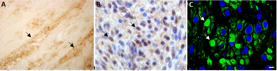
Figure 1: GPER Distribution in Myometrium. (A): GPER- ir cells (black arrows) in myometrium connective tissue (Diaminobenzidine immunoperoxidase (DABIP)). (B): GPER-ir (DAB) cells with nuclear reaction (black arrows). Nuclei have been counterstained with haematoxylin. (C-D): Confocal images showing GPER-ir cells (green immunofluorescence (GIF)) with nuclear reaction (white arrows). Nuclei have been counterstained with DAPI. Scale bar: 5µm.
Distribution of GPER Immunoreactivity in Leiomyomas
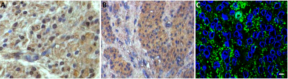
Leiomyoma do not show GPER immunoreactivity in connective tissue. In muscle cells, a cytoplasmic location is predominantly observed, increasing in fast-growing leiomyomas (Figure 2). A Leiomyoma, B, and C, fast growing leiomyoma).
The quantification of GPER-ir cells showed how the subcellular location of the receptor was mainly nuclear in myometrium, while in leiomyoma it was mainly cytoplasmic, being the number of GPER-ir cells with cytoplasmic reaction slightly higher in fast-growing leiomyoma (Table 1). These values reflect the different ratio Nu/Cy observed in the different tissues. Statistical analysis showed significant differences in the number of GPER-ir cells with nuclear reaction between different tissues analysed, myometrium, leiomyoma and fast growing leiomyoma, while the number of GPER-ir cells with cytoplasmic reaction showed significant differences only between myometrium and leiomyoma. Likewise, the Nu/Cy ratio shows significant differences between the myometrium and the leiomyoma (Table 1).
| Subcellular location | Mean ± SE¹ | Ratio Nu/Cy² | |
|---|---|---|---|
| Myometrium | Nuclei | 74,3333 ± 4,3879a | 3,18a,b |
| Myometrium | Cytoplasms | 23,3333 ± 2,8413b,c | 3,18a,b |
| Typical Leiomyoma | Nuclei | 46,2667 ± 5,0966a | 0,97a |
| Typical Leiomyoma | Cytoplasms | 47,8667 ± 5,1601b | 0,97a |
| Fast-growing Leiomyoma | Nuclei | 20,1333 ± 1,6899a | 0,36b |
| Fast-growing Leiomyoma | Cytoplasms | 56,4000 ± 4,7577c | 0,36b |
Table 1: Means ± SE of GPER-ir cells (nuclei and cytoplasms) per unit area (1000 µm2) in myometrium and two types of leiomyomas s
In this work, we demonstrate that the main differences in muscle cells expressing GPER in myometrium and leiomyoma affect subcellular location rather than cell number, as shown by the data on the total number of GPER-ir cells and the ratio Nu/Cy. This may reflect different biological responses to estrogens mediated by GPER in these cells, and a relationship with the estrogenic status, since the time of the menstrual cycle, the body mass index (BMI) and the degree of cell proliferation were taken into account in samples selection. On the other hand, we show for the first time GPER immunoreactivity in connective tissue. This allows us to suggest a possible role for this receptor as a modulator of growth factors synthesis by muscle cells that maintain the physiological state of the myometrium and a possible implication in the pathogenesis of leiomyomas. Likewise, the presence of GPER in myometrial connective tissue suggests the involvement of this receptor in the alterations that the extracellular matrix of this tissue presents in this type of tumours [5].
References
-
Flake GP, Andersen J, Dixon D (2003) Etiology and pathogenesis of uterine leiomyomas: a review Environ Health Perspect 111(8): 1037-1054.
-
Prossnitz ER, Barton M (2014) Estrogen biology: new insights into GPER function and clinical opportunities Mol Cell Endocrinol 389(1-2): 71-83.
-
Pandey DP, Lappano R, Albanito L, Madeo A, Maggiolini M, et al. (2009) Estrogenic GPR30 signalling induces proliferation and migration of breast cancer cells through CTGF. EMBO J 28(5): 523-532.
-
Tian R, Wang Z, Shi Z, Li D, Wang Y, et al. (2013) Differential expression of G-protein-coupled estrogen receptor-30 in human myometrial and uterine leiomyoma smooth muscle. Fertil Steril 99(1): 256-263.
-
Leppert PC, Baginski T, Prupas C, Catherino WH, Pletcher S, et al. (2004) Comparative ultrastructure of collagen fibrils in uterine leiomyomas and normal myometrium Fertil Steril 82(3): 1182-1187.
- The Muculent Bleb-Mucinous Cystic Neoplasm-Hepatobiliary Region
- Insulin Sensitizers as Anti-Aging Agents: Unveiling Synergies with Albumin, GLP-1RA, Klotho Protein, and Metformin in the Quest to Combat Aging
- Reprogramming of GLP-1 Response at Prediabetes for the Prevention of Type 2 Diabetes: The Role of Albumin and GLP-1 Receptor Agonists
- The Mingled Allies-Combined Hepatocellular Carcinoma and Cholangiocarcinoma
- Compilation and Embodiment-Leydig Cell Tumour Testis
- Glucolipotoxicity: A Novel Different Perspective on the Causes of Cancer