Schisandrin B Suppressed the Proliferation of MM Cell Lines Via IL-6
Background: Multiple myeloma (MM) is a hematologic malignancy. Schizandrin B (Sch B) is one of the main dibenzocyclooctadiene lignans which is found in schisandra chinensis. Sch B has been previously demonstrated to exhibit antitumor properties. However, the effect of Sch B on MM remains nuclear. Materials and Methods: The impact of Sch B on the proliferation of MM cell lines was determined by CCK-8 assay. The cell cycle and apoptosis of MM cell lines were evaluated by flow cytometry. RT-PCR to identify the effect of Sch B on the chemokines CCL2, CCL3 and CCL14 in MM cell lines. IL-6 and VEGF concentrations of serum and cell-free supernatants were tested by ELISA. Result: Our studies revealed that Sch B suppressed the proliferation of MM cell lines via IL-6. Sch B induced cell cycle of MM cell lines arrest at G0/G1 phase and increased its apoptosis. Sch B suppressed the mRNA expression of the chemokines CCL2, CCL3and CCL14 in MM cell lines. Conclusion: Sch B suppressed the proliferation of MM cell lines via IL-6. These findings suggest that Sch B may be a promising traditional Chinese medicine for MM.
Introduction
Multiple myeloma (MM) is the second most common hematological malignancy. Its characteristic is terminally differentiated plasma cells in the bone marrow and monoclonal immunoglobulin accumulation in the blood or urine [1]. MM is typically diagnosed between the ages of 65- 74 years [2]. MM accounts for 1.8% of all new cancer cases and 18% of all hematologic malignancies in the United States. In 2022, an estimated 34,470 cases will be diagnosed and the malignancy will result in 12,640 deaths [3]. Over the last two decades, many novel treatments have been developed for MM, including proteasome inhibitors, immunomodulatory drugs, monoclonal antibodies and CAR-T-cell [4]. However, MM remains an incurable disease, and the current survival is approximately 6-8 years [5]. The 5-year relative survival rate across all stages combined is 54% [6]. Other cures may be needed for a small minority of patients.
Schisandrin B (Sch B) is a traditional Chinese herbal medicine. Sch B possess diversified pharmacokinetic propriety such as antioxidant, anti-inflammation, cardioprotection, antitumor and neuroprotection [7, 8, 9]. Sch B was reported against a wide variety of human cancer cell lines. However, the activity of Sch B has not been tested in MM.
In the present study, our datas suggest that Sch B suppressed the proliferation of MM cell lines via IL-6. Sch B induced cell cycle of MM cell lines arrest at G0/G1 phase and increased its apoptosis. Finally, Sch B suppressed the mRNA expression of the chemokines CCL2, CCL3 and CCL14 in MM cell lines.
Materials and Methods
Cells and Cell Culture
The human MM cell lines (MM.1S, U266) were bought from Fenghuishengwu Co., Ltd. Two cell lines were cultured in RPMI-1640 supplemented with fetal bovine serum (10%) in a humidified incubator at 37 ̊C with 5% CO2. In general, 4-6 passages of cell lines were used for experiments.
Drug Preparations
Sch B, IL-6 and anti-IL-6 were purchased from Nanchang Zhenshan Biotechnology Co., Ltd. A total of 20 mg Sch B was dissolved in 1.5 ml DMSO. Sch B was stored at ‐80°C and used for subsequent use by diluting immediately prior to application.
CCK-8 Assay
MM.1S or U266 were seeded and cultured at a density of 4 × 10³/well in 96‐well microplates. The cells were treated with various concentrations of Sch B (0, 3.125, 6.25, 12.5, 25, 50, 100μM) for 24, 48 and 72 hours respectively. 10 μL of CCK‐8 reagent was added to each well and then cultured for 2 hours. The absorbance was analysed at 450 nm using a microplate reader (Bio‐Rad, Hercules, CA, USA).
Cell Cycle Analysis
MM.1S or U266 were respectively seeded in 6-well plates at a density of 5 × 10⁵ cells/ml and treated with Sch B at a concentration of 25 μmol/L for 48 h. The cells were harvested, washed and fixed. They were incubated with 30 μg/ml of propidium iodide and 40 μg/ml of Rnase. Then, Cell cycle were analyzed by the FACSCalibur cytometer and the Modfit LT software.
Cell Apoptosis Assay
MM.1S or U266 were respectively seeded in different 6-well plates at a density of 1 × 10⁵ cells/ml. The cells were treated with Sch B at a concentration of 25 μmol/L for 48 h. The cells were washed twice with ice-cold PBS. Subsequently, apoptosis was assayed using the FITC Annexin V Apoptosis Detection Kit I, following the manufacturer’s instructions. The cells were assessed using flow cytometric analysis.
Enzyme-Linked Immunosorbent Assay (ELISA)
IL-6 and VEGF concentrations of serum and cell-free supernatants were tested by Human IL-6 and VEGF ELISA assay kits (Peprotech) according to manufacturer’s protocol.
Real-Time Quantitative (RT-PCR)
MM.1S or U266 was collected. RNA of MM.1S, U266 was extracted with E.Z.N.A. Total RNA Kit I (OMEGA). cDNA synthesis was done with the MLV RT kit (Invitrogen). Polymerase chain reaction (PCR) analyses were performed by Platinum SYBR Green qPCR SuperMix-UDG w/ROX on an Applied Biosystems 7300 Real-Time PCR System. The CCL2, CCL 3 and CCL 14 mRNA was expressed with ΔΔCt values. The primer of human CCL2 is 5’-TGTCCCAAAGAAGCTGTGATC-3’ and 5’-ATTCT TGGGTTGTGGAGTGAG-3’. The primer of human CCL3 is 5’-CGGCAGA TTCCACAGAATTTC-3’ and 5’-AGGTCGCTGACATATTTCTGG -3’. The primer of human CCL14 is 5’-ACATCTCACAAAGCATCCCG-3’and 5’-TCATGCAATC CTGAACTCCC-3’.
Statistical Analysis
The data were presented as mean ± standard deviation. Comparison between groups were performed using a one- way ANOVA. P<0.05 was considered to indicate a statistically significant difference. Statistical analyses were performed using GraphPad Prism version 7.0.
Results
Sch B Suppressed the Proliferation of MM Cell Lines
The impacts of Sch B on the proliferation of MM cell lines were determined by CCK-8 assay. As shown in Figure 1A,1B, Sch B significantly inhibited the proliferation of two MM cell lines (MM.1S,U266). The inhibition rates were increased with the increasing incubation time and doses of Sch B when MM.1S and U266 were treated with Sch B (0,3.125,6.25,12.5,25,50,100μM) for 24, 48 and 72 hours, respectively (Figure 1C,1D). It was demonstrated that the inhibitory effects of Sch B on MM cell lines were in a dose and time-dependent manner.
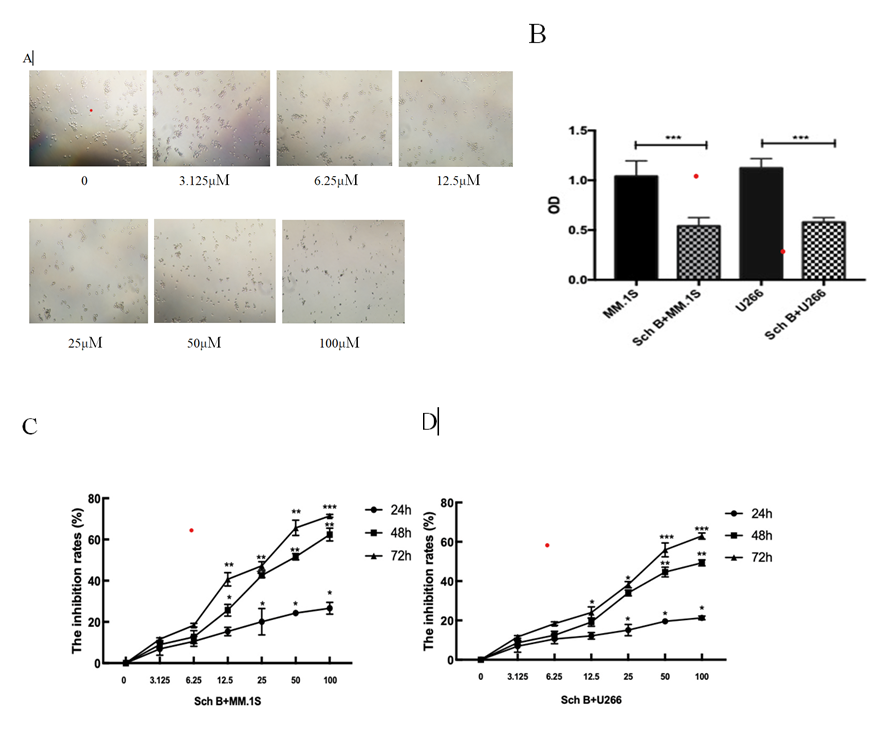
Figure 1: Sch B suppressed the proliferation of MM cell lines. (A) MM.1S; (B)The proliferation was evaluated by CCK- 8 assay;(C) Sch B suppressed the proliferation of MM.1S in a dose-and time-dependent manner;(D) Sch B suppressed the proliferation of U266 in a dose-and time-dependent manner. Data represent one of the three independent experiments, each performed in triplicate. ∗∗∗𝑃 < 0.001, ∗∗𝑃 < 0.01, ∗𝑃 < 0.05.
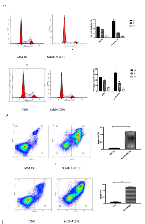
Sch B Induced the Cell Cycle Arrest at G0/G1 Phase and the Apoptosis of MM Cell Lines
The effects of Sch B on the cell cycle and apoptosis of MM cell lines were evaluated. MM.1S and U266 were treated with Sch B for 48 hours. The distributions of different phases and apoptosis of MM cell lines were analyzed by flow cytometry. As shown in Figure 2A, compared with control, when Bch B was added into MM.1S and U266, MM.1S and U266 in the G1/G0 phase were significantly increased and cells in G2/M and S phases were significantly reduced. The percentages of apoptotic cells in the MM.1S and U266 treated with Sch B were also significantly higher than control groups (Figure 2B). Thus, Sch B induced cell cycle arrest in G1/G0 phase and increased apoptosis in both MM cell lines.
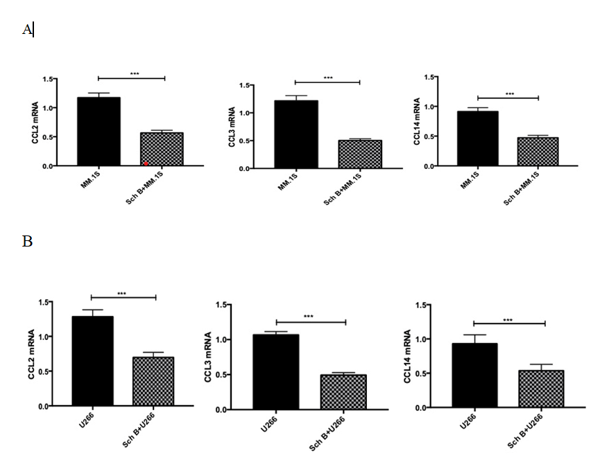
Sch B Suppressed the Mrna Expression of the Chemokines CCL2, CCL3, CCL14 in MM Cell Lines
We used RT-PCR to identify the effect of Sch B on the chemokines CCL2, CCL3 and CCL14 in MM cell lines. As shown in Figure 3, compared with control groups, the mRNA expressions of CCL2, CCL3, and CCL14 were reduced in MM.1S and U266 treated with Sch B. Collectively, Sch B suppressed the mRNA expression of the chemokines CCL2, CCL3and CCL14 in MM cell lines.
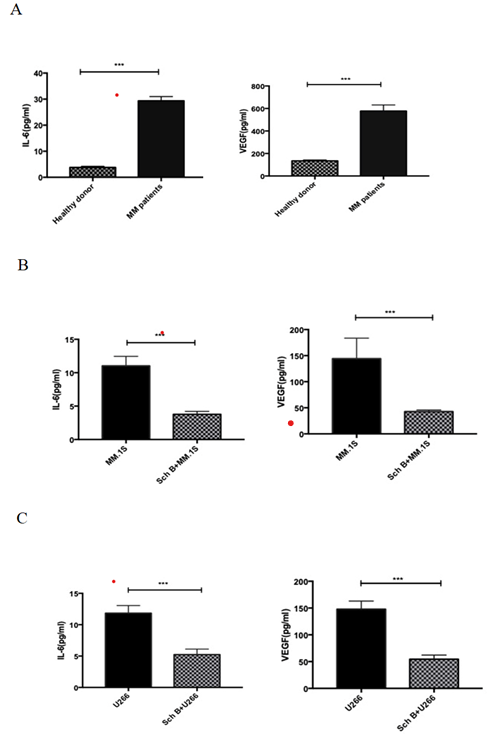
Sch B Inhibited MM Cell Lines Secreting IL-6 and VEGF
We measured the production of IL-6 and VEGF to investigate the effect of Sch B on MM cell lines. As shown in Figure 4, there were high levels of IL-6 and VEGF in MM patients, MM.1S and U266, But the concentration of IL-6 and VEGF was decreased enormously when Sch B was added into MM.1S and U266. Thus, Sch B inhibited MM cell lines secreting IL-6 and VEGF.
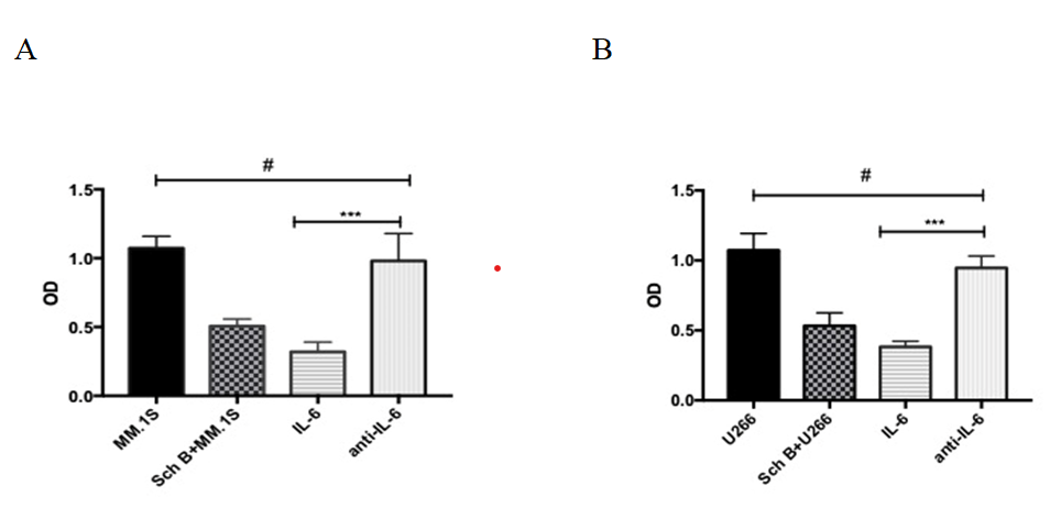
Sch B Suppressed the Proliferation of MM Cell Lines Via IL-6
To investigate the mechanism of Sch B suppressing the proliferation of MM cell lines. As shown in Figure 5A, we found that the OD of MM.1S was decreased by Sch B. When IL-6 was added, the OD of MM.1S was significantly reduced. However, when anti-IL-6 was added, the inhibition efficacy of Sch B in MM.1S was disappeared. This phenomenon was observed in proliferation of U266 (Figure 5B). Thus, Sch B suppressed the proliferation of MM cell lines via IL-6.
Discussion
In this present study, we have successfully demonstrated a previously uncharacterized fact that Sch B suppressed the proliferation of MM cell lines via IL-6.
MM is a common hematological malignancy. The diagnosis of MM is made from anemia, renal complications, bone lesions, hypercalcemia, monoclonal proteins and bone marrow plasmacytosis. The pathogenesis of MM is not elucidated. Tumor microenvironment supports MM cell growth and survival. The direct contacts between MM cells and the surrounding stroma upregulate the expression of interleukin-6(IL-6), VEGF, IGF-1 and GDF15, as well as increased expression of anti-apoptotic proteins [10, 11]. CXCR4/CXCL12 axis can regulate homing, adhesion, invasion, migration and mobilization of MM cells out of the bone marrow [12]. CXCL12 also increases VEGF and IL-6 secretion [13, 14]. IL-6 play an important role in the pathogenesis of MM. IL-6 mediates the proliferation of MM and prevent the MM cells from apoptosis [15, 16]. In addition, MM patients with high BM macrophages infiltration have poor prognosis [17]. Chemokines CCL3, CCL2, and CCL14 were highly expressed by myeloma and BM cells, and the levels of CCL14 and CCL3 in myeloma BM positively correlated with the percentage of BM-infiltrating macrophages [18].
Sch B is well-known for its antitumor effect. It was reported that Sch B can induce inhibition of lung cancer, cholangiocarcinoma, gallbladder cancer and gastric cancer cells [19, 20, 21, 22]. Sch B has multiple functions against cancer through cell cycle arrest, apoptosis, ROS production and autophagy [23]. Sch B might induce cell cycle arrest in all the phases of the cell cycle in the cancer cell. For example, Sch B induces prostate cancer cell arrest at S phase by inhibition of cyclin E/CDK2 [24]. Sch B inhibited the proliferation of human lung adenocarcinoma A549 cells in a dose- dependent manner and induced cell cycle arrest at G0/G1 phase by down-regulating the expression of cyclin D1, cyclin- dependent kinase (CDK)4, and CDK6, up-regulating p53 and p21 expression[19]. Sch B induced the apoptosis of human hepatoma SMMC-7721 cells and gallbladder cell [25, 26]. Sch B inhibited the levels of TNF-a, IL-6, IL-1β and PGE2 upregulated by LPS [27]. In this study, we found that Sch B suppressed the proliferation of MM cell lines in a dose-and time-dependent manner and induced cell cycle arrest in G1/ G0 phase and increased apoptosis in both MM cell lines. Sch B suppressed secreting IL-6, VEGF and the mRNA expression of the chemokines CCL2, CCL3, CCL14 in MM cell lines. Most important, Sch B suppressed the proliferation of MM cell lines via IL-6.
Conclusion
This study reports for the first time that Sch B suppressed the proliferation of MM cell lines via IL-6.
Acknowledgment
The author would like to thank Professor Zhizhe Chen and Xiongpeng Zhu for editing the manuscript.
Conflict of Interest
The authors have no competing financial interests to declare.
Founding
It is founded by Jiangxi Provincial Health Commission Science and Technology plan (202210137).
References
-
Xia J, Zhang J, Wu X, Du W, Zhu Y, et al. (2022) Blocking glycine utilization inhibits multiple myeloma progression by disrupting glutathione balance. Nat Commun 13(1): 4007.
-
Vo JN, Wu YM, Mishler J, Hall S, Mannan R, et al. (2022) The genetic heterogeneity and drug resistance mechanisms of relapsed refractory multiple myeloma. Nat Commun 13(1): 3750.
-
Mikhael J, Bhutani M, Cole CE (2023) Multiple Myeloma for the Primary Care Provider: A Practical Review to Promote Earlier Diagnosis Among Diverse Populations. Am J Med 136(1): 33-41.
-
Bazarbachi AH, Al Hamed R, Malard F, Harousseau JL, Mohty M (2019) Relapsed refractory multiple myeloma: a comprehensive overview. Leukemia 33(10): 2343- 2357.
-
Hideshima T, Mitsiades C, Tonon G, Richardson PG, Anderson KC (2007) Understanding multiple myeloma pathogenesis in the bone marrow to identify new therapeutic targets. Nat Rev Cancer 7(8): 585-598.
-
Kumar SK, Rajkumar SV, Dispenzieri A, Lacy MQ, Hayman SR, et al. (2008) Improved survival in multiple myeloma and the impact of novel therapies. Blood 111(5): 2516- 2520.
-
Ma MY, Wei N, Yang JR, Ding TG, Song AP, et al. (2023) Schisandrin B promotes senescence of activated hepatic stellate cell via NCOA4-mediated ferritinophagy. Pharm Biol 61(1): 621-629.
-
Yang P, Liu P, Li J (2022) The Regulatory Network of Gastric Cancer Pathogenesis and Its Potential Therapeutic Active Ingredients of Traditional Chinese Medicine Based on Bioinformatics, Molecular Docking, and Molecular Dynamics Simulation. Evid Based Complement Alternat Med 2022: 5005498.
-
Li JN, Lu Y, Wang DW, Quan F, Chen X, et al. (2019) Schisandrin B prevents ulcerative colitis and colitis- associated-cancer by activating focal adhesion kinase and influence on gut microbiota in an in vivo and in vitro model. European Journal of Pharmacology 854: 9-21.
-
García-Ortiz A, Rodríguez-García Y, Encinas J, Maroto- Martín E, Castellano E, et al. (2021) The Role of Tumor Microenvironment in Multiple Myeloma Development and Progression. Cancers (Basel) 13(2): 217.
-
Tanno T, Lim Y, Wang Q, Chesi M, Bergsagel PL, et al. (2014) Growth differentiating factor 15 enhances the tumor-initiating and self-renewal potential of multiple myeloma cells. Blood 123(5): 725-733.
-
Alsayed Y, Ngo H, Runnels J, Leleu X, Singha UK, et al. (2007) Mechanisms of regulation of CXCR4/SDF-1 (CXCL12)-dependent migration and homing in multiple myeloma. Blood 109(7): 2708-2717.
-
Aggarwal R, Ghobrial IM, Roodman GD (2006) Chemokines in multiple myeloma. Exp Hematol 34(10): 1289-1295.
-
Hideshima T, Chauhan D, Hayashi T, Podar K, Akiyama M, et al. (2002) The biological sequelae of stromal cell- derived factor-1alpha in multiple myeloma. Mol Cancer Ther 1(7): 539-544.
-
Zhong L, Xu ZY, Jin X, He Y, Zhang JB, et al. (2020) miR- 451a suppression of IL-6R can inhibit proliferation and increase apoptosis through the JAK2/STAT3 pathway in multiple myeloma. Oncol Lett 20(6): 339.
-
Wang JP, Sheng LX, Lai YL, Ouyang GF, Xu ZJ (2022) Effects of physical activity on clinical and inflammatory markers in diagnosing multiple myeloma patients. Front Physiol 13: 1094470.
-
Suyani E, Sucak GT, Akyurek N, Sahin S, Baysal NA, et al. (2013) Tumor-associated macrophages as a prognostic parameter in multiple myeloma. Ann Hematol 92(5): 669-677.
-
Li Y, Zheng YH, Li TS, Wang Q, Qian JF, et al. (2015) Chemokines CCL2,3,14 stimulate macrophage bone marrow homing, proliferation and polarization in multiple myeloma. Oncotarget 6(27): 24218-24229.
-
Lv XJ, Zhao LJ, Hao YQ, Su ZZ, Li JY, et al. (2015) Schisandrin B inhibits the proliferation of human lung adenocarcinoma A549 cells by inducing cycle arrest and apoptosis. Int J Clin Exp Med 8(5): 6926-6936.
-
Yang XH, Wang SA, Mu YC, Zheng YX (2016) Schisandrin B inhibits cell proliferation and induces apoptosis in human cholangiocarcinoma cells. Oncol Rep 36(4): 1799-1806.
-
Xiang SS, Wang XA, Li HF, Shu YJ, Bao RF, et al. (2014) Schisandrin B induces apoptosis and cell cycle arrest of gallbladder cancer cells. Molecules 19(9): 13235-13250.
-
He L, Chen H, Qi Q, Wu N, Wang Y, et al. (2022) Schisandrin B suppresses gastric cancer cell growth and enhances the efficacy of chemotherapy drug 5-FU in vitro and in vivo. Eur J Pharmacol 920: 174823.
-
Chen YQ, Kong Y, Wang QL, Chen J, Chen H, et al. (2021) Schisandrin B Attenuates Airway Inflammation by Regulating the NF-kB/Nrf2 Signaling Pathway in Mouse Models of Asthma. J Immunol Res 2021: 8029963.
-
Nasser MI, Han T, Adlat S, Tian Y, Jiang N (2018) Inhibitory effects of Schisandrin B on human prostate cancer cells. Oncology Reports 41(1): 677-685.
-
Wu YF, Cao MF, Gao YP, Chen F, Wang T, et al. (2004) Down-modulation of heat shock protein 70 and up- modulation of Caspase-3 during schisandrin B-induced apoptosis in human hepatoma SMMC-7721 cells. World Journal of Gastroenterology 10(20): 2944-2948.
-
Xiang SS, Wang XA, Li HF, Shu YJ, Bao RF, et al. (2016) Schisandrin B induces apoptosis and cell cycle arrest of gallbladder cancer cells. Molecules 19(9): 13235-13250.
-
Liu N, Zheng JX, Zhuang YS, Zhou ZK, Zhao JH, et al. (2017)Anti-Inflammatory Effects of Schisandrin B on LPS-Stimulated BV2 Microglia via Activating PPAR-γ. Inflammation 40(3): 1006-1011.
- The Muculent Bleb-Mucinous Cystic Neoplasm-Hepatobiliary Region
- Insulin Sensitizers as Anti-Aging Agents: Unveiling Synergies with Albumin, GLP-1RA, Klotho Protein, and Metformin in the Quest to Combat Aging
- Reprogramming of GLP-1 Response at Prediabetes for the Prevention of Type 2 Diabetes: The Role of Albumin and GLP-1 Receptor Agonists
- The Mingled Allies-Combined Hepatocellular Carcinoma and Cholangiocarcinoma
- Compilation and Embodiment-Leydig Cell Tumour Testis
- Glucolipotoxicity: A Novel Different Perspective on the Causes of Cancer