Paravesical Hernia, An Extremely Rare Cause of Small Bowel Obstruction: Case Report
We report a case of a paravesical internal hernia with a secondary low-grade small bowel obstruction in a 60-year-old man without previous history of surgery.
Morales-Cárdenas A¹*, Duran-Torres Marcela1, Cogollo-Delgado J², Pérez Juan M¹ and Caviedes Gabriel1
¹Department of Diagnostic Radiology, Rosario University, Colombia ²Department of Diagnostic Radiology, Pontifical Xavierian University, Colombia Keywords: Internal Hernia; Paravesical Hernia; Bowel Obstruction; Computed Tomography
Introduction
Internal hernias are characterized by the protrusion of intra-abdominal contents through a retroperitoneal fossa or a defect in the abdominal cavity. The location of the defect defines the subtype of the hernia, of which the least frequent is paravesical.
In paravesical hernias, the herniated contents protrude through the median and medial umbilical ligaments.
Given the nonspecific radiological findings, in the few previous case reports the diagnosis was always done by surgery. This is the first case with suggestive findings on CT.
Case Presentation
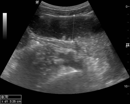
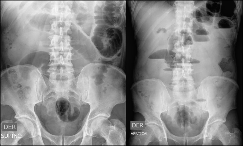
60-yo-male with one week of small bowel obstruction symptoms including abdominal pain, vomiting, and constipation. An initial abdominal US was performed, revealing dilated small bowel loops (Figure 1) and an abdominal x-ray showed multiple air-fluid levels (Figure 2).
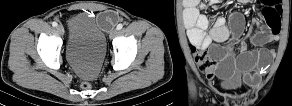
Due to poor response to medical treatment with NG tube placement an abdominal CT was performed, demonstrating several dilated small bowel loops with a transitional area in “closed-loop” configuration in the distal ileum, localized in the left paravesical space, suggestive of an internal hernia, more specifically a left paravesical hernia (Figure 3). A laparotomy was done and the diagnosis was surgically confirmed.
Discussion
Internal hernias are defined as the protrusion of internal structures through a retroperitoneal, peritoneal, or mesenteric defect. The incidence is low (less than 1%) and is responsible for 5,8% of the bowel obstructions [1, 2, 3, 4, 5, 6].
According to the location of the defect, internal hernias are subclassified in paraduodenal, pericecal, transmesenteric, transomental, through the Winslow foramina, through the sigmoid mesocolon, supravesical and paravesical, of which the least frequent is the latter [3, 5].
In paravesical hernias, the defect is located between the median and medial umbilical ligaments. There are four subtypes based on the course of the herniated organs:
anterior, posterior, right, or left lateral**. The most frequent clinical manifestation is bowel obstruction [1].
The radiological findings of the internal paravesical hernias include a cluster of bowel loops and mesenteric fat adjacent to the bladder with proximal dilatation, deformity of the bladder walls adjacent to the hernia and congestive mesenteric vessels [4, 2]. According to our case, a “closed- loop” adjacent to the bladder also could be found.
Conclusion
Paravesical hernias are extremely rare internal abdominal hernias, which usually require surgery for the diagnosis. Timely diagnosis based on the radiological findings leads to rapid treatment and, therefore, a better prognosis for patients, avoiding vascular complications.
References
-
Sardiwalla II, Phakula ML, Zimba MT, Koto ZM (2016) Small bowel obstruction secondary to paravesical hernia. Int J Surg Case Rep 26: 156-158.
-
Martin LC, Merkle EM, Thompson WM (2006) Review of internal hernias: radiographic and clinical findings. AJR Am J Roentgenol 186(3): 703-717.
-
Salar O, El-Sharkawy AM, Singh R, Speake W (2013) Internal hernias: a brief review. Hernia 17(3): 373-377.
-
Takeyama N, Gokan T, Ohgiya Y, Satoh S, Hashizume T, et al. (2005) CT of Internal Hernias. Radio Graphics 25(4): 997-1015.
-
Blachar A, Federle MP, Dodson SF (2001) Internal hernia: clinical and imaging findings in 17 patients with emphasis on CT criteria. Radiology 218(1): 68-74.
-
Selçuk D, Kantarci F, Oğüt G, Korman U (2005) Radiological evaluation of internal abdominal hernias. Turk J Gastroenterol 16(2): 57-64.
- Ultrasound Guided Therapeutic Nerve Blocks
- Cyclops Lesion Without ACL Reconstruction: A Rare Case in a Patient with Intact Anterior Cruciate Ligament and Tibial Plateau Fracture
- Dosimetric Comparison between Two Dose Calculation Algorithms in SBRT Treatment of Lung Cancer in Ring-based and C-arm Radiation Therapy Equipment
- Adolescent Testicular Adrenal Rest Tumors: A Case Report and Review of the Literature
- Giant Intrathoracic Lipoma: A Rare Presentation
- Image of a Right Renal Angiomyolipoma Complicated by Hemorrhage