A Case Report of an Isolated Calvarial Defect in the Region of the Lambdoid Suture: A Rare Bone Manifestation of Neurofibromatosis Type 1
The Calvarial bone defects in the lambdoid suture region are a rare bone manifestation of neurofibromatosis type 1. They may be isolated or associated with an adjacent structural lesion, such as a dural ectasia or plexiform neurofibroma. The underlying pathophysiology for the development of these types of skull defects is still unclear. We present the case of a Calvarial defect in the region of the lambdoid suture in a 7-year-old boy with a medical history of neurofibromatosis type 1 who was referred for a brain scan to explore a palpable skull defect in right parieto-occipital region , noticed by his doctor.
Introduction
Neurofibromatosis type 1, also known as Von Recklinghausen’s disease, is the most common type of neurofibromatosis, characterized by neurocutaneous and bone tropisms. Bone manifestations can often be revealing of the condition. The calvarial defect in the lambdoid suture region is a rare manifestation of neurofibromatosis type 1, and its discovery is usually incidental.
Case Report
A 7-year-old boy with a medical history of neurofibromatosis type 1 was referred for a head CT to explore a palpable skull defect in the right parieto-occipital region posterior to the right ear, noticed by his doctor. The head CT shows a well-defined, rounded bone defect in the region of the right lambdoid suture (Figure 1). There are non-cerebral parenchyma abnormalities.
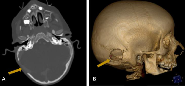
Figure1: Head CT in bone window (A) with three-dimension reconstruction (B) shows a well-defined, rounded bone defect involving the left lambdoid suture (yellow arrow).
Discussion
Neurofibromatosis (NF) is a group of genetic disorders that primarily affect the cell growth of neural tissues. Neurofibromatosis type 1, also known as Von Recklinghausen’s disease, is the most common type of NF, and accounts for around 90% of all cases [1]. The disease commonly begins at a young age in children and involves multiple systems including the bone system. It is diagnosed using a set of well-defined criteria, including clinical and imaging findings [2]. These criteria include the presence of at least six brown spots, two neurofibromas or one plexiform neuroma, two Lisch nodules, axillary or inguinal lentigines, an optic pathway glioma, a distinctive bone lesion, and a first-degree family history. When at least two of these criteria are met, the diagnosis is made.
Bone manifestations have been reported in approximately 50% of neurofibromatosis type 1 patients [3]. The most common abnormalities are scoliosis, sphenoid wing dysplasia, short stature, tibial pseudarthrosis and macrocephaly. Calvarial defect is a very rare bone manifestation of NF1, and its most obvious site is the lambdoid suture. It may be isolated, as in our case, or associated with a plexiform neurofibroma or dural ectasia. The pathogenesis of calvarial defect in patients with NF1 is not clearly identified. One hypothesis proposes that the mutation in the NF-1 gene causes inherent abnormalities in bone development due to a deficiency of neurofibromin protein, alongside impaired mesodermal and neuroectodermal development [4]. Others suggest that bone defects related to NF-1 may be acquired due to elevated external pressure from underlying plexiform neurofibromas and meningiomas, resulting in bone corrosion [5]. This calvarial defect is generally asymptomatic. The diagnosis is suspected clinically and confirmed by a CT scan. There are no guidelines for the management of this calvarial defect in neurofibromatosis type 1. A systematic follow -up is recommended.
Conclusion
NF-1 is a multisystem autosomal dominant disorder, affecting the skin, bones, soft tissues, and nervous system. Calvarial defects are rare bone manifestations but distinct findings in patients with neurofibromatosis Type 1. It is important for the radiologist to be aware of this entity, as in some cases it can reveal the disease.
References
-
Feldman DS, Jordan C, Fonseca L (2010) Orthopaedic manifestations of neurofibromatosis type 1. J Am Acad Orthop Surg 18(6): 346-357.
-
Aoki S, Barkovich AJ, Nishimura K, Kjos BO, Machida T, et al. (1989) Neurofibromatosis types 1 and 2: cranial MR findings. Radiology 172 (2): 527-534.
-
Cimino PJ, Gutmann DH (2018) Neurofibromatosis type 1. Handb Clin Neurol 148: 799-811.
-
Mislow JM, Proctor MR, Mcneely PD, Greene AK, Rogers GF (2007) Calvarial defects associated with neurofibromatosis type 1. Report of two cases. J Neurosurg 106(6S): 484-489.
-
Almahmood H, Al Sayed S, Agab W (2024) Lambdoid Suture Defect in a 12-year-old Neurofibromatosis Patient. Cureus 16(2): e54567.
- Ultrasound Guided Therapeutic Nerve Blocks
- Cyclops Lesion Without ACL Reconstruction: A Rare Case in a Patient with Intact Anterior Cruciate Ligament and Tibial Plateau Fracture
- Dosimetric Comparison between Two Dose Calculation Algorithms in SBRT Treatment of Lung Cancer in Ring-based and C-arm Radiation Therapy Equipment
- Adolescent Testicular Adrenal Rest Tumors: A Case Report and Review of the Literature
- Giant Intrathoracic Lipoma: A Rare Presentation
- Image of a Right Renal Angiomyolipoma Complicated by Hemorrhage