Distribution of Sesamoid Bones in the Foot and Hand Radiographs; A Descriptive Cross-Sectional Study in a Tertiary Care Center in Nepal
Objective: The aim of this study was to present data about the prevalence and distribution of sesamoid bones of the hands and feet in Nepalese population by using radiographs, to determine the number of the sesamoid bones of the foot and hand, commonest site of occurrence and to correlate the findings between gender and age. Methodology: This prospective quantitative study was performed in the Department of Radiology and Imaging, TUTH. The radiographs of foot and hand were reviewed to evaluate the presence or absence of sesamoid bones and patient demographics were also noted. Data collection was done for a period of three months and random probability sampling method was used. Results: This study resulted that in foot radiographs, the most common sites of sesamoid bones was first metatarsophalangeal joint (1st MTPJ) where two sesamoid bones were found, whereas single sesamoid bone was found in other sites. The percentage of sesamoid bones in first interphalangeal joint (1st IPJ), second metatarsophalangeal joint (2nd MTPJ), third metatarsophalangeal joint (3rd MTPJ), fourth metatarsophalangeal joint (4th MTPJ) and fifth metatarsophalangeal joint (5th MTPJ) were 7.9%, 15.4%, 0 %, 0.4%, and 26.7% respectively. The number of sesamoid bones ranged from 2- 5 in single foot with frequency of occurrence of 2 bones being highest (58.3%). The percentage of sesamoid bones in foot was found to be relatively higher in female than male. In the analysis of hand radiographs, the most common sites of sesamoid bones was 1st MCPJ ( single sesamoid bone in 80.33% and a pair of sesamoid bones in 19.67% ) .The percentage of sesamoid bones in 1st IPJ, 2nd MCPJ, 3rd MCPJ, 4th MCPJ and 5th MCPJ were 37.33%, 58.67% ,5.33% , 5.33% and 59.33% respectively. The number of sesamoid bones ranged from 1 - 6 in single hand with frequency of occurrence of 3 bones being higher (32%). The percentage of sesamoid bones in hand was relatively higher in male than female. Conclusion: The distribution and frequency of sesamoid bones differ according to sites, gender and age. This study improves the knowledge on distribution of sesamoid bones of the foot and hand and also provides anatomical data that could help clinicians in diagnosis and management of various sesamoid bone disorders in patients who present with pain and discomfort in the foot and hand.
Sandip Sah* and Sudil Paudyal
Abbreviations: TUTH: Tribhuvan University Teaching Hospital.
Introduction
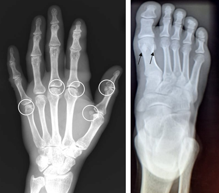
The word Sesamoid bone is attributed to “Galen” who described small bones of hands and feet which resembled the seed of the “Sesamum Indicum” an ancient East Indian plant used for purging by the Greeks [1]. Sesamoid bones are small nodules with a round or oval shape that are embedded within tendon [2]. Sesamoid bones can be found in places where a tendon crosses a joint, such as the hand, wrist, knee, and foot. Sesamoid bones provide a variety of purposes. They preserve the tendon while also increasing its mechanical action by retaining it in place. Certain Sesamoid bones, such as the patella, are present in all people. The great toe’s Sesamoid bones, which are small and appear inconsequential, can be the site of crippling pathology. Any structural irregularity in them can induce gait pain, which is why their research is so important. When symptoms appear, it’s easy to overlook the pathologic states of these bones, and the patients are diagnosed. There could be two or more ossification centers, which could converge or not. As a result, bi, tri, and even quadripartite Sesamoid can be found in adults [3]. Most Sesamoid begins as cartilaginous nodules that undergo endochondral ossification during early to late childhood [4]. Ossification commences first at the age of 10 years in females and 11 years in males and is completed by the age of 13 years and 14 years respectively [1]. The clinical importance of Sesamoid bones are; Reiter’s syndrome, Sesamoid arthritis, fractures and sesamoiditis. Sesamoid bones have been seen with periostitis in Reiter’s syndrome [4]. The purpose of this study was to examine the plain antero-posterior radiographs and oblique radiographs of foot and hand in order to determine the distribution of Sesamoid bones in different parts of foot and hand (Figure 1).
Methodology
This was a quantitative cross–sectional study performed in Department of Radiology and Imaging, Tribhuvan University Teaching Hospital (TUTH). The materials required for this study were radiographs of hand and foot, computer software from digital x-ray machine (Shimadzu Corporation). The AP and oblique radiograph of hand and foot were collected from the DR x-ray machine. The presence and absence of Sesamoid bones was evaluated. Proforma
was made to evaluate the distribution of Sesamoid bones on the radiograph. The results were analysed and corrected using SPSS version 26. Chi-square test was used to analyse the data. P-value < 0.05 was considered as statistically significant. Ethical approval was taken from the Institutional Review Committee of Institute of Medicine.
Results
Out of 540 cases, 240 cases of foot and 300 cases of hand radiographs were studied. Among them 251 (46.48%) were female and 289 (53.52%) were male. In the foot radiographs, the mean age (± SD) of male patients was 39.63±16.36 years, the range being 16-80 years; in females the mean age was 36.72±13.58 years, the range being 17-73 years. Majority of them, 25 % (60) belongs to age group 16 to 25 years, 24.58%(59) belongs to age group 26 to 35 years, 20.83% (50) belongs to 36 to 45 years, 13.33%(32) belongs to 46 to 55 years, 11.67% (28) belongs to age groups 56 to 65 years, 3.33% (8) belongs to 66 to 75 years and 1.25%(3) belongs to above 76 years. The percentage of Sesamoid bones in the first MTPJ (the most common location of Sesamoid bones in foot) was found to be 100% of the total cases. At these joint two Sesamoid bones was found whereas single Sesamoid bone was found in rest sites. The prevalence of 1st IPJ, 2nd MTPJ, 3rd MTPJ, 4th MTPJ, 5th MTPJ was 7.9%, 15.4%, 0%, 0.4% and 26.7% respectively. The minimum number of Sesamoid bone in single foot is 2 and maximum number is 5. The percentage and frequency of Sesamoid bones in single foot- 2 is 58.3%(140), 3 is 33.3%(80), 4 is 7.9%(19), and 5 is 0.4%(1). The Sesamoid bones were statistically more frequent at the 2nd MTPJ in female (22.9%) than male (9.2%) with p value = 0.003. The percentage of Sesamoid bones was found to be higher in female at the 1st IPJ and 5th MTPJ compared with male; however, these finding were not statistically significant (p= 0.859 and p = 0.571) respectively. There is no any difference between male and female at 1st MTPJ and 3rd MTPJ which is shown below in Table 1. The distribution and frequency of Sesamoid bones in bilateral foot was found similar and equal in 24 patients. So, no difference was found between right and left foot in the patients.
| Gender | 1st IPJ | 1st MTPJ | 2nd MTPJ | 3rd MTPJ | 4th MTPJ | 5th MTPJ |
|---|---|---|---|---|---|---|
| Female (n=109) | 8.30% | 100% | 22.90% | 0% | 0% | 28.45 |
| Male (n=131) | 7.60% | 100% | 9.20% | 0% | 0.80% | 25.20% |
| p- value | 0.859 | - | 0.003 | - | 0.361 | 0.571 |
Table 1: The distribution and frequency of Sesamoid bones in bilateral foot was found similar and equal in 24 patients. So, no di
In the hand radiographs, the mean age (±SD) of male patients was 35.95 ±14.7 years, the range being 16-79 years; in females the mean age was 41.28±14.6 years, the range being 16-72 years. Majority of them 24.33%(73) belongs to age group 16 to 25 years, 25% (75) belongs to 26 to 35 years, 21.33% (64) belongs to age group 36 to 45 years, 14% (42) belongs to age group 46 to 55 years, 11.33% (34) belongs to age group 56 to 65 years, 3% (9) belongs to age group 66 to 75 years and 1% (3) belongs to age group above 76. The percentage of Sesamoid bones in the 1st MCPJ (the most common location of sesamoid bones in hand) was found to be 100% of the total cases. At this joint, single sesamoid bone was found in 80.33% and a pair of sesamoid bones in 19.67%. The percentage of sesamoid bones at 1st IPJ, 2nd MCPJ, 3rd MCPJ, 4th MCPJ and 5th MCPJ was found to be 37.33% , 58.67% ,5.33%, 5.33% and 59.33% respectively. The minimum number of sesamoid bone in single hand is 1 and maximum number is 6. The number of sesamoid bones ranges from 1-6 in single hand. The percentage and frequency sesamoid bone in single hand- 1 is 14%(42), 2 is 24%(72), 3 is 32%(96), 4 is 23.3%(70), 5 is 5.7%(17), 6 is 1%(3). The sesamoid bones were statistically more frequent at the 2nd MCPJ and 3rd MCPJ in male than female with p= 0.009, and p=0.004 respectively. The percentage of sesamoid bones was found to be higher in male than female at 1st IPJ and 5th MCPJ; however , these finding were not statistically significant ( p= 0.809 and p = 0.000) respectively. The percentage of single sesamoid bone is higher in male than female whereas pair of sesamoid bone is higher in female which is shown in Table 2. The distribution and frequency of sesamoid bones in bilateral hand was found similar and equal in 32 patients. So, no difference was found between right and left hand in the patients.
| Gender | 1st IPJ | 1stMCPJ(1SB) | 1stMCPJ(2SB) | 2nd MCPJ | 3rd MCPJ | 4th MCPJ | 5th MCPJ |
|---|---|---|---|---|---|---|---|
| Female (n=142) | 36.60% | 76.80% | 23.20% | 52.10% | 1.40% | 4% | 48.60% |
| Male (n=158) | 38.00% | 83.50% | 16.50% | 64.60% | 8.90% | 7.60% | 69.00% |
| P - value | 0.809 | 0.141 | 0.141 | 0.029 | 0.004 | 0.066 | 0 |
Table 2: The distribution and frequency of sesamoid bones in bilateral hand was found similar and equal in 32 patients. So, no di
Discussion
This study was done to ascertain the prevalence of sesamoid bones in the foot and hand using radiographs. The result obtained showed that the two sesamoid bone in the first meta-tarsophalangeal joint of foot was the most common site accounting for the prevalence of 100% in both male and female. This value was higher than an early study done on the Nigerians population by Udoaka AI, et al. [5] in 2013 whose value of prevalence was 81% in 1st MTPJ. This may be the result of geographical variation and ethnic group. The result obtained in this study was similar on comparing with another study done by Yammine K [6] in 2015, who found 100% prevalence. Also, Goldberg I, et al. [7] in 1987 recorded 100% occurrence of sesamoid bones in 1st MTPJ. A study done in Sudan by Dr. Talha AE, et al. [8] in 2016 found that the prevalence occurrence sesamoid bone was 100% in the first MTPJ .This study was similar with study of Goldberg I, et al. [7] and Talha AE, et al. [8].
The result obtained in this study showed that the prevalence of sesamoid bone at first IPJ is 7.9% but it was found to be 22.4% in a study done by Yammine K in 2015. The prevalence of sesamoid bone at first IPJ in this study was somewhat low. The respective percentage of occurrence of sesamoid bones at the MTPJ of 2nd, 3rd, 4th, and 5th are 15.4%, 0%, 0.4% and 26.7% respectively. However, in Yammine K [6] study, the prevalence at MCPJ for 2nd, 3rd, 4th and 5th was 1.9%, 0.32%, 0.9% and 13% respectively. This study showed higher result at 5th MCPJ. The variation found was due to racial and geographical regions. There was no significant difference between right and left foot radiograph in frequency and distribution of sesamoid bone however a study done in Sudan by Dr. Talha AE, et al. [8] in 2016 found difference in right and left foot radiograph .Thus, this study was differ from study of Dr. Talha AE, et al. [8] may be the result of racial variation. The results were correlated with the age and sex. The results showed that the prevalence was higher in female than male at 1st IPJ, 2nd MTPJ and 5th MTPJ and lower at 4th MTPJ but similar at 1st MTPJ. The number of sesamoid bones ranges from 2-5 in single foot and the frequency of number of sesamoid bone 2 was found higher(58.3%)and lower in frequency of sesamoid bone 5(0.4%). However, number of sesamoid bone identified in a single foot was 2-7 in the study done by Dr. Talha AE, et al. [8] in 2016.
In the study of hand radiographs, the result showed that the sesamoid bone of 1st MCPJ was the most common accounting for the prevalence of 100% (80.33% one sesamoid bone and 19.67 % two sesamoid bone). The respective percentage of occurrence of sesamoid bones at 1st IPJ, 2nd MCPJ, 3rd MCPJ, 4th MCPJ and 5th MCPJ was 37.33%, 58.67%, 5.33%, 5.33% and 59.33% respectively . In Turkey by Kose O, et al. [2] in 2012 1st MCPJ in all subjects have 100% bilaterally sesamoid bones and the prevalence of 2nd MCPJ, 3rd MCPJ, 4th MCPJ 5th MCPJ and 1st IPJ was 42.8%, 1.6% , 0.1% , 72.5% and 21.8% respectively. This study was dissimilar with study in Turkey. This may be the result of geographical and racial group. Another study done by Lam GYT, et al. [9] in 2017 in china, sesamoid bones were found at the first MCPJ at a rate of 100%, whereas the prevalence at the second MCPJ, third MCPJ, fourth MCPJ, fifth MCPJ and first IPJ was 59.0%, 2.93%, 0%, 47.6% and 28.0%, respectively. Two sesamoid bones were always found at the first MCPJ, whereas one sesamoid bone was identified at other joints. This study is similar 1st MCPJ, but somewhat differ at other site of sesamoid bone. Also another study were done in Sudan by Mohammed W, et al. [4] in 2018 found that sesamoid bones were always found at the 1st MCPJ and percentage of sesamoid bone at 1st IPJ, 2nd MCPJ, 3rd MCPJ, 4th MCPJ and 5th MCPJ were 7%, 47.36%, 7%, and 14% respectively. This study was similar at 1st MCPJ but differ at other sites of sesamoid bones.
The number of sesamoid bone range from 1-6 in single hand. The minimum number of sesamoid bone was 1 and maximum number of sesamoid bone was 6. The frequency of sesamoid bones of 1, 2, 3, 4, and 6 was 14%, 24%, 32%, 23.3%, 5.7%, 1% respectively. However the range of sesamoid bone in single hand was 1- 4 in the study by Mohammed W, et al. [4] in 2018 and the frequency of one sesamoid bone was found higher (45.6%) and lower (1.8%) in 4 sesamoid bone . This study was quite different than study of Mohammed W, et al. [4]. There is no significant difference between frequency and distribution of sesamoid bone between both hands in a single patient which is similar to the study done by Lam GYT, et al. [9] in 2017 in china and also similar with study in Sudan by Mohammed W, et al. [4] in 2018. This may be the result of study done in similar age group.
The sesamoid bones were statistically more frequent at the 1st IPJ, 2nd MCPJ and 3rd MCPJ, 5th MCPJ in male than female. The percentage of single sesamoid bone is higher in male than female whereas pair of sesamoid bone is higher in female. In the study done by Kose O, et al. [2] in 2012 in Turkish, 1st IPJ, 2nd MCPJ, 3rd MCPJ and 5th MCPJ have higher prevalence in females than male. Thus, this study was different from study of Kose O, et al. [2] in 2012 in Turkish. This may be the result of geographical variations. The most common and almost constant sesamoid bone in foot and hand is 1st MTPJ and 1st MCPJ respectively. The prevalence of sesamoid bone is higher in female than male in foot but lower in hand. The number of sesamoid bone ranges from 2-5 in single foot and in single hand from 1- 6 sesamoid bones. The present study represents the report on the prevalence and distribution of sesamoid bone of foot and hand in Nepalese people. The results of the study seem to differ considerably from similar studies in other countries. This indicated that the prevalence of sesamoid bone of the foot and hand vary in different population. This will improve the knowledge on sesamoid bone of the foot and hand. It also provides anatomical data that could help clinicians in diagnosis and management of sesamoid bone disorder, a problem of misdiagnosis, mistreatment and missed diagnosis often occur which are often occurring in patients who present with pain and discomfort in the foot and hand.
The new information that can be inferred from this study is that distribution of sesamoid bone is higher in female than male in foot and there is no any difference between bilateral feet in the distribution of sesamoid bone. Sesamoid bones can be found in 1st MTPJ in every individual. Similarly, the distribution of sesamoid is higher in male than female in hand and there is no any difference between bilateral hands in the distribution of sesamoid bone. Sesamoid bone can be found in 1st MCPJ in every individual. The distribution of sesamoid bone was found to be different from other studies. This may be due to geographical variation and ethnicity [10, 11, 12, 13, 14, 15, 16, 17].
Conclusion
Since the sesamoid is an insignificant bone which is not normally taken care of. Different pathological conditions such as stress fractures, turf toe and sesamoiditis may cause various types of problems to the individuals. Researches are being done on the percentage of prevalence of sesamoid bones but not in other aspects of them. That’s why, further study can be done on effect of various pathological conditions of the sesamoid bones which can cause problems to the individuals and correlate them with age, sex etc. The study can also be done on correlating different problems on foot and hand of the individuals with the presence or absence of sesamoid bones.
References
-
Archana A, Sanakkayala S, Nalluri HB, Nagothu RS (2017) Study of Incidence and Ossification of Sesamoid Bones in Hands of South Indian Population. Int J Anat Res 5(1): 3505-3509.
-
Kose O, Guler F, Turan A, Canbora K, Akalin S (2012) Prevalence and distribution of sesamoid bones of the hand. A radiographic study in Turkish subjects. Int J Morphol 30(3): 1094-1099.
-
Ghimire I, Maharjan S, Pokharel GB, Subedi K (2017) Evaluation of occurrence of sesamoid bones in the lower extremity radiographs. Journal of Chitwan Medical College 7(20): 11-14.
-
Mohammed W, Abdulghani YS, Elbadawy K, Ali TO (2017) A Radiographic Study of Prevalence of Sesamoid Bones of the Hand in Adult Sudanese. Indian Journal of Applied Research 7(5): 636-639.
-
Udoaka AI, Didia BC (2013) Prevalence and types of accessory ossicles and sesamoid bones of the feet of adult Southern Nigerians. Research Journal of Medical Sciences 7(1): 25-27.
-
Yammine K (2015) The sesamoids of the feet in humans: a systematic review and meta-analysis. Anat Sci Int 90(3): 144-160.
-
Goldberg I, Nathan H (1987) Anatomy and pathology of the sesamoid bones The hand compared to the foot. Int Orthop 11(2): 141-147.
-
Talha AE, Abbas AA, Abdelghany YS, Yousif F, Mahdi ME (2016) A radiographic study of the prevalence and distribution of sesamoid bones of the foot in adult Sudanese. World Journal of Pharmaceutical and Medicinal Research 2(4): 164-176.
-
Lam GYT, Chow ECS, Ng B, Ho CHG, Chan CW (2017) Distribution of Sesamoid Bones in the Hand A Study in the Chinese Population. J Orthop Trauma Rehabil 23: 45- 48.
-
Pappas DG, Cure JK (2002) Diagnostic imaging. Otolaryngol Clin North Am 35(6): 1317-1363.
-
Whitley AS, Jefferson G, Holmes K, Sloane C, Anderson C, et al. (2015) Clark’s Positioning in Radiography. 13th (Edn.), CRC Press, London, pp: 592.
-
Shabibi AA, Sirasanagandla SR, Thuhli ZA, Dhuhli HA, Mushaiqri MA, et al. (2020) Radiological study on sesamoid bones of the foot among Omani subjects. Oman Med J 35(4): e163.
-
Bushong SC, Stewart CB (2005) Radiologic Science for Technologists: Physics, Biology and Protection. pp: 1-29.
-
Bushberg JT, Anthony SJ, Edwin ML, John MB (2012) The essential physics of medical imaging. 3rd (Edn.), Lippincott Williams & Wilkins, Philadelphia, pp: 1030.
-
Guberman RM (2018) Radiology and Imaging. In: Rock GP, et al. (Eds.), Pocket Foot and Ankle Medicine and Surgery, 1st (Edn.), Lippincott Williams & Wilkins, Philadelphia, US, pp: 27-36.
-
Dharap AS, Al-Hashimi H, Kassab S, Abu-Hijleh MF (2007) Incidence and ossification of sesamoid bones in the hands and feet a radiographic study in an Arab population. Clin Anat 20(4): 416-423.
-
Msamati BC, Igbigbi PS (2001) Radiographic appearance of sesamoid bones in the hands and feet of Malawian subjects. Clin Anat 14(4): 248-53.
- Ultrasound Guided Therapeutic Nerve Blocks
- Cyclops Lesion Without ACL Reconstruction: A Rare Case in a Patient with Intact Anterior Cruciate Ligament and Tibial Plateau Fracture
- Dosimetric Comparison between Two Dose Calculation Algorithms in SBRT Treatment of Lung Cancer in Ring-based and C-arm Radiation Therapy Equipment
- Adolescent Testicular Adrenal Rest Tumors: A Case Report and Review of the Literature
- Giant Intrathoracic Lipoma: A Rare Presentation
- Image of a Right Renal Angiomyolipoma Complicated by Hemorrhage