Measurement of Liver Size by Ultrasound Unveils Large Livers in Overweight Children
Purpose: To analyze the relationship between Body Mass Index (BMI) percentile and liver size and to predict the probabilities of fatty liver and hepatomegaly for overweight and obese boys and girls among Mexican-American children. Methods: One thousand two hundred fourteen reports of children visiting a South Texas pediatric clinic from 2011 to 2018 were analyzed. Ultrasonography was requested for patients whenever the patient was gaining excessive weight and the readings for alkaline phosphatase levels were 2 SD above the normal population; or when liver enzymes were elevated, Aspartate Aminotransferase (AST) above 50/46, Alanine Aminotransferase (ALT) above 47/41, and Gammaglutamyl transferase (GGT) above 32/28 for boys and girls respectively. The data was analyzed using linear regression and logistic regression models. Results: The results of the analysis support that the probability of fatty liver and hepatomegaly increase exponentially as BMI percentile increases. There is also a positive linear relationship between liver size and BMI percentile. Conclusion: The logistic regression analysis predicts that as BMI percentile increases the probability of fatty liver and hepatomegaly increases.
Francisco J Cervantes1, Margarita Faz2, Fernando G Quintana3* and Hongwei Wang3
Science Center 311, Laredo, TX 78041, USA, Tel: 956 326-2589; Email: fquintana@tamiu.edu and logistic regression models.
hepatomegaly increases.
Keywords: Liver; Ultrasound; Overweight; Children
Measurement of Liver Size by Ultrasound Unveils Large Livers in Overweight Children Scan: Computed Tomography Scan; FL: Fatty Liver; H: Hepatomegaly; U/S: Ultrasound Scan.
Introduction
There has been a constant increase of children obesity in many countries of the world. This is a cause of alarm in Diabetes Obes Int J
the society. The prevalence of childhood obesity in USA is a serious problem, putting both children and adolescents at risk for poor health. According to the Centers for Disease Control and Prevention (CDC), an 18.5% or 13.7 million children in USA were obese from 2015 to 2016 [1]. Comparing the prevalence of children obesity in USA from 1999 to 2016, it shows that obesity has increased from 13.9% to 18.5% during this period [1]. In 2005 we showed that, in Laredo TX, at 3 to 4 years of age one third of children were either overweight or obese, at 5 to 9 years one out of two and by 18 years of age 56% of girls and 61% of boys were either overweight (20 and 17% respectively) or obese (36 and 44% respectively) [2] .
Research and clinical results indicate that there is an association between obesity and nonalcoholic fatty liver diseases (NAFLD), which will be the focus of this paper [3, 4, 5, 6, 7]. NAFLD causes no symptoms, but continued damage to the liver can lead to nonalcoholic steatohepatitis (NASH) and this can develop into cirrhosis.
The study of the relation between obesity, fatty liver, and hepatomegaly is of interest for many, as a result of the high prevalence of obesity in children [8, 9, 10]. The aim of this study is to analyze the relationship between BMI and liver size in overweight and obese children visiting a pediatric clinic located in South Texas. Moreover, the aim of this study is also to analyze the relationship between BMI percentile and liver size and to predict the Radiology reports of one thousand two hundred fourteen liver ultrasounds of 699 Mexican-American children visiting a South Texas pediatric clinic from 2011 to 2018 were collected and assessed. The children’s summaries by gender and age for BMI, fatty liver, and hepatomegaly can be found in Table 1.
probabilities of fatty liver and hepatomegaly for overweight and obese boys and girls visiting the facility.
Measuring liver size has always been difficult; it can be established by percussion and palpation which is very difficult, if not impossible, when the patient has an excess of abdominal fat [11]. Ultrasound is the most accessible, non-invasive, cost efficient and reliable method of measuring liver size, other imaging techniques are currently available, such as MRI, CT Scan, and X Ray, but the former are expensive and the latter exposes patients to radiation [12, 13]. Liver biopsy remains the gold standard for NAFLD diagnosis [14].
The normal range of liver size has been reported by various authors and according to our local radiologist, it is standard procedure to measure the sagittal length of the right lobe of the liver, yet the reports that we receive do not mention liver size, or if the liver size is normal or not, and the frequency of fatty liver reported not always correlates with the frequency of hepatomegaly [15, 16, 17].
Methodology
| Age range (years) | N | Age Average ( years)* | BMI average* | Fatty liver % | Hepatomegaly % | ||||||||||||
|---|---|---|---|---|---|---|---|---|---|---|---|---|---|---|---|---|---|
| Boys | |||||||||||||||||
| 0 – 4.9 | 26 | 3.54 ± 0.16 | 21.51 ± 0.74 | 4% | 4% | ||||||||||||
| 5 – 9.9 | 194 | 8.25 ± 0.81 | 24.94 ± 0.29 | 29% | 19% | ||||||||||||
| 10 – 12.9 | 235 | 11.44 ± 0.06 | 27.62 ± 0.36 | 39% | 27% | ||||||||||||
| 13 – 15.9 | 193 | 14.23 ± 0.06 | 29.21 ± 0.49 | 36% | 27% | ||||||||||||
| 16 – 19 | 113 | 16.89 ± 0.12 | 30.96 ± 0.64 | 39% | 24% | ||||||||||||
| Girls | |||||||||||||||||
| 0 – 4.9 | 34 | 2.98 ± 0.19 | 21.90 ± 0.65 | 21% | 3% | ||||||||||||
| 5 – 9.9 | 149 | 8.00 ± 0.10 | 25.72 ± 1.39 | 26% | 13% | ||||||||||||
| 10 – 12.9 | 120 | 11.36 ± 0.08 | 26.20 ± 0.56 | 18% | 21% | ||||||||||||
| 13 – 15.9 | 93 | 14.29 ± 0.08 | 28.32 ± 0.63 | 26% | 13% | ||||||||||||
| 16 – 19 | 57 | 17.03 ± 0.11 | 29.16 ± 0.94 | 21% | 9% |
Table 1: Boys and Girls Age, BMI, Fatty liver and Hepatomegaly Ultrasonography was requested for patients whenever the patient
* Mean± Standard error Table 1: Boys and Girls Age, BMI, Fatty liver and Hepatomegaly Ultrasonography was requested for patients whenever the patient was gaining excessive weight and the readings for alkaline phosphatase levels were 2 times the standard Fernando G Quintana, et al. Measurement of Liver Size by Ultrasound Unveils Large Livers in Overweight Children. Obes Int J 2019, 4(4): 000210.
deviation (2 SD) above the normal population or when liver enzymes were elevated, AST above 50/46, ALT above 47/41, and GGT above 32/28 for both boys and Copyright© Fernando G Quintana, et al.
girls, respectively. BMI percentiles were calculated using the CDC BMI-for-age growth charts. The image readings were made at three different facilities assigned to two groups of radiologists. If the Ultrasound report did not mention the liver size, the image was reviewed and the measure documented by the technician was obtained. IBM-SPSS was used to process the statistical analysis of the data. Linear regression and logistic regression17 were used to model the relation between age and liver size, and to model the relation between BMI and fatty liver and the relation between BMI and hepatomegaly. The data analysis was processed under TAMIU IRB approval # 2012-01-03. Fatty liver (FL) and hepatomegaly (H) were diagnosis given by the radiologists based on their interpretation of the ultrasound, fatty liver is represented as an increased echogenicity of the liver and hepatomegaly is applied when they compare the liver size in question to the radiology standards for children 4,5.
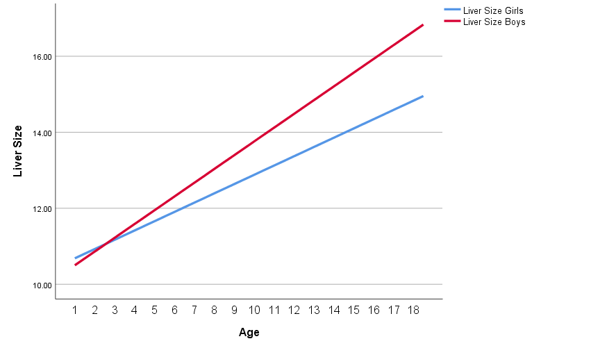
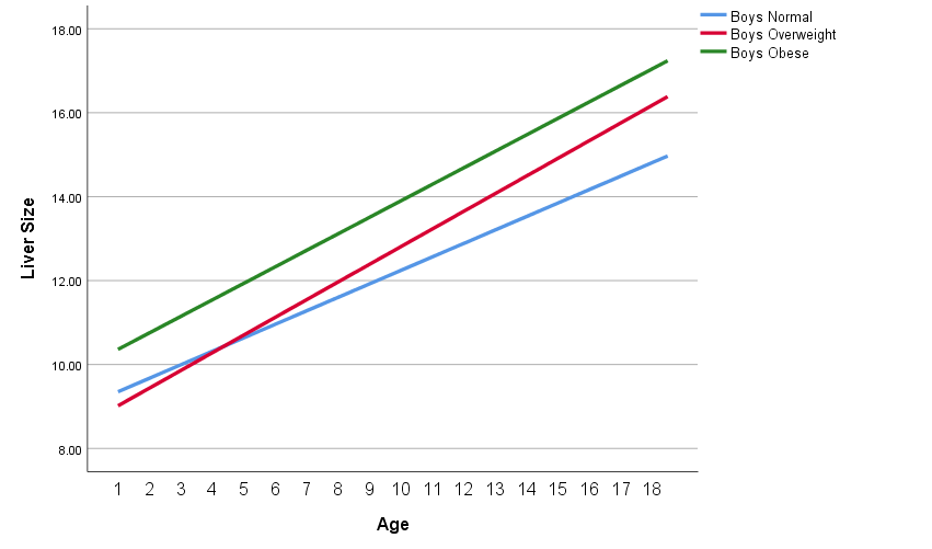
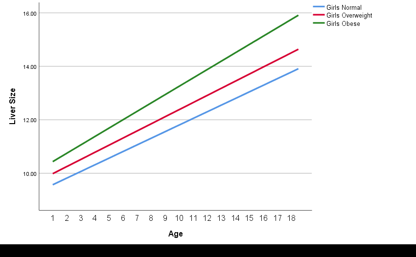
Figure 1 compares liver size by age between boys and girls. Liver size is always larger in boys when compared with girls (girls, a = 10.56, b = 0.24, p < 0.05; boys, a = 10.32, b = 0.36, p < 0.05). Figure 2 compares liver size by age among normal, overweight and obese boys (boys normal, a = 9.19, b = 0.32 p < 0.05; boys overweight, a = 8.08, b = 0.42, p < 0.05; boys obese, a = 10.16, b = 0.39, p < 0.05). Liver size is larger for obese boys when compared with overweight boys and overweight boys have larger liver size than normal boys after 4.5 years of age and gaps remaining increased thereafter. Figure 3 compares liver size by age among normal, overweight, and obese girls (girls normal, a = 9.45, b = 0.25, p < 0.05; girls overweight, a = 9.85, b = 0.27, p < 0.05; girls obese, a = 10.28, b = 0.31, p < 0.05). Liver size is larger for obese girls when compared with overweight girls and overweight girls liver size is always larger than liver size for normal girls.
Results and Analysis
Fernando G Quintana, et al. Measurement of Liver Size by Ultrasound Unveils Large Livers in Overweight Children. Obes Int J 2019, 4(4): 000210.
Copyright© Fernando G Quintana, et al.
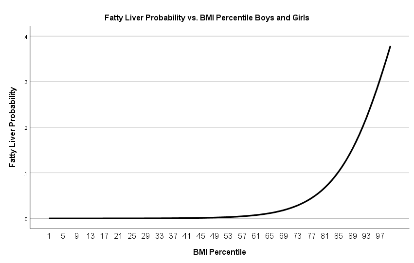
The data was analyzed using logistic regression. The results were as follows. a) boys and girls logistic regression of BMI percentile vs. Fatty Liver (n = 1218, a = -11.83, Wald=49.00, df = 1, p < 0.05; b = 0.12, Wald = 43.39, df = 1, p <0 .05), b) boys and girls logistic regression of BMI Percentile vs. Hepatomegaly (n = 1218, a = -9.42, Wald = 33.47, df = 1, p<0.05; b = .085, Wald =
25.73, df = 1, p < 0.05). Figure 4 describes the probability of fatty liver for boys and girls by BMI percentiles. The probability of fatty liver increases exponentially as BMI percentile increases. Similarly, the probability of hepatomegaly increases exponentially as BMI percentile increases.
Discussion and Conclusion
The results of the logistic regression analysis in this study support that there is a relationship between fatty liver and BMI percentile for the population of boys and girls, and that there is a relationship between hepatomegaly and BMI percentile for the population of Fernando G Quintana, et al. Measurement of Liver Size by Ultrasound Unveils Large Livers in Overweight Children. Obes Int J 2019, 4(4): 000210.
boys and girls. Ezzat WM, et al. [8] reported that enlarged liver was correlated with BMI (r = 0.35, p = 0.0001). Arslan N, et al. [6] found that increased liver echogenicity resembling fatty liver is correlated with higher BMI. These findings support the results obtained in this study. Tominaga K, et al. [7] clinical results confirmed that there is direct relationship between degree of obesity and fatty Copyright© Fernando G Quintana, et al.
liver among Japanese children supporting that the obesity and liver size are correlated.
ethnicity, being the Hispanic (11.8%) the highest prevalence; his study points that NAFLD increases with age from 0.7% in 2 to 4 years old to 17% by teen years; this findings are reproduced in our study, in another study Shwimmer JB [19], identified Hispanics as higher risk for NAFLD.
In a large postmortem study, Schwimmer JB, et al. [18] reported the highest rate of fatty liver in obese children (38% of 742) and it differed significantly by race and
| BMI percentile vs. fatty liver | BMI percentile vs. hepatomegaly | ||||||
|---|---|---|---|---|---|---|---|
| Boys and Girls | n = 1161 | Boys and Girls | n = 1161 | ||||
| a = -11.83 | Wald = 49.00 | a = - 9.42 | Wald = 33.47 | ||||
| b = 0.12 | Wald = 43.00 | b = 0.085 | Wald = 25.73 | ||||
| Boys | n = 840 | Boys | n = 840 | ||||
| a = -22.98 | Wald = 51.00 | a = -16.58 | Wald = 31.79 | ||||
| b = 0.23 | Wald = 51.00 | b = 0.16 | Wald = 29.43 | ||||
| Girls | n = 450 | Girls | n = 450 | ||||
| a = -8.1 | Wald = 18.00 | a = -5.67 | Wald = 12.04 | ||||
| b = 0.07 | Wald = 14.00 | b = 0.041 | Wald = 5.65 |
Table 2: Logistic regression of BMI percentile vs. fatty liver and hepatomegaly.
* df = 1, p < 0.05 Table 2: Logistic regression of BMI percentile vs. fatty liver and hepatomegaly.
Our results indicate that liver size increases for boys and girls as the child becomes overweight and obese and this relationship is maintained as the child ages. The logistic regression analysis predicts that as BMI percentile increases the probability of fatty liver and hepatomegaly increases. For obese children the predicted logistic regression probability for fatty liver is 0.4. It has been estimated that more than one third of the children in USA are overweight or obese [1]. This implies that the predicted number of children with liver disease is very high. Therefore, if we consider that it can be calculated that there are 73.8 million children in USA today [1]. We can also predict using the logistic regression results that the number of children with liver disease today may be as high as 24.6 million. If this trend continues for the years to come, the adult population with liver diseases could be an overwhelming burden on the healthcare system and NASH will be the main cause of liver transplant which is considered expensive and could be fatal [20].
Measurement of the liver by U/S unveils undiagnosed large livers, which obviously relates to an increase of BMI and magnifies the importance of early detection of fatty liver and/or hepatomegaly (even if the radiologist doesn’t label FL or H). We ask for liver U/S, when overweight or obese children had abnormal liver enzymes, which point to liver involvement, most likely NAFLD and/or NASH.
NAFLD and NASH are asymptomatic and by Ultrasound, very hard, if not impossible, to separate from each other. We feel that the best approach is to follow Fernando G Quintana, et al. Measurement of Liver Size by Ultrasound Unveils Large Livers in Overweight Children. Obes Int J 2019, 4(4): 000210.
these children with serial liver enzymes every 6 - 12 months to evaluate function and liver Ultrasound in a yearly basis to follow changes in size. Conflict of Interest: The authors have indicated that they have no potential conflicts of interest to disclose. Funding Source: All phases of this study were supported by Laredo Pediatrics & Neonatology PA Financial Disclosure: The authors have indicated that they have no financial relationships to disclose.
Persons who have Made a Substantive Contribution to the Study
- Francisco J Cervantes, conceptualized, designed, coordinated the data collection, analyzed the initial data and reviewed the manuscript.
- Fernando Quintana, performed the statistical analysis, created the first manuscript and prepared and reviewed the final manuscript.
- Margarita Faz, created the data collection tool, collected data, reviewed the manuscript and ensured it met all the requirements for publication
- Hongwei Wang, performed together with Fernando Quintana, the statistical analysis of the data, reviewed the manuscript and prepared the final manuscript.
Copyright© Fernando G Quintana, et al.
- All authors participated in the analysis and discussion of the results, approved the final manuscript for submission and agreed to become accountable for all aspects of the work.
- The authors wish to acknowledge Dr. Rohitha Goonatilake, TAMIU Professor, for his editorial contribution as a statistician.
- The abstract of this work was presented at the 78th Scientific Sessions of the American Diabetes Association at Orlando, FL on June 25th, 2018.
References
-
Hales CM, Carrol MD, Fryar CD, Cynthia L Ogden (2017) Prevalence of obesity among adults and youth: United States, 2015-2016. NCHS data Brief, No. 288, National Center for Health Statistics.
-
Cervantes FJ, Faz CM (2005) Overweight in children and results of diet modification: Poster presentation. Border Health Association 63rd Annual Meeting, 2005 Laredo TX, Mexico, USA.
-
Yan Y, Hou D, Zhao X, Liu J, Cheng H, et al. (2017) Childhood adiposity and nonalcoholic fatty liver disease in adulthood. Pediatrics 139(4): e20162738.
-
Cervantes FJ, Quintana FG (2016) Statistical relationship between obesity, age, and gender with nonalcoholic fatty liver disease in a group of overweight and obese children visiting a south texas pediatric clinic. Poster presentation, American Diabetes Association 76th Scientific Sessions_,_ New Orleans LA.
-
Cervantes FJ, Quintana FG (2018) Obesity and liver function in a group of children visiting a pediatric clinic in south texas. Poster presentation, American Diabetes Association 78th Scientific Sessions, Orlando FL.
-
Arslan N, Büyükgebiz B, Oztürk Y, Cakmakçi H (2005) Fatty liver in obese children: prevalence and correlation with anthropometric measurements and hyperlipidemia. Turkish journal pediatrics 47(1): 23- 27.
-
Tominaga K, Kurata JH, Chen YK, Fujimoto E, Miyagawa S, et al. (1995) Prevalence of fatty liver in Japanese children and relationship to obesity. An epidemiological ultrasonographic survey. Digestive Diseases and Sciences 40(9): 2002-2009. Fernando G Quintana, et al. Measurement of Liver Size by Ultrasound Unveils Large Livers in Overweight Children. Obes Int J 2019, 4(4): 000210.
-
Ezzat WM, Ragab S, Abdallah N, Yasser A Elhosary, Abeer M Nour Eldin, et al. (2012) Frequency of non- alcoholic fatty liver in overweight/obese children and adults: Clinical sonography picture and biochemical assessment. Journal of Genetic Engineering and Biotechnology 10(2): 221-227.
-
Della Corte C, Alisi A, Saccari A, De Vito R, Vania A, et al. (2012) Nonalcoholic fatty liver in children and adolescents: An overview. Journal of Adolescent Health 51(4): 305-312.
-
El-Koofy N, Anwar GM, El-Raziky MS, El-Hennawy AM, El-Mougy FM, et al. (2012) The association of metabolic syndrome, insulin resistance and non- alcoholic fatty liver disease in overweight/obese children. Saudi J Gastroenterology 18(1): 44-49.
-
Wolf DC (1990) Evaluation of the size, shape, and consistency of the liver. Clinical Methods: The History, Physical, and Laboratory Examinations, 3rd (Edn.), Butterworths, Boston, pp: 478-481.
-
Awai HI, Newton KP, Sirlin CB, Behling C, Schwimmer JB (2014) Evidence and recommendations for imaging liver fat in children, based on systematic review. Clinical Gastroenterology and Hepatology 12(5): 765-773.
-
El-Koofy N, El-Karaksy H, El-Akel W, Helmy H, Anwar G, et al. (2012) Ultrasonography as a non-invasive tool for detection of nonalcoholic fatty liver disease in overweight/obese Egyptian children_._ European Journal of radiology 81(11): 3120-3123.
-
Dumitrascu DL, Neuman MG (1957) Non-alcoholic fatty liver disease: An update on diagnosis. Clujul Med 91(2): 147-150.
-
Konuş OL, Ozdemir A, Akkaya A, Erbaş G, Celik H, et al. (1998) Normal liver, spleen, and kidney dimensions in neonates, infants, and children: Evaluation with sonography. American Journal of Roentgenology 171(6): 1693-1698.
-
Safak AA, Simsek E, Bahcebasi T (2005) Sonographic assessment of the normal limits and percentile curves of liver, spleen, and kidney dimensions in healthy school‐aged children. Journal of Ultrasound in Medicine 24(10): 1359-1364.
-
Bewick V, Cheek L, Ball J (2005) Statistics review 14: Logistic regression. Critical Care 9(1): 112-118. Copyright© Fernando G Quintana, et al.
-
Schwimmer JB, Deutsch R, Kahen T, Lavine JE, Stanley C, et al. (2006) Prevalence of fatty liver in children and adolescents. Pediatrics 118(4): 1388-1393.
-
Schwimmer JB, McGreal N, Deutsch R, Finegold MJ, Lavine JE, et al. (2005) Influence of gender, race, and Fernando G Quintana, et al. Measurement of Liver Size by Ultrasound Unveils Large Livers in Overweight Children. Obes Int J 2019, 4(4): 000210. ethnicity on suspected fatty liver in obese adolescents Pediatrics 115: e561-e565.
-
Intermountain Medical Center (2018) Economic burden of fatty liver disease in US is $32 billion annually, new study finds. Science Daily. Copyright© Fernando G Quintana, et al.
- Investigation of Polymorphisms in PPAR-Ɣ and TRHR Genes and their Impact on Turkish Diabetic and Obese Individuals
- The Impact of Aircraft Noise Exposure on the Efficacy of Empagliflozin Therapy in an Animal Model of Obesity
- Rooibos Mitigates Metabolic and Inflammatory Dysfunctions in Mice Fed a High-Carbohydrate Diet
- Synergistic Effect of Combined Leaf Extract of Vernonia amygdalina, Ocimum gratissimum, and Zingiber officinale Tuber on Phytochemical Profile, Antioxidant Activity, Serum Insulin, and Biochemical Parameters in Streptozotocin-Induced Diabetic Rats
- Investigation of Cardiovascular Responses to Aerobic Exercise in Obese University Students
- A Look at the Phase Angle Obtained by Electrical Bioimpedance