Evaluation of Health Status and Histo-Architecture of Heterobranchus Longifilis and Clarias Buthupogon Obtained from Asa River, Nigeria
Gross impact of water pollution and water borne pollutants on aquatic organisms and man has become a great concern to public health in the recent time. Therefore, the aim of this study was to evaluate the effects of pollutants from the polluted Asa River on the liver histology of Clarias buthupogon and Heterobranchus longifilis. The sample included a total of 55 individuals (28 for C. buthupogon and 27 for H. longifilis).There was a recorded moderate to intense alterations of liver tissues. The most notable alterations were congestion of central vein, degeneration of hepatocyte, sinusoidal distortion, cellular inflammation and necrosis. However, all histological alterations recorded in the liver tissues were most likely to be caused by increased concentrations of certain pollutants from industrial, domestic and agricultural premises. Furthermore, these results represent an additionalreason to proceed with a detailed monitoring of the Asa River and the wildlife therein, for the sake of public health.
Introduction
Declinein the populations of important fish species have become asevere problem in many Nigerian rivers over the past fewdecades and the reasonsfor this remained unclear. Water pollution has been suspected to be possible contributors [1]. Asa River in Nigeria isa typical example of a river in which fish populations have declined over the past years [2]. The freshwaterecosystemis threatened by increasing levels of various pollutants originating from anthropogenic activities, urban, agricultural and industrial discharges. A considerable number of chemicals have already been released into the environment and persist in sediment, water and biota [3] .
Antropogenic activity has had a significant impact on the hydrologic regime, water and sediment quality of river Asa, main sources of pollution to this riverare irrigation channels which are notmaintained properly, sewage effluents, effluents from industrial premises, fish ponds and livestock farms and so on Ogundiran & Fawole [3, 4].
Histopathological biomarkers are valuable indicators of the harmful effects ofpollutants and potential pathogens. These markers are intermediate biomarkers interms of ecological importance, response time and level of biological organisation,and as such are very suitable for the assessment of potentially harmful efects ofvarious pollutants [5]. Histopathological evaluation is a sensitive tool in toxicantimpact assessment to indicate the effects of toxicants on fishhealth and also allows for early warning signs of disease andinjury in cells, tissues, or organs. Such structural changes infish as biomarkers in various tissues in different species havealso been studied by many researchers [6, 7, 8, 9]. Therefore, this present study aimed at evaluating the histo-architectural alterations in gill and liver tissues of Clarias buthupogon and Heterobranchus longifilis obtained from Asa River, Nigeria. Materials and Methods Samples of Clarias buthupogon and Heterobranchus longifilis were collected separately from the downstream portion of Asa River using standard fishing device. The samples include a total number of 55 fish, selected from the pool of fish collection, 28 for Clarias buthupogon and 27 for Heterobranchus longifilis. They were transported in pre-treated plastic containers to laboratory for histological analysis. The samples were sacrificed and the liver tissue obtained was immediately fixed in 10% formaldehyde. After 24 hours, the fixed tissues were taken for histological investigation using the modified method of Bernet, et al. [1]. Sections were made at 5-6μm thickness and stained with Haematoxyl in and Eosin (H&E), stained slides were examined under light microscope and photographed (Labomed). A qualitative histological assessment was done to identify histological alterations in the liver tissues of the sampled fish population. These results were assessed and analysed using a protocol developed by Takashima & Hibiya [10], Bernet, et al. [1].
Results and Discussion
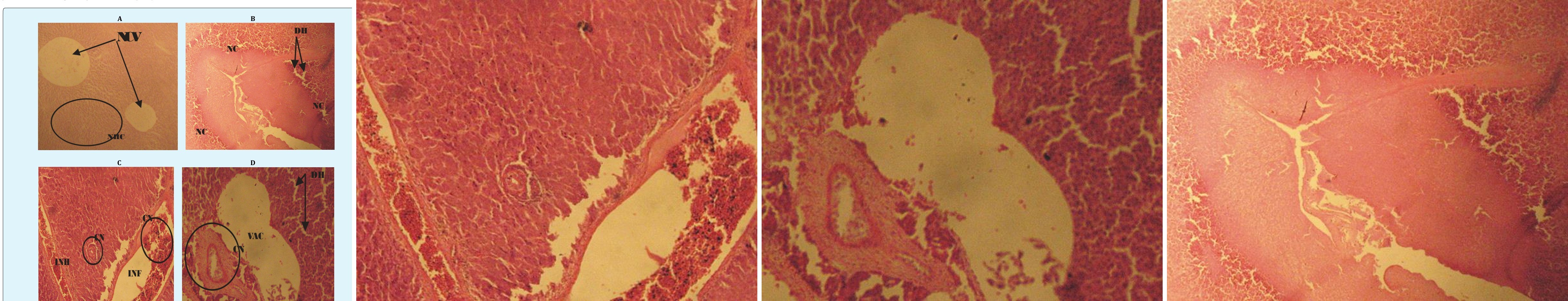
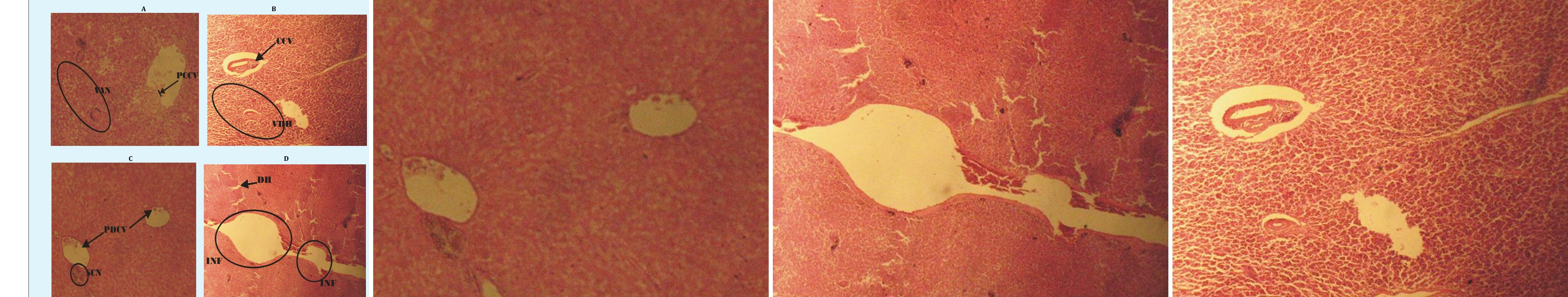
Liver is vital organ that is most affected by the pollutants in the water due to its role in the detoxification and biotransformation processes. It also serves as an integrator for biochemical and physiological functions, and caries out key functions in excretion of xenobiotics. Histological alterations recorded in the liver of the studied species were in agreement with many studies that examined the effects of different pollutants on fish liver [11, 12]. They alsoobserved degeneration of the hepatocytes and focal necrosis in the liver of C. gariepinus being exposed to be in consonance with the present study.
Although, liver histological changes are not specific to pollutants, several studies have established a casual relationship. Varied degree of histological alterations was documented in this work; ranging from congestion of central vein, hepatocytes degeneration. Distortion of sinuses, inflammation, heamolysis, vacuolation and cellular necrosis. This finding is in conformity to several other findings world-wide [7, 12]. The marked histological alterations observed in this study may be due to the additive or cumulative effects of increased metal concentrations in the liver. These results agreed with the findings of Authman & Abbas [13], which submitted that the liver has an important detoxification role of endogenous wastes products as well as externally derived toxins such as heavy metals mixtures. The liver section of almost all the specimens examined in the work showed dilation and destruction of central vein. The hepatocytes revealed fatty degeneration with pronounced vacoulation and necrosis and manifestation of hemorrhage. Hemorrhagic lesions were abundant and this may be due to high pollution index or probably because of the inflammation of liver tissue in between hepatocytes (Figures 1 & 2).
Fatty degeneration of hepatocytes in the liver may also be attributed to an oxygen deficiency as a result of gill degeneration or the vacuolar dilation and intravascular hemolysis observed in the blood vessels [14]. Many authors have also reported similar histological alterations in fish livers that were exposed to metals [2, 7]. Other studies revealed that vacoulation, inflammation and congestion were early stages in the hepatic degeneration, thus, these could be used as histological biomarker of different level of exposure [2, 15, 16]. Hepatic necrosis as generally observed in this work, has also been established to be the order of the day in fishes collected from contaminated ecosystem with metals [17]. The significantly high values of heavy metals in the liver could be linked to the occurrence of heterogeneous parenchyma in the liver of the two fish species in response to the metal of these fish to the polluted water of Asa River.
Therefore, it is possible to use liver hepatocytotic alterations as a biomarker to assess the impact of heavy metals or other pollutants toxicity on fish health and production. This study however, investigated the non- suitability of fish from polluted sites with respect to anthropogenic discharge. Consequently, this study has been able to establish the fact that, exposure of fish to even low concentration of toxicant in the aquatic phase can induce various toxicological effects and histological degradations which depend on the period of exposure, physiological status of the resident species, volume of water (in terms of seasonal variation) and concentrations of such pollutants.Obtained results showed that the histological alterations in the gill and liver tissues aremost likely caused by increased concentrations of certain pollutants. Accordingto the previous water quality studies of the Asa River, metals concentrations were found at an elevated level. This information verifies that histopathological changes are valuable biomarkers for field evaluation, especiallyin tropical regions that are naturally affected by variety ofenvironmental variations. It should be highlighted thathistopathology is able to assess the initial effects and reactionsto acute exposure to chemical stressors.
Conclusively, C. buthupogon and H. longifilis in Asa River are indeed responding to some stressors whose exact nature could be anthropogenic. According to the previous water quality studies of the Asa River, concentrations of iron, copper, cobalt, arsenic, lead, chromium and chlorine were at elevated levels and these substances could be responsible for the observed histopathological alterations. Present study represents an additional reason to proceed with a detailed monitoring of this river and the wildlife within it. Therefore, consumption of river foods from Asa River should be discouraged and urgent water monitoring system is required in the river.
Acknowledgments
We hereby acknowledge the technical and laboratory staffs of the Pure and Applied Biology Department, Ladoke Akintola University of Technology, Ogbomoso, Nigeria for their kind support in terms of materials supplies.
References
-
Bernet D, Schmidt H, Meier W, Burkhardt-Holm P, Wahli T (1999) Histopathology in fish: Proposal for a protocol toassess aquatic pollution. J Fish Disease 22(1): 25-34.
-
Ogundiran MA, Fawole OO, Adewoye SO, Ayandiran TA (2010) Toxicological impact of detergent effluent on juvenile of African Catfish _(Clarias gariepinus)_ Buchell, 1822). Agriculture and Biology Journal of North America, pp: 2151-7525.
-
Ogundiran MA, Fawole OO (2017) Spatio-Seasonal Distribution and Condition factors of _Clarias_ _buthupogon_ and _Heterobranchus longifilis_ from Asa River, Nigeria. International Journal of Fisheries and Aquatic Studies 5(2): 343-349.
-
Ogundiran MA, Fawole OO (2014) Relative abundance and length-weight relationship of _Clarias buthupogon_ and _Heterobranchus longifilis_ in Asa River, Ilorin, Kwara State, Nigeria. International Journal of Zoology and Research 4(4): 87-98.
-
Van der Oost R, Beyer J, Vermeulen NPE (2003) Fish bioaccumulation and biomarkers in environmental risk assessment: A review. Environ Toxicol Pharmacol 13(2): 57-149.
-
Magar RS, Bias UE (2013) Histopathological impact on theovary of the fresh water fish, _Channa punctatus_. Int Res Jrnl Envi Sci 2(3): 59-61.
-
Ogundiran MA, Fawole OO, Adewoye SO, Ayandiran TA (2009) Pathologic lesions in the gills structures of Clarias gariepinus on exposure to sub lethal concentrations of soap and detergent effluents.Journal of Cell and Animal Biology 3(5): 078- 082.
-
McDonald DG, Wood CM (1993) Branchial acclimation tometals, In: Fish Ecophysiology, Eds., Rankin JC and Jensen FB, Chapman and Hall, London, pp: 297-321.
-
Greco AM, Gilmour KM, Fenwick JC, Perry SF (1995) The effects of soft water acclimation on respiratory gastransfer in the rainbow trout, Oncorhynchus mykiss. J Experimental Biol 198: 2557-2567.
-
Takashima F, Hibiya T (1995) An atlas of fish Histology. Normal and pathological features, SecondEditionKodansha ltd., Tokyo, Japan.
-
Ptashynski M, Pedlar R, Evan R, Baron C, Klaver-Kamp J (2002) Toxicology of dietary nickel in lake white fish (coregomus clupeaformis). Aquat Toxicol 58: 229- 247.
-
Fanta E, Rios F, Romao S, Vianna A, Frieberger S (2003) Histopathology of the fish Corydoras paleatus contaminated with sublethal levels of organophosphorus in water and food. Ecotoxicol. Environ Safe 54(2): 119-130.
-
Authman MM, Abbas HH (2007) Accumulation and distribution of copper and zinc in both water and some vital tissues of two fish species (Tilapa zilli and Mugil cephalus) of Lake Qarun, Fayoum Province, Egypt. Pak J Biol Sci 20(13): 2106-2122.
-
Mohammed FA (2001) Impacts of environmental pollution in the southern region of Lake Manzalah, Egypt, on the histological structures of the liver and intestine of Oreochromisniloticus and Tilapia zillii. J Egyptian Acad Soc Environ Develop 2: 25-42.
-
Stentiford GD, Longshaw M, Lyons BP, Jones G, Green M, et al. (2003) Histpathological biomarkers in estuarine fish species for the assessment of biological effects of contaminants. Marine Environment Research 55(3-4): 137-159.
-
Au DWT (2004) The application of histo- cytopathologicalbiomarkers in marine pollution monitoring: a review. Mar Pollut Bull 48(9-10): 817- 834.
-
Olojo EA, Olurin KB, Mbaka G, Oluwamimo AD (2005) Histopathology of the gill and liver tissues of the African catfish Clarias gariepinus exposed to lead. Afr J Biotechnol 4: 117-122.
- The Role of Podocyte Apoptosis and the Involvement of SIRT1 in Diabetic Nephropathy
- Dealcoholization of Beer by Osmotic Distillation for the Beverage Industry
- Biopolymer-Based Edible Packaging- Biomaterials, Methods, and Applications in Food Industry: An Updated Review
- Influence of Bioprocessing Methods on 'China Rice' (Gawal R1), and Soyabean Supplementation on the Quality of Complementary Food
- Cassava (Manihot esculenta) Varietal Growth, Yield and Cyanide Content Performance in Three Sites in the South- Eastern Semi Arid Regions of Kenya
- Food Waste Treatment, Recycling, Management and Production of Value-Products-An Update on Methodologies and Current Trends