Antioxidants Capacity of Milk, Probiotics and Postbiotics: A Review
This paper explores the antioxidant capacity of milk, probiotics, and postbiotics. Antioxidants are compounds that can protect cells from damage caused by free radicals, which are unstable molecules that can cause oxidative stress. Milk is a rich source of antioxidants, including vitamins A and E, and some evidence suggests that milk consumption may be associated with a reduced risk of chronic diseases. Probiotics are live microorganisms that can confer health benefits when ingested, and some strains have been shown to have antioxidant properties. Postbiotics are non-viable microorganisms or their metabolites, and recent research has suggested that they may have antioxidant effects. This paper reviews the current evidence on the antioxidant capacity of milk, probiotics, and postbiotics, and discusses the potential implications for human health. Overall, the findings suggest that these three sources may have antioxidant effects, which could contribute to their health-promoting properties. However, further research is needed to fully understand the mechanisms underlying these effects and to determine their clinical relevance.
Introduction
Several milk-derived peptides have been found to possess antioxidant (AO) activity. The ability of peptides to interact with radical species or to inhibit oxidative reactions could lead to the development of novel food ingredients relevant in health promotion and disease prevention [1]. Milk and milk products included the good source of nutritional components like valuable proteins, lipids, and lactose as well as micro- and macronutrients. It is well known that the milk nature is a complete food for the growth and development of mammals. In recent years, there has been an increasing interest of consumers in functional food, which not only meets the primary function, but also has some benefits on human health [2]. Apart from of macro components such as proteins, lipids, lactose, and micro- and macronutrients in milk, there are variety of other components that occur during milk processing or intestinal digestion (enzyme hydrolysis) and are called bioactive components since they provide various health benefits [3]. Milk is a highly consumed product worldwide due to its nutrient content containing valuable macro and micro-nutrients such as carbohydrates, proteins, fat, minerals and some vitamins [4].
Nevertheless, this issue is controversial since there are many milk components that promote health benefits including oleic acid, conjugated linoleic acid, omega-3 fatty acids, proteins, vitamins, minerals and bioactive compounds, and various milk proteins and their peptides have been suggested to possess anti-cancer activity [5, 6, 7]. Milk-based beverages contain several components in addition to the basic components with beneficial physiological functions, such as peptides, oligosaccharides, enzymes, vitamins, and minerals. These components can be used as functional ingredients for the development of functional and nutraceutical food products with certain health benefits [8].
Antioxidants in Food
In the food and pharmaceutical industries, antioxidants are used to prevent deterioration, rancidity and discoloration caused by oxidation during processing and storage [9]. Reactive oxygen species and free radicals can be produced in food products by exogenous chemicals and endogenous metabolic processes.
Natural antioxidants currently have taken significant attention for their potential effects on health promotion and disease prevention. The main action of antioxidants consists of minimizing cellular damage exerted by free radicals [10]. Antioxidants locate in fruits and vegetables, specially vitamin C, vitamin E, vitamin A, is as well as enzymes such as catalase, glutathione peroxidase, glutathione reductase, and superoxide dismutase. studies have shown that Antioxidants are able to prevent a number of diseases such as cancer, coronary heart disease, delay ageing, boost the body’s immune system, neurological disorders, and neurodegenerative diseases [8]. It is well known that lipid peroxidation occurring in food products causes deteriorations in food quality (e.g. rancid flavour, unacceptable taste and shortening of shelf life). In addition, it has been recognized that oxidative stress plays a vital role in a number of age specific diseases [11, 12]. Radical scavenging is the main mechanism by which antioxidants act in foods. The radical scavenging assays primarily operate by direct measurement of hydrogen donation or electron transfer from the potential antioxidant to a free radical in simple ‘‘lipid free’’ systems [12].
The oxidative processes in our body produce highly reactive compounds called free radicals or these may also enter our body from the environment [13].
Antioxidant Importance
It is well known that reactive species such as free radicals or non-radical oxidants reactive oxygen, nitrogen, and sulphur species (RSS), including the recently discovered RSS, cause oxidative damage to lipids, proteins, and DNA [14]. According to Cheeseman, et al. [15], free radicals are typically electrically charged and have a tendency to oxidise other substances in order to balance themselves out [15]. The three main classes of oxidants that are produced within the body are reactive oxygen species (ROS), reactive nitrogen species (RNS), and reactive sulphur species (RSS) [16]. Free radicals like superoxide radicals (O2), hydroxyl radicals (OH), and non-radical oxidants like hydrogen peroxide (H2O2) and hypochlorous acid are the main ROS players (HOCl). Nitric oxide (NO) and peroxynitrite (ONOO) are major RNS in contrast to others. Nucleic acids, sugars, lipids, and proteins are the primary targets of these oxidants [16, 17, 18]. The alterations of organic biomolecules, such as the polyunsaturated fatty acids in membrane lipids, oxidation of proteins, DNA strand breakage, RNA oxidation, mitochondrial depolarization, and apoptosis, resulting in the harmful effects of these ROS and RNS free radicals, such as O2, OH, H2O2, and ONOO [16].
Under normal circumstances, scavenging enzymes like superoxide dismutase (SOD), catalase (CAT), glutathione peroxidase (GPx), etc., or chemicals that block the activities of oxidant-generating enzymes like xanthine oxidase, such as polyphenols, are responsible for clearing reactive species [19]. The major roles of antioxidants in human health are reported to prevent the generation of oxidizing species or diminish the effects of dangerous metabolic or xenobiotic oxidants. So the body is prevented from acute or chronic maladies and/or restoration of the cellular/tissue damage [20].
Antioxidant Mechanism of Probiotic
Probiotic could exert antioxidant capacity by different pathways. Probiotic would possess Antioxidant Enzymes System and Antioxidant Metabolites. Superoxide and Catalase comprise antioxidant enzymatic system of probiotic. sod is one of best known enzyme produced by the mitochondria possess antioxidant capacity that catalyses the superoxide radical’s dismutation (or partitioning) into common molecular oxygen (O2) and hydrogen peroxide in alternating fashion (H2O2). However, SOD due to its short circulatory half-life is suffering narrow-ranging bioavailability in the therapeutic studies. Lactobacillus fermentum strains E-3 and E-18 were able to express Mn- SOD under oxidative stress condition. Catalase (CAT) by breaking down hydrogen peroxide contributes to the cellular defences against free radical production and delays the Fenton reaction from producing hydroxyl radicals [21]. Although LAB are typically CAT-negative [22], de LeBlanc and colleagues demonstrated that a CAT-producing Lactococcus slactis could prevent 1, 2-dimethylhydrazine-induced colon cancer in rats. In addition, CAT-producing Lactobacillus casei BL23 strains created through genetic engineering were able to stop or lessen the severity of intestinal diseases brought on by ROS. Furthermore, probiotics are able to increase effectually the activities of antioxidases and promote the host’s antioxidant system. Studies in pigs demonstrated that dietary Lactobacillus fermentum supplementation might improve hepatic CAT, muscle SOD, Cu and Zn-SOD, and serum SOD and GPx compared to the control group. Wang et al research’s also inferred that Bacillus amyloliquefaciens SC06 rose CAT and GST gene expressions and the CAT activity in intestinal porcine epithelial cells-1 (IPEC-1) [23]. According to Amaretti and his collegues’ study, the highest restriction of inolenic acid peroxidation (TAALA) and ascorbate autoxidation (TAAAA), the TEAC are belonged to the strains Bifidobacterium animalis subsp. lactis DSMZ 23032, Lactobacillus acidophilus DSMZ 23033, and Lactobacillus brevis DSMZ 23034 [24]. In addition, Feng and his colleagues observed exopolysaccharide from Lactococcus lactis Z-2 had antioxidant capacity on hepatopancreas [25]. Intracellular cellular contents of probiotic including tertbutyl, hydroperoxide, ferrous ions, and intracellular polysaccharides could possess antioxidant properties [26]. The antioxidant capacity of intact cells of probiotic and postbiotic has been related to cell wall components such as cell surface polysaccharides and proteins, and amino acids [27, 28, 29]. Based on investigation of Cuevas-Gonzále, et al. [30]. The intracellular content of Lactobacillus fermentum J10, Lactobacillus pentosus J27, and Lactobacillus paracasei CRL431 had capacity to diminish the disturbance of the antioxidant system and diminish cell damage in erythrocytes exposed to acrylamide [30].
Metal ion chelation is another mechanism cause to exert antioxidant activity of probiotic [31, 32, 33]. Lin and Yen in 1999 declared firstly that LAB strains have The metal ion (ferrous and cupric ions) chelating ability [34]. Researchers discovered that Lactobacillus casei KCTC 3260 has a strong antioxidant capacity By chelating Fe2+ or Cu2+, 43 In the same way, tpostbiotic of Lactobacillus helveticus CD6 exhibited higher Fe2+ ion chelation [31]. the causes of metal ion chelation in probiotic bacteria are not entirely understood, however [35].
Antioxidant Properties of Milk
Milk antioxidants could exert possible health effects by acting as defense compounds against oxidative stress in the consumer, as well as prevent milk lipid oxidation and/ or maintain the oxidative stability of the product [36]. due to its containing sulfur amino acids cysteine, vitamins A, E, carotenoids, enzyme systems, superoxide dismutase, catalase and glutathione peroxidase, milk has a magnetic antioxidant capacity [37]. The major antioxidants in milk fat are composed by conjugated linoleic acid (CLA), vitamins A and E, β-carotene and coenzyme Q10. Other compounds also with antioxidant properties include vitamin D3, phospholipids, ether lipids and, possibly, 13-methyl-tetradecanoic acid [38, 39]. Dairy products showed antioxidant activity and have been considered as important dietary components that contribute to the total intake of antioxidants. Milk proteins (especially caseins) are the most important radical scavenger compounds [40].
Some dairy products like milk, skim milk or whey and some proteins from these products, such as lactoferrin, α-lactalbumin, β-lactoglobulin, caseins or specific endogenous milk enzymes, have been reported to possess antioxidant properties [41, 42]. It should be noted the role of milk phospholipids in the antioxidative properties of milk is related to acting synergistically with a-tocopherol [43].
The oxidation process lead to strong off-flavors and in deterioration of the nutritional quality of milk. then, The oxidative stability of milk and dairy products is the result of a delicate balance between the anti- and pro-oxidative processes in milk [44]. Probiotic milk is known as one of the best foods functional food produced by the probiotic bacteria [45]. Lactobacillus, Bifidobacterium, accharomyces, Streptococcus, Enterococcus, Escherichia, and Bacillus are comprised [46]. probiotic bacteria due to their ability to produce lactic acid by fermentation, caused lactose to be hydrolyzed by β-galactosidase enzyme to make galactose and glucose monosaccharides, as a result, the pH of milk dropped, and the growth condition is negatively changed for microorganisms over the LAB. However, Numerous studies admit that there are health advantages for which cell viability is not necessary or in which the advantage is not at all mediated by the microbial cell [47]. Then, postbiotics is described as non-viable microbial cells, their metabolites and cell fragments could possess health promoting effect without any side-effect of probiotic [48]. LAB were shown to have beneficial effects on maintaining and treating ulcerative colitis (UC), metabolic diseases, The growth, immune system, and oxidative status of sea bream, Pagrus major, were also improved by Lactobacillus rhamnosus or/and Lactobacillus lactis in fishes [49, 50]. Bifidobacterium was effective in boosting antitumor immunity [51] and treating women’s irritable bowel syndrome [52]. Due to their stability as spore-forming bacteria and capacity to produce a wide range of enzymes, including protease, amylase, and lipase, Bacillus species are preferred in the feed industry [53]. Many studies have indicated the antioxidant capacity of probiotics and intracellular content of probiotics. According to the studies, other forms of probiotic like a culture supernatant, intact cells, and intracellular cell-free extracts are able to scavenge hydroxyl radicals and superoxide anion in vitro and vivo studies. Romero-Luna and her colleagues reported culture supernatant of S. cerevisiae C41 and S. boulardii neutralized free radicals of ddph by 63.03% and 43.60% respectively [21]. Also Mangala Lakshmi Ragavan and Nilanjana Das indicated L. starkeyi VIT-MN03 had highest antioxidant capacity (76%) compared to K. lactis VIT-MN02 (68%), S. fibuligera VIT-MN04 (55%), Y. lipolytica VIT-MN01 (16%) and B. custersianus VIT-
MN05 (8%) in DPPH assay [22]. To defend cells from the damage that oxidative stress causes, LAB strains can boost the activity of antioxidant enzymes or modulate and relieve circulatory oxidative stress [54]. Probiotic LAB has been shown to have antioxidant properties both in vitro and in vivo, but the mechanism by which they control oxidative stress tolerance is not fully understood7. In probiotic LAB, an aerobic environment may encourage the production of toxic oxygen byproducts like ROS, reactive nitrogen species (RNS), and reactive sulphur species [24, 55]. Lactic acid bacteria (LAB) were examined to determine whether they could shield HT29 cells from oxidative DNA damage. Lactic acid can be produced by these bacteria, which are typically linked to dairy products, and is a key by product of the fermentation of carbohydrates [56]. A vital class of microorganisms employed in the food business is the lactic acid bacteria. Oxidative stress was induced using plumbago and hydrogen peroxide [55].
It should be noted that the bioaccessibility of antioxidants is a very vital factor [57]. Studies indicate that probiotic bacteria’s dead parts may have better bioaccessibility [58]. Based on the study, Lyophilized cells of Lactococcus lactis subsp. cremoris absorbed about 10 (U mL-1) dpph and heat killed cell was reported 6(U mL-1) dpph while intact cell were shown just 1.30 (U mL-1) [59]. In addition, cell free extract of Probionebacterium freudenreichii and Lactobacillus retueria had the highest antioxidant capacity by (97.75 %) and (96.74 %) respectively [60]. Aside from developing a healthy gut and promoting growth performance, a preliminary study from this laboratory revealed that the postbiotics produced by L. plantarum have high antioxidant activities [42]. Similarly, bacterial cultures of L. Plantarum were reported to exhibit high antioxidative activities [47, 48].
Natural Antioxidants in Milk
Bioactive Peptides
Several studies have confirmed that peptides released from milk protein hydrolysates have antioxidant activity [61, 62]. Bioactive peptides (BPs) have been defined as specific protein remains that have a positive influence on physiological and metabolic functions or condition of the body and may have ultimate beneficial effects on human health. BPs can be delivered to consumers in conventional foods, dietary supplements, functional foods, or medical foods [63]. These bioactive peptides possess very important biological activities and functionalities, including antimicrobial, antihypertensive, antioxidative, anticytotoxic, immunomodulatory, opioid and mineral-carrying activities [64, 65]. Milk proteins are known as very important sources of bioactive peptides [66]. The health benefits of these peptides are classified as cytomodulatory, mineral binding, antimicrobial, immunomodulatory, blood-pressure lowering (Angiotensin-converting enzyme ACE- inhibitory), antithrombotic, antioxidant and opioopioid-like addition to cholesterol-lowering and mineral absorption/bioavailability enhancers [66, 67]. Bioactive peptides have been classified as specific fragments of protein that have a positive impact on body functions or conditions and may ultimately influence health [64, 68]. The release of bioactive peptides from milk proteins in the gastrointestinal tract results from the action of digestive enzymes such as pepsin and pancreatic enzymes (trypsin, chymotrypsin, carboxy- and aminopeptidases) [69, 70]. In most studies the primary classes of bioactive milk peptides, based on their specific physiological functions:
- Antimicrobial Peptides.
- ACE Inhibitors.
- Antithrombotic Peptides.
- Opioid Milk Peptides.
- Immunomodulatory peptides.
- Mineral binding peptides.
- Antidiabetic peptides [71, 72].
Recently, the bioactive peptides derived from milk has reported to possess different functional properties such as stimulation of immune system, digestion, absorption of nutrients in body, prevention of obesity, and prevention of development of metabolic disorders [73]. Bioactive peptides can be derived from milk proteins, either casein or whey proteins. Production of bioactive peptides offers ingredients for a low-cost production and positive nutritional image associated with fermented milk. Generally, bioactive peptides can be produced by three main ways: 1: enzymatic hydrolysis with digestive enzymes; 1; fermentation of milk with proteolytic starter cultures; 3 proteolysis with enzymes derived from microorganisms or plants. Also, some studies suggest the combination of these three mentioned models to be effective in generating functional bioactive peptides [74, 75]. Antioxidant activity of bioactive peptides can be attributed to their radical scavenging, inhibition of lipid peroxidation and metal ion chelation properties of peptides. It also has been proposed that peptide structure and its amino acid sequence can affect its antioxidative properties [76]. Peptides derived from αs-casein have free radical scavenging activity and inhibit enzymatic and non- enzymatic lipid peroxidation [42]. These dipeptides can promote the synthesis of glutathione, which is an important antioxidant for cellular protection and repair processes. At present, proteins and peptides with biological activity constitute one of the most important categories within the functional food sector. Peptides with biological activity can be generated from milk proteins [77, 78]. Milk proteins have shown antioxidant activity for the scavenging of reactive oxygen species. Studies have shown that casein inhibited the lipoxygenase-catalyzed lipid autoxidation. Free amino acids cannot quench the free radicals and for the scavenging of free radicals, primary structure of casein molecules acts as scavenger [79]. Moreover, the antioxidant activity of whey protein hydrolysates depends on peptide molecular weight, with low-molecular-weight peptides (0.1–2.8 kD) showing the strongest in vitro radical scavenging activity [80].
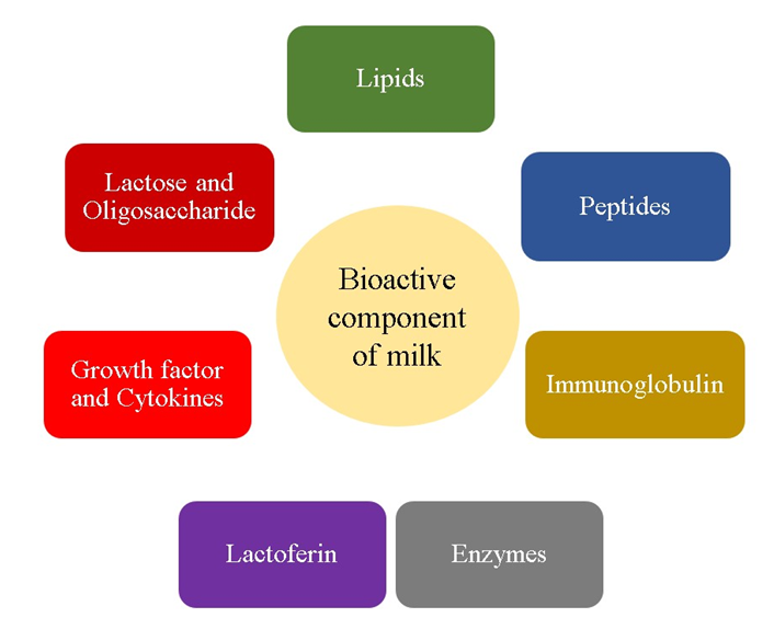
Figure1: Schematic review of major bioactive components in milk.
Proteins
Caseins: CN’s have a relatively open or disordered structure which makes them highly susceptible to proteolysis. The proteins of milk are divided into caseins and whey proteins. Caseins include αs1-casein, αs2-casein, β-casein, and κ-casein and make up 80% of the total protein content. The other 20% consists of whey proteins, including β-lactoglobulin, α-lac- talbumin, serum albumin and immunoglobulins. The distinction between the two groups is based on solubility at pH 4.6 [81]. Caseins are the major protein of bovine and ovine milks present in the form of macro-molecular aggregates. Due to the difference in phosphate content, various casein fractions are present in milk, for example, phosphate content of α, β and κ caseins are 10, 5 and 1 mol per casein mole and phosphate can provide antioxidant activity to the casein micelles [82]. Proteins (casein fractions and whey proteins) and peptides released by proteolytic enzymes have antioxidant properties. Model studies conducted with the involvement of prooxidants – ferric ions, ascorbic acids and lipoxygenase – demonstrated that all casein fractions can inhibit the autoxidation of arachidonic acid. Selected b-casein sequences (169–176 and 33–48) inhibit the oxidation of oleic acid in vitro [42]. The antioxidant properties of caseins can be modulated by dephosphorylation of the protein chain. Dephosphorylated casein and β-casein are more hydrophobic, and they inhibit chemically induced peroxidation of linoleic acid more effectively than their native forms. Higher hydrophobicity increases the scope of interactions with linoleic acid. Casein scavenges free radicals more effectively when the peroxidation of linoleic acid is chemically induced (with 2,20-azobis (2-amidinopropane dihydrochloride) than when oxidation is initiated enzymatically [83]. Proteolysis affects casein’s ability to inhibit lipid peroxidation. Regardless of concentration, casein hydrolysates demonstrate antioxidant activity due to a higher content of amino acids with antioxidant properties, including histidine, proline, lysine and tyrosine. Free radicals are deactivated by peptides containing hydrophobic amino acids (proline, histidine, tyrosine and tryptophan) and selected free amino acids (tyrosine and cysteine). A peptide composed of six amino acids (Tyr–Phe–Tyr–Pro–Glu–Leu) is particularly effective in eliminating peroxide radicals. Its antioxidant activity is highly influenced by the C-terminus [65, 84]. Casein fragments containing valine (Val) or leucine (Leu) in the N-terminus are also highly effective in scavenging superoxide radicals. Peptide hydrophobicity was not found to be correlated with inhibition of peroxidation, which implies that it is not the sole cause of antioxidant activity. It is believed that the antioxidant activity of peptides is significantly enhanced by tyrosine (Tyr), a strong proton donor [83]. Furthermore, Shazly, et al. [85] who studied antioxidant peptides from buffalo and bovine casein reported buffalo casein hydrolyzed (CBH) by pepsin contrasted to bovine casein hydrolysates (CNH) contained more hydrophobic amino. also, oxygen radical absorbance capacity and Hydroxyl radical scavenging capacity for CBH hydrolyzed by alcalase and CNH hydrolyzed by trypsin were reported 81.16% and 84.55% and 2.45 and 2.23 mM BHA respectively. Consequently, alcalase-CBH and trypsin-CNH are offered in functional foods and pharmaceuticals as an appropriate antioxidants [85]. In addition, it is reported that a-casein added into liposomal suspension (1 g/L ) was able to inhabit entirely a mixture of ferrous sulfate and ascorbate, 50 and 500 m M final concentration, respectively [82]. Li, et al. [86] who studied bio peptides hydrolyzed from goat casein (GMC) by utilizing a combination of neutral and alkaline proteases (GMCH) indicated that GMCH has a higher inhibition effect on lipid peroxidation compared with tertbutylhydroquinone and phytogermine. it should be noted that the high antioxidant activity of GMCH is specifically related to 5 novel oligopeptides( Val-Tyr-Pro-Phe, Phe-Gly- Gly-Met-Ala-His, Phe-ProTyr-Cys-Ala-Pro, Tyr-Val-Pro-Glu- Pro-Phe, and TyrPro-Pro-Tyr-Glu-Thr-Tyr) which were first observed in GMCH. so, its observed due to the 5 oligopeptides , GMCH antioxidant capacity compared with GMC increased 3.59 to 380 times [86]. Also, it’s reported LHSMK which is The synthetic peptide from casein hydrolyzed by pepsin not only has the antioxidant activity and free radical scavenging ability exhibited a dose-dependent increase, but also could regulate antioxidant enzymes activity in oxidative-damage cells [87]. Bamdad, et al. [88] who produced bio peptides driven casein by the combination of High Hydrostatic Pressure with protease enzyme indicated that casein exposed to flavourzyme and High Hydrostatic possessed the highest antioxidant activity compared with atmospheric pressure- enzymatic hydrolysis. In addition, Liquid chromatography with tandem mass spectrometry presented the amino acid sequences of biopeptides produced by flavourzyme and High Hydrostatic are reported approximate 60 % proline, valine, and leucine [88]. Whey: Whey has a globular structure and is composed of five major fractions: β-lactoglobulin (β-LG), α-lactalbumin (α- LA), bovine serum albumin, immunoglobulins and a number of minor proteins such as lactoferrin and lactoperoxidase. Each whey protein faction has different physicochemical properties, which can be modified by enzymatic hydrolysis [89]. Mann, et al. [90] investigated that WPC hydrolyzed by using corolase peptidase compared with flavouzyme, alcalase possessed greater radical scavenging activity [90]. One of the peptides, Trp–Tyr–Ser–Leu–Ala–Met–Ala–Ala– Ser–Asp–Ile which is observed in the β-lactoglobulin A to have more radical scavenging activity than BHA [91]. Preparation of antioxidant enzymatic hydrolysates from alphalactalbumin and beta-actoglobulin. Furthermore, Contreras del Ma, et al. [92] informed the sequence of peptides from β-lactoglobulin fragment (92-100) and β-lactoglobulin fragment hydrolyzed by flavourzyme were VLVLDTDYK and IDALNEK that possessed antioxidant capacity Production of Antioxidant hydrolysates from whey protein hydrolysate with thermolysin [93]. In addition, using ultrasound treatment led to increasing the antioxidant capacity of whey protein, results revealed that ultrasound treatment improved the proteolysis of β-lactoglobulin by both pepsin and trypsin and enhanced the antioxidant capacity of the protein and its proteolytic products [92]. It is reported that the antioxidant capacity of whey protein is related to the degree of hydrolysis, study showed protein whey has higher Trolox equivalent antioxidant capacity (TEAC) at %13 hydrolysis degree [94]. Lactoferrin (LF) is an iron-binding glycoprotein of the transferrin family with a high molecular weight (80 kDa). It is present in many biological fluids and is widely distributed in colostrum and milk [95]. Lactoferrin delivers a host of health benefits, and it is the most valuable protein in the human diet. Lactoferrin binds to iron and increases its bioavailability while blocking its pro-oxidant action. Despite the above, the antioxidant potential of lactoferrin decreases proportionally to the degree of iron saturation. The lactoferrin content of milk is estimated at 0.02–0.35 g/L and it is higher in colostrum [96]. The biological activity of lactoferrin is determined by various technological factors: parameters of the heat treatment, the fat content and the degree of saturation of iron [97, 98]. Lactoferrin is not highly susceptible to digestive enzymes, trypsin and chymotrypsin, and it maintains its biological properties when ingested with food [99]. It also exerts immunostimulatory effects by preventing pathogen colonization in the gastrointestinal tract and promotes the growth of beneficial gut microbiota. Lactoferrin attenuates the inflammatory response, increases the cytotoxicity of natural killer cells in vitro and inhibits the release of oxygen radicals by leucocytes at sites of inflammation [100]. Lactoferrin is synthesized by the epithelial cells of the external mucous membranes, present also in some bodily fluids, such as tears and saliva, although the highest levels are detected in milk secretions [101]. Rachman, et al. [100] showed that lactoferrin concentration varies with lactation days i.e. on the 1st day of lactation it was observed that lactoferrin was more than the following lactation days. Thus, it can be seen that lactation period, age and other maternal characteristics plays an important role in the lactoferrin concentration. Lactoferrin concentration varies with breeds too [102]. Lactoferrin is present in mammalian secretions such as milk, tears, saliva, seminal fluids, vaginal fluids, nasal mucosa, bronchial mucosa as well as in some white blood cells and secondary granules of neutrophils [7, 103]. The alteration of the activity of lactoferrin in milk could have an impact on the shelf life of raw milk and also on the development of neonates [104]. Lactoferrin is considered to be an important host defense molecule and has a diverse range of physiological activities such as antibacterial, antiprotozoal, antifungal, antiviral, anticancer, antioxidant, antiinflammatory and immunomodulatory [103, 105].
Sacharides
Oligosacharides: Lactose is the main carbohydrate in milk and accounts for 54% of the total non- fat milk solids [106]. Lactose consists of one glucose unit and one galactose unit, connected by a glycosidic linkage in β-config- uration, described as β-D-galactopyranosyl-(1→4)-D-glucopyranose) [107]. Lactose is synthesised by the proteins α-lactalbumin and galactosyltransferase, which catalyses the reaction whereby lactose is formed from glucose and uridine- diphosphate-ga- lactose (UDP galactose) [108]. Galacto- Oligosaccharides are mixture of substances produced from lactose or lactulose composed of 3–8 molecules of galactose and glucose. Glucose is present at the terminal position and remaining saccharide and disaccharide comprised of two galactose units [109]. GOSs are nondigestible in nature and are established as prebiotic ingredient as they enhance the growth and number of beneficial bacteria like Bifidobacteria, Lactobacillus, etc. and decrease the growth and number of harmful bacteria like Clostridia, Enterobacter, etc in GIT. GOSs are beneficial to host because the role of beneficial bacteria in gut health has already been proved. It plays role in immune function indicated by remarkable increase in phagocytosis, NK cell activity, production of anti-inflammatory cytokine interleukin-10, significant reduction in the production of pro-inflammatory cytokines and recovery of innate immune cells in the elderly persons for the enhancement of gastrointestinal health and immune function in elderly persons. Stable gut microflora also plays an important role in metabolic as well as in overall health. It has also been reported to offer anti- anxiety benefits. It is also associated with colorectal cancer [110].
Enzymes
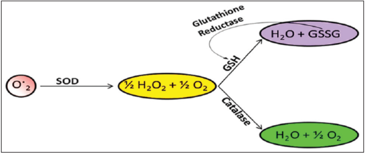
Superoxide Dismutase: SOD catalyzes the removal of free O2− and safe¬guards the cells against harmful effects through the following reaction.
2 2 2 2 2O +2H H O +O → Catalase, GSHPx, or other reducing agents con¬vert H2O2 to H2O, hydrogen peroxide formed from O2 −, and oxidases are eliminated by catalases and peroxidases [110]. Cytosolic Cu/ Zn-SOD, mitochondrial Mn-SOD, and extracellular-SOD are the major forms of SOD [111]. SOD can inhibit LPO. In cow milk, SOD is exclusively present in skim milk fraction, with a concentration of 0.15 mg/l-2.4 mg/l. Human milk has 2.0-2.3 times more concentration of SOD as compared to cow milk [112].
Glutathione Peroxidase: GSHPx is a selenium encompassing enzyme that provides protection against lipid peroxidation. It catalysis the breakdown of H2O2 and organic hydroperoxides (R-OOH) by glutathione (γGlu.Cys.Gly) as per following chemical reaction [113]:
2 ROOH+2GSH ROH+GSSG+H O → More than 90% of GSHPx exists in milk as extra cellular enzyme and it is only enzyme which fixes selenium (about 30% of the total. Its concentration varies among the mammals and concentration is in the order of human > caprine > bovine [114]. Concentration of GSHPx in cow milk ranges from 12 to 30 U/mL and its activity is mainly dependent upon the concentration of selenium. Antioxidant activity and selenium content decreases with the progression of lactation [115]. Peroxidase: Peroxidases are frequently used in the studies of metabolic reactions, enzymatic functions, protein structures [116]. Prosthetic groups of peroxidase are protoheme and be connected to the apoprotein loosely in contrast to many hemoprote [117]. Lactoperoxidase (LPO; EC 1.11.1.7) belongs to the peroxides family of enzymes and is secreted from mammary, salivary, and other mucosal glands that is acting as a natural antibacterial agent. It has the ability to catalyze certain molecules, including the reduction of hydrogen peroxide [118]. Lactoperoxidase is an enzyme present in milk, whey, and colostrum. It has a molecular weight of about 77,500 Da. Lactoperoxidase is involved in the defense mechanism of the host against bacteria which makes it biologically significant [119]. Lactoperoxidase is capable of inhibit the growth of wide range of microorganisms through an enzymatic reaction which involves H2O2 and thiocyanate ions as cofactors. These cofactors along with lactoperoxidase constitute the lactoperoxidase (LP) system. Antimicrobial action of this LP system is based on the formation of hypothiocyanite ions via enzyme activation. Hypothiocyanite ions react with the bacterial membranes and also disrupt the functioning of certain metabolic enzymes. In some countries, H2O2 addition is not permitted hence in situ development of hydrogen peroxide is done via addition of glucose oxidase which is permitted. The source of thiocyanate ion can be either natural or added as sodium or potassium thiocyanate. LP system prevents the growth of Gram-positive bacteria and kills the Gram-negative bacteria [120]. The general formula of the reaction catalyzed by peroxidases is shown below [121].
2 2 ROOH+AH ROH+H O+A → The H2O2 formed during the metabolism having oxidizing property must be quickly removed. The catalase and peroxidase enzymes exhibiting antioxidant properties play this role in cells [122]. Lactoperoxidase (LP) is a member of the peroxidase family, a group of natural enzymes, widely distributed in nature and found in plants and animals, including humans. Its primary function is to catalyze the oxidation of certain molecules, at the expense of hydrogen peroxide, in order to generate reactive products with a wide antimicrobial activity [119]. Milk contains some essential antimicrobial factors such as lactoperoxidase, lysozyme, immunoglobulin, and lactoferrin [123]. Lactoperoxidase system (LPS) is a natural antibacterial system having both bactericidal and bacteriostatic to a wide variety of microorganisms primarily used to prevent undue bacterial multiplication thereby mitigating milk spoilage and associated financial loss [124, 125]. The system has three primary components named as LP, hydrogen peroxide (H2O2), and thiocyanate (SCN). LP catalyzes H2O2-dependent oxidation of thiocyanate (SCN-) to hypothiocyanite (OSCN-). The latter ion is a potent antimicrobial agent against gram- negative and gram-positive bacteria, fungi, and viruses [126]. Moreover, it has, antiviral activity, degradation of various carcinogens and protection of animal cells against peroxidative effects [119]. The most commonly used method to stop or retard the deterioration of milk from the farmer to the industry is cooling [127]. However, in many parts of the world, this is not possible for various reasons, such as lack of available capital, lack of electricity, less developed road systems, high operational costs, frequent break downs of equipment, lack of spare parts and difficulties in repair of equipment in rural areas [126]. Prevailing high ambient temperatures often further complicate the problem of milk collection in these areas [128]. This causes a considerable loss of fresh milk qualities, and in many regions only a minor part of the production reaches the dairy industry in an acceptable condition for use as human food. In many regions most of the evening milk is spoiled after storage over-night [129].
Catalase: Milk catalase is a heme protein, molecular weight of catalase is 200 kDa, with isoelectric pH of 5.5, and it is stable in a wide range of pH 5-10 and rapidly loses activity when it is out of this pH range [114]. The most of catalyzes contain heme, also catalase causes the dismutation of H2O2, consequently, one is converted to O2, and the other two are converted to two molecules of H2O [130]. A polarographic method showed that aver-age catalase activity in cow milk was 1.95 U/ml [131]. Although, the concentration of catalase in human milk is approximately 10 times greater than cow milk [132]. Endogenous antioxidant enzymes - superoxide dismutase, catalase, and glutathione antioxidant mechanisms [133].
Co-Enzymes: Milk fat contains small amounts of coenzyme Q10. Coenzyme Q10 transports electrons in the electron transport chain and participates in ATP synthesis, which improves energy efficiency in cells and tissues. The heart muscle is most sensitive to coenzyme Q10 deficiency. Coenzyme Q10 is a highly active antioxidant, and its reduced form (ubiquinol) protects cell membranes and low Density Lipoprotein (LDLs) against peroxidation more effectively than β-tocopherol and b-carotene. Ubiquinol prevents the initiation and progression of peroxidation of lipid and phospholipid PUFAs in mitochondrial membranes. By binding with proteins, coenzyme Q10 stabilises mitochondrial membranes and ensures their optimal liquidity [134]. Ubiquinol promotes the antioxidant effects of vitamin E which inhibits lipid peroxidation in membranes only at the propagation stage. Ubiquinol also participates in the regeneration of the reduced form of vitamin E (reduces the tocopheryl radical to tocopherol). Despite the above, the antioxidant potential of coenzyme Q10 is not determined by vitamin E. It is believed that direct antioxidant effects on cells can be attributed to the reduced form of coenzyme Q10, whereas indirect effects follow from the regeneration of vitamin E [135, 136].
| Unfermented Product | CoQ10(mg/g) | Fermented Product | CoQ10(mg/g) |
|---|---|---|---|
| Milk 3.5% Fat | 1.3 | Yogurt 0 % fat | Up 0.1 |
| Milk 3.5% Fat | 0.7-1.2 | Yogurt 1.5% fat | 0.7-1.4 |
| Uht Milk 3.5% Fat | 1.7 | Fermented milk 1.6% fat | 0.5 |
| Uht Milk 1.6% Fat | 1.2 | Fermented milk 3.2% fat | 0.5-0.9 |
| Uht Milk 0.5% Fat | 0.5 | Kefir 3.5% fat | 0.9 |
| Cream 35 % Fat | 0.9 | Kefir 1.6% fat | 0.7 |
| Cream 20-22 % Fat | 0.5-0.9 | Emmentaler cheese | 1.3 |
| Butter | 7.1 | Edam cheese | 12 |
Table 1: Amounts of coenzyme Q10 in dairy products.
Milk fat contains small amounts of coenzyme Q10 as represented in Table 1. Coenzyme Q10 transports electrons in the electron transport chain and participates in ATP synthesis, which improves energy efficiency in cells and tissues [137]. Ubiquinol promotes the antioxidant effects of vitamin E which inhibits lipid peroxidation in membranes only at the propagation stage. Ubiquinol also participates in the regeneration of the reduced form of vitamin E (reduces the tocopheryl radical to tocopherol). Despite the above, the antioxidant potential of coenzyme Q10 is not determined by vitamin E. It is believed that direct antioxidant effects in cells can be attributed to the reduced form of coenzyme Q10, whereas indirect effects follow from the regeneration of vitamin E [135, 136]. Coenzyme Q10 is a highly strong antioxidant, and its reduced form (ubiquinol) protects cell membranes. LDLs against peroxidation more effectively than α-tocopherol and β-carotene. Ubiquinol prevents the initiation and progression of peroxidation of lipid and phospholipid polyunsaturated fatty acids (PUFAs) in mitochondrial membranes. By binding with proteins, coenzyme Q10 stabilises mitochondrial membranes and ensures their optimal liquidity [134].
Co-factor: Manganese is a specific enzyme co-factor involved in the synthesis of mucopolysaccharides, and a non-specific co-factor for many other enzymes. There are several known manganese metaloenzymes like arginase, glutamine synthetase, phosphoenlopyruvate decarboxilase and manganese superoxide dismutase [138].
Enriched Milk with Postbiotic
Postbiotics, as previously stated, are the byproducts of probiotic bacteria, such as metabolites including proteins, fatty acids, peptides, bacteriocins, enzymes, organic acids, and hydrogen peroxide, which have been shown to have beneficial effects on human health. Enriching milk with postbiotics can potentially enhance its nutritional value and provide additional health benefits. To begin with, postbiotics have been shown to have a positive impact on gut health by promoting the growth of beneficial bacteria and reducing inflammation in the gut, as well as, enhanced immune. In addition, postbiotics may help improve digestion and nutrient absorption, leading to better overall digestive health in practically patient suffering from lactose intolerance without change in sensory properties of milk by enzyme lactase obtained from postbiotics. Furthermore, antioxidant properties of postbiotics into milk may help reduce cell stress and the risk of chronic diseases associated with inflammation. To end, postbiotics may help improve the bioavailability of certain nutrients in milk, making them easier for the body to absorb and utilize, although it should be studied profoundly [139, 140]. Overall, enriching milk with postbiotics can potentially provide an added layer of health benefits, making it a more valuable and functional food product.
Conclusion
In conclusion, the antioxidant capacity of milk, probiotics, and postbiotics is an area of active research that has received growing interest in recent years. Milk is a rich source of antioxidants, including vitamins A and E, which have been shown to have potential health benefits. Milk and dairy products, which are basic foods for human development, can be beneficial for the oxidative defence of consumers by several mechanisms. Probiotics and postbiotics also have the potential to confer health benefits, including antioxidant effects, which could contribute to their overall health-promoting properties. Postbiotics, as metabolites or non-living forms of probiotics with many health effects which have been used in recent years for various purposes, including assessing their antimicrobial, anticancer, and antioxidant activities. Excellent results have been reported in this regard. Each postbiotics compound (proteins, fatty acids, peptides, bacteriocins, enzymes, organic acids, and hydrogen peroxide) has specific antioxidant mechanisms. Postbiotics are achieved through the activity of probiotic bacteria (fermentation) or are produced on a laboratory scale. Previous research has shown that postbiotics have clinical (safety), technological (sustainability) and economical (low costs of production) advantages over probiotics. In this regard possibility of producing of enriched milk by postbiotics as a functional food. However the evidence on the antioxidant capacity of the combination of milk with postbiotics is still limited and more research is needed to fully understand the underlying mechanisms and determine their clinical relevance. Additionally, the effects of these sources on different populations and under various conditions should be further explored.
Conflict Interest
There are limited studies due to the uniqueness of issues. It should be more studied.
Acknowledgment
Not funded.
References
-
Tadesse SA, Emire SA (2020) Production and processing of antioxidant bioactive peptides: A driving force for the functional food market. Heliyo 6(8): e04765.
-
Fuquay JW, Fox PF, McSweeney PLH (2011) Encyclopedia of Dairy Sciences. 3rd(Edn.), Bioactive peptides.
-
Nongonierma AB, FitzGerald RJ (2015) Bioactive properties of milk proteins in humans: A review. Peptides 73: 20-34.
-
Claeys WL, Cardoen S, Daube G, Block JD, Dewettinck K, et al. (2013) Raw or heated cow milk consumption: Review of risks and benefits. Food control 31(1): 251- 262.
-
Duarte DC, Nicolau A, Teixeira JA, Rodrigues LR (2011) The effect of bovine milk lactoferrin on human breast cancer cell lines. Journal of dairy science 94(1): 66-76.
-
Rodrigues LR, Teixeira JA (2016) Potential applications of whey proteins in the medical field. Engineering Aspects of Milk and Dairy Products, pp: 221-252.
-
Rodrigues, L, Teixeira J, Schmitt F, Paulsson M, Månsson HL (2008) Lactoferrin and cancer disease prevention. Critical reviews in food science and nutrition 49(3): 203- 217.
-
Özer BH, Kirmaci HA (2010) Functional milks and dairy beverages. International Journal of Dairy Technology 63(1): 1-15.
-
Nirmala C, Bisht MS, Bajwa HK, Santosh O (2018) Bamboo: A rich source of natural antioxidants and its applications in the food and pharmaceutical industry. Trends in Food Science & Technology 77: 91-99.
-
Rahman K (2007) Studies on free radicals, antioxidants, and co-factors. Clinical interventions in aging 2(2): 219- 236.
-
Halliwell B (2001) Role of free radicals in the neurodegenerative diseases: therapeutic implications for antioxidant treatment. Drugs & aging 18(9): 685-716.
-
Pihlanto A (2006) Antioxidative peptides derived from milk proteins. International dairy journal 16(11): 1306- 1314.
-
Jiang J, Xiong YL (2016) Natural antioxidants as food and feed additives to promote health benefits and quality of meat products: A review. Meat science 120: 107-117.
-
Pallavi S, Jha AB, Dubey RS, Pessarakli M (2012) Reactive oxygen species, oxidative damage, and antioxidative defense mechanism in plants under stressful conditions. J Bot 2012: 1-26.
-
Cheeseman KH, Slater TF (1993) An introduction to free radical biochemistry. British medical bulletin 49(3): 481-493.
-
Lü JM, Lin PH, Yao Q, Chen C (2010) Chemical and molecular mechanisms of antioxidants: experimental approaches and model systems. Journal of cellular and molecular medicine 14(4): 840-860.
-
Carocho M, Ferreira IC (2013) A review on antioxidants, prooxidants and related controversy: Natural and synthetic compounds, screening and analysis methodologies and future perspectives. Food and chemical toxicology 51: 15-25.
-
Craft BD, Kerrihard AL, Amarowicz R, Pegg RB (2012) Phenol‐based antioxidants and the in vitro methods used for their assessment. Comprehensive Reviews in Food Science and Food Safety 11(2): 148-173.
-
Ali SS, Ahsan H, Zia MK, Siddiqui T, Khan FH (2020) Understanding oxidants and antioxidants: Classical team with new players. Journal of food biochemistry 44(3): e13145.
-
Azab AE, Adwas AA, Elsayed ASI, Quwaydir FA (2019) Oxidative stress and antioxidant mechanisms in human body. J Appl Biotechnol Bioeng 6(1): 43-47.
-
Romero-Luna HE, Hernández-Sánchez H, Ribas-Aparicio RM, Cauich-Sánchez PI, Dávila-Ortiz G (2019) Evaluation of the probiotic potential of Saccharomyces cerevisiae Strain (C41) isolated from Tibicos by in vitro studies. Probiotics and antimicrobial proteins 11(3): 794-800.
-
Ragavan ML, Das N (2020) In vitro studies on therapeutic potential of probiotic yeasts isolated from various sources. Current Microbiology 77(10): 2821-2830.
-
Izuddin WI, Humam AM, Loh TC, Foo HL, Samsudin AA (2020) Dietary postbiotic Lactobacillus plantarum improves serum and ruminal antioxidant activity and upregulates hepatic antioxidant enzymes and ruminal barrier function in post-weaning lambs. Antioxidants 9(3): 250.
-
Amaretti A, di Nunzio M, Pompei A, Raimondi S, Rossi M, et al. (2013) Antioxidant properties of potentially probiotic bacteria: in vitro and in vivo activities. Applied microbiology and biotechnology 97(2): 809-817.
-
Feng J, Cai Z, Chen Y, Zhu H, Chang X, et al. (2020) Effects of an exopolysaccharide from Lactococcus lactis Z-2 on innate immune response, antioxidant activity, and disease resistance against Aeromonas hydrophila in Cyprinus carpio L. Fish shellfish immunology 98: 324- 333.
-
Ou CC, Chiu YH, Lin SL, Chang YJ, Huang HY, et al. (2012) Hepatoprotective effect of lactic acid bacteria in the attenuation of oxidative stress from tert-butyl hydroperoxide. Journal of food and drug analysis 20(1): 10.
-
Aguilar-Toalá JE, Estrada-Montoya MC, Liceaga AM, Garcia HS, González-Aguilar GA, et al. (2019) An insight on antioxidant properties of the intracellular content of Lactobacillus casei CRL-431. LWT 102: 58-63.
-
Li S, Zhao Y, Zhang L, Zhang X, Huang L, et al. (2012) Antioxidant activity of Lactobacillus plantarum strains isolated from traditional Chinese fermented foods. Food chemistry 135(3): 1914-1919.
-
Aguilar-Toalá J, Hall FG, Urbizo-Reyes UC, Garcia HS, Vallejo-Cordoba B, et al. (2020) In silico prediction and in vitro assessment of multifunctional properties of postbiotics obtained from two probiotic bacteria. Probiotics and antimicrobial proteins 12(2): 608-622.
-
Cuevas-González P, Aguilar-Toalá JE, García HS, González- Córdova AF, Vallejo-Cordoba B, et al. (2020) Protective effect of the intracellular content from potential probiotic bacteria against oxidative damage induced by acrylamide in human erythrocytes. Probiotics and Antimicrobial Proteins 12(4): 1459-1470.
-
Ahire JJ, Mokashe NU, Patil HJ, Chaudhari BL (2013) Antioxidative potential of folate producing probiotic Lactobacillus helveticus CD6. Journal of food science and technology 50(1): 26-34.
-
Gutteridge JM, Richmond R, Halliwell B (1979) Inhibition of the iron-catalysed formation of hydroxyl radicals from superoxide and of lipid peroxidation by desferrioxamine. Biochemical Journal 184(2): 469-472.
-
Gaisawat MB, Iskandar MM, MacPherson CW, Tompkins TA, Kubow S (2019) Probiotic supplementation is associated with increased antioxidant capacity and copper chelation in C. difficile-infected fecal water. Nutrients 11(9): 2007.
-
Lin MY, Yen CL (1999) Antioxidative ability of lactic acid bacteria. Journal of agricultural and food chemistry 47(4): 1460-1466.
-
Halliwell B, Murcia MA, Chirico S, Aruoma OI (1995) Free radicals and antioxidants in food and in vivo: what they do and how they work. Critical Reviews in Food Science & Nutrition 35(1-2): 7-20.
-
Donovan SM (2006) Role of human milk components in gastrointestinal development: current knowledge and future needs. The Journal of paediatrics 149(5): S49-S61.
-
Usta B, Yılmaz-Ersan L (2013) Antioxidant enzymes of milk and their biological effects. Ziraat Fakültesi Dergisi, Uludağ Üniversitesi 27(2): 123-130.
-
German JB, Dillard CJ (2006) Composition, structure and absorption of milk lipids: a source of energy, fat-soluble nutrients and bioactive molecules. Critical reviews in food science and nutrition 46(1): 57-92.
-
Baldi A, Pinotti L (2008) Lipophilic microconstituents of milk. Bioactive components of milk 2008: 109-125.
-
Clausen MR, Skibsted LH, Stagsted J (2009) Characterization of major radical scavenger species in bovine milk through size exclusion chromatography and functional assays. Journal of Agricultural and Food Chemistry 57(7): 2912-2919.
-
Lindmark-Månsson H, Åkesson B (2000) Antioxidative factors in milk. British journal of Nutrition 84(S1): 103- 110.
-
Rival SG, Boeriu CG, Wichers HJ (2001) Caseins and casein hydrolysates. 2. Antioxidative properties and relevance to lipoxygenase inhibition. Journal of Agricultural and Food chemistry 49(1): 295-302.
-
Gavella M, Kveder M, Lipovac V, Jurasin D, Filipovi- Vincekovic N (2007) Antioxidant properties of ganglioside micelles. Free radical research 41(10): 1143-1150.
-
Nielsen JH, Lund-Nielsen T, Skibsted LH (2004) Higher antioxidant content in organic milk than in conventional milk due to feeding strategy. In: Newsletter from Danish Research Centre for Organic Farming 3: 1-2.
-
Savaiano DA, Hutkins RW (2021) Yogurt, cultured fermented milk, and health: A systematic review. Nutrition reviews 79(5): 599-614.
-
Yeung PSM, Sanders ME, Kitts CL, Cano R, Tong PS (2002) Species-specific identification of commercial probiotic strains. Journal of Dairy Science 85(5): 1039-1051.
-
Collado MC, Vinderola G, Salminen S (2019) Postbiotics: facts and open questions. A position paper on the need for a consensus definition. Beneficial microbes 10(7): 711-719.
-
Moradi M, Molaei R, Guimarães JT (2021) A review on preparation and chemical analysis of postbiotics from lactic acid bacteria. Enzyme and Microbial Technology 143: 109722.
-
Dawood MAO, Koshio S, Ishikawa M, El-Sabagh M, Esteban MA, et al. (2016) Probiotics as an environment- friendly approach to enhance red sea bream, Pagrus major growth, immune response and oxidative status. Fish & Shellfish Immunology 57: 170-178.
-
Dawood MAO, Koshio S, Ishikawa M, Yokoyama S, El Basuini MF, et al. (2016) Effects of dietary supplementation of Lactobacillus rhamnosus or/and Lactococcus lactis on the growth, gut microbiota and immune responses of red sea bream, Pagrus major. Fish & Shellfish Immunology 49: 275-285.
-
Sivan A, Corrales L, Hubert N, Williams JB, Aquino- Michaels K, et al. (2015) Commensal Bifidobacterium promotes antitumor immunity and facilitates anti–PD- L1 efficacy. Science 350(6264): 1084-1089.
-
Whorwell PJ, Altringer L, Morel J, Bond Y, Charbonneau D, et al. (2006) Efficacy of an encapsulated probiotic Bifidobacterium infantis 35624 in women with irritable bowel syndrome. Official journal of the American College of Gastroenterology| ACG 101(7): 1581-1590.
-
Lei K, Li YL, Yu DY, Rajput IR, Li WF (2013) Influence of dietary inclusion of Bacillus licheniformis on laying performance, egg quality, antioxidant enzyme activities, and intestinal barrier function of laying hens. Poultry science 92(9): 2389-2395.
-
Dowarah R, Verma AK, Agarwal N, Singh P, Singh BR (2018) Selection and characterization of probiotic lactic acid bacteria and its impact on growth, nutrient digestibility, health and antioxidant status in weaned piglets. PloS one 13(3): e0192978.
-
Mishra V, Shah C, Mokashe N, Chavan R, Yadav H, et al. (2015) Probiotics as potential antioxidants: a systematic review. Journal of agricultural and food chemistry 63(14): 3615-3626.
-
Ljungh A, Wadstrom T (2006) Lactic acid bacteria as probiotics. Current issues in intestinal microbiology 7(2): 73-90.
-
Gagnon M, Savard P, Rivière A, LaPointe G, Roy D (2015) Bioaccessible antioxidants in milk fermented by Bifidobacterium longum subsp. longum strains. BioMed Research International 2015: 169381.
-
Hoffmann A, Kleniewska P, Pawliczak R (2021) Antioxidative activity of probiotics. Archives of Medical Science: AMS 17(3): 792-804.
-
Ramalho JB, Soares MB, Spiazzi CC, Bicca DF, Soares VM, et al. (2019) In vitro probiotic and antioxidant potential of Lactococcus lactis subsp. cremoris LL95 and its effect in mice behaviour. Nutrients 11(4): 901.
-
Afify AEMR, Romeilah RM, Sultan SIM, Hussein MM (2012) Antioxidant activity and biological evaluations of probiotic bacteria strains. International Journal of Academic Research 4(6): 1-9.
-
Nongonierma AB, FitzGerald RJ (2013) Dipeptidyl peptidase IV inhibitory and antioxidative properties of milk protein-derived dipeptides and hydrolysates. Peptides 39: 157-163.
-
O’Keeffe MB, FitzGerald RJ (2014) Antioxidant effects of enzymatic hydrolysates of whey protein concentrate on cultured human endothelial cells. International Dairy Journal 36(2): 128-135.
-
Vargas-Bello-Pérez E, Márquez-Hernández RI, Hernández-Castellano LE (2019) Bioactive peptides from milk: Animal determinants and their implications in human health. Journal of Dairy Research 86(2): 136- 144.
-
Kitts DD, Weiler K (2003) Bioactive proteins and peptides from food sources. Applications of bioprocesses used in isolation and recovery. Current pharmaceutical design 9(16): 1309-1323.
-
Mohanty DP, Mohapatra S, Misra S, Sahu PS (2016) Milk derived bioactive peptides and their impact on human health–A review. Saudi journal of biological sciences 23(5): 577-583.
-
Korhonen H (2009) Milk-derived bioactive peptides: From science to applications. Journal of functional foods 1(2): 177-187.
-
Plaisancié, P, Claustre J, Estienne M, Henry G, Boutrou R, et al. (2013) A novel bioactive peptide from yoghurts modulates expression of the gel-forming MUC2 mucin as well as population of goblet cells and Paneth cells along the small intestine. The Journal of nutritional biochemistry 24(1): 213-221.
-
Atanasova J, Ivanova I (2010) Antibacterial peptides from goat and sheep milk proteins. Biotechnology & Biotechnological Equipment 24(2): 1799-1803.
-
Vermeirssen V, Van Camp J, Verstraete W (2004) Bioavailability of angiotensin I converting enzyme inhibitory peptides. British Journal of Nutrition 92(3): 357-366.
-
Picariello G, Ferranti P, Fierro O, Mamone G, Caira S, et al. (2010) Peptides surviving the simulated gastrointestinal digestion of milk proteins: Biological and toxicological implications. Journal of Chromatography B 878(3-4): 295-308.
-
Silva SV, Malcata FX (2005) Caseins as source of bioactive peptides. International dairy journal 15(1): 1-15.
-
El-Sayed M, Awad S, Wahba A, El Attar A, Yousef MI, et al. (2016) In Vivo anti-diabetic and biological activities of milk protein and milk protein hydrolyaste. Adv Dairy Res 4(2): 1-6.
-
Li-Chan EC (2015) Bioactive peptides and protein hydrolysates: research trends and challenges for application as nutraceuticals and functional food ingredients. Current Opinion in Food Science 1: 28-37.
-
Egger L, Ménard O, Baumann C, Duerr D, Schlegel P, et al. (2019) Digestion of milk proteins: Comparing static and dynamic in vitro digestion systems with in vivo data. Food Research International 118: 32-39.
-
Mann B, Athira S, Sharma R, Kumar R, Sarkar P, et al. (2019) Bioactive peptides from whey proteins, in Whey proteins. Elsevier, pp: 519-547.
-
Sarmadi BH, Ismail A (2010) Antioxidative peptides from food proteins: A review. Peptides 31(10): 1949-1956.
-
Brandelli A, Daroit DJ, Corrêa APF (2015) Whey as a source of peptides with remarkable biological activities. Food Research International 73: 149-161.
-
FitzGerald RJ, Murray BA, Walsh DJ (2004) Hypotensive peptides from milk proteins. The Journal of nutrition 134(4): 980S-988S.
-
Suetsuna K, Ukeda H, Ochi H (2000) Isolation and characterization of free radical scavenging activities peptides derived from casein. The Journal of nutritional biochemistry 11(3): 128-131.
-
Peng X, Xiong YL, Kong B (2009) Antioxidant activity of peptide fractions from whey protein hydrolysates as measured by electron spin resonance. Food chemistry 113(1): 196-201.
-
Kelly A, Larsen LB (2010) 1- Milk biochemistry. In: Griffiths MW (Ed.), Improving the safety and quality of milk, Elsevier, pp: 3-26.
-
Cervato RC, Benvenuto CG (1999) Studies on the antioxidant activity of milk caseins. International Journal of food sciences and nutrition 50(4): 291-296.
-
Rival SG, Fornaroli S, Boeriu CG, Wichers HJ, (2001) Caseins and casein hydrolysates, Lipoxygenase inhibitory properties. Journal of Agricultural and Food Chemistry 49(1): 287-294.
-
Irshad I, Kanekanian AD, Masud T, Peters A (2015) Antioxidant activity of bioactive peptides derived from bovine casein hydrolysate fractions. Journal of Food Science and Technology 52(1): 231-239.
-
Shazly AB, Haibo Mu, El-Aziz MA, Zeng M, Liu Z, et al. (2019) Release of antioxidant peptides from buffalo and bovine caseins: Influence of proteases on antioxidant capacities. Food chemistry. 274: 261-267.
-
Li Z, Jiang A, Yue T, Wang J, Wang Y, et al. (2013) Purification and identification of five novel antioxidant peptides from goat milk casein hydrolysates. Journal of Dairy Science 96(7): 4242-4251.
-
Liu F, Liu M, Zang T, Zhao X, Wang X, et al. (2021) Production and transepithelial transportation of casein- derived peptides and identification a novel antioxidant peptide LHSMK. LWT 151: 112194.
-
Bamdad F, Shin SH, Suh JW, Nimalaratne C, Sunwoo H (2017) Anti-inflammatory and antioxidant properties of casein hydrolysate produced using high hydrostatic pressure combined with proteolytic enzymes. Molecules 22(4): 609.
-
Walzem RL, Dillard CJ, German JB (2002) Whey components: millennia of evolution create functionalities for mammalian nutrition: what we know and what we may be overlooking. Critical reviews in food science and nutrition 42(4): 353-375.
-
Mann B, Kumari A, Kumar R, Sharma R, Prajapati K, et al. (2015) Antioxidant activity of whey protein hydrolysates in milk beverage system. Journal of food science and technology 52(6): 3235-3241.
-
Hernández‐Ledesma B, Miralles B, Amigo L, Ramos M, Recio I (2005) Identification of antioxidant and ACE‐ inhibitory peptides in fermented milk. Journal of the Science of Food and Agriculture 85(6): 1041-1048.
-
Ma S, Wang C, Guo M (2018) Changes in structure and antioxidant activity of β-lactoglobulin by ultrasound and enzymatic treatment. Ultrasonics Sonochemistry 43: 227-236.
-
Kokkinidou S, Peterson DG (2013) Response surface methodology as optimization strategy for reduction of reactive carbonyl species in foods by means of phenolic chemistry. Food & function 4(7): 1093-1104.
-
Naik L, Mann B, Bajaj R, Sangwan RB, Sharma R, et al. (2013) Process optimization for the production of bio-functional whey protein hydrolysates: adopting response surface methodology. International Journal of Peptide Research and Therapeutics 19(3): 231-237.
-
Tsakali E, Petrotos K, Chatzilazarou A, Stamatopoulos K, Goulas P, et al. (2014) Determination of lactoferrin in Feta cheese whey with reversed-phase high-performance liquid chromatography. Journal of Dairy Science 97(8): 4832-4837.
-
Liang, Y, Wang X, Wu M, Zhu W (2011) Simultaneous isolation of lactoferrin and lactoperoxidase from bovine colostrum by SPEC 70 SLS cation exchange resin. International Journal of Environmental Research and Public Health 8(9): 3764-3776.
-
Wakabayashi H, Yamauchi K, Takase M (2006) Lactoferrin research, technology and applications. International Dairy Journal 16(11): 1241-1251.
-
Considine T, Patel HA, Anema SG, Singh H, Creamer LK (2007) Interactions of milk proteins during heat and high hydrostatic pressure treatments—a review. Innovative Food Science & Emerging Technologies 8(1): 1-23.
-
Adlerova, L, Bartoskova A, Faldyna M (2008) Lactoferrin: a review. Veterinarni Medicina 53(9): 457-468.
-
Grażyna C, Hanna C, Adam A, Magdalena BM (2017) Natural antioxidants in milk and dairy products. International Journal of Dairy Technology 70(2): 165- 178.
-
Sebastien F, Robert WE (2003) Lactoferrin-A multifunctional protein with antimicrobial properties. Molecular immunology 40(7): 395-405.
-
Rachman AB, Maheswari RR, Bachroem MS (2015) Composition and isolation of lactoferrin from colostrum and milk of various goat breeds. Procedia Food Science 3: 200-210.
-
Iigo M, Alexander D, Long N, XU J, Fukamachi K, et al. (2009) Anticarcinogenesis pathways activated by bovine lactoferrin in the murine small intestine. Biochimie 91(1): 86-101.
-
Campanella L, Martini E, Pintore M, Tomassetti M (2009) Determination of lactoferrin and immunoglobulin G in animal milks by new immunosensors. Sensors 9(3): 2202-2221.
-
Parhi P, Mohanty C, Sahoo SK (2012) Nanotechnology-based combinational drug delivery: an emerging approach for cancer therapy. Drug discovery today 17(17-18): 1044-1052.
-
Saxelin M, Korpela R, Mayra-Makinen A (2003) Introduction: classifying functional dairy products. Functional dairy products, pp: 1-16.
-
Abrahamson A (2015) Galactose in dairy products.
-
Walstra P, Wouters J, Geurts T (2006) Milk components. Dairy science and technology 2: 17-108.
-
Mahoney RR (1998) Galactosyl-oligosaccharide formation during lactose hydrolysis: a review. Food chemistry 63(2): 147-154.
-
Bruno-Barcena JM, Azcarate-Peril MA (2015) Galacto-oligosaccharides and colorectal cancer: Feeding our intestinal probiome. Journal of functional foods 12: 92-108.
-
Rajendran P, Nandakumar N, Rengarajan T, Palaniswami R, Gnanadhas EN, et al. (2014) Antioxidants and human diseases. Clinica chimica acta 436: 332-347.
-
Fox PF, Guinee TP, Cogan TM, McSweeney PLH (2017) Chemistry of milk constituents, in Fundamentals of cheese science. Springer, pp: 71-104.
-
Kendall A,Woolcock A, Brooks A, Moore GE (2017) Glutathione peroxidase activity, plasma total antioxidant capacity, and urinary F2‐isoprostanes as markers of oxidative stress in anemic dogs. Journal of Veterinary Internal Medicine 31(6):1700-1707.
-
Hameed A, Hussain M, Akhtar S (2017) Lactation Responses toward Milk Indigenous Enzymes. In: Sekkin S (Ed.), Livestock Science: InTech, pp: 91-108.
-
Matos C, Ribeiro M, Guerra A (2015) Breast feeding: Antioxidative properties of breast milk. Journal of Applied Biomedicine 13(3): 169-180.
-
Hiraga S, Hiraga S, Sasaki K, Ito H, Ohashi Y, et al. (2001) A large family of class III plant peroxidases. Plant and Cell Physiology 42(5): 462-468.
-
Dumontet C, Rousset B (1983) Identification, purification, and characterization of a non-heme lactoperoxidase in bovine milk. Journal of Biological Chemistry 258(23): 14166-14172.
-
Bjorck L (2009) Antibacterial effect of the lactoperoxidase system on psychrotrophic bacteria in milk. Journal of Dairy Research 45(1): 109-118.
-
Kussendrager KD, Hooijdonk ACV (2000) Lactoperoxidase: physico-chemical properties, occurrence, mechanism of action and applications. British Journal of Nutrition 84(1): 19-25.
-
Seifu E, Buys EM, Donkin E (2005) Significance of the lactoperoxidase system in the dairy industry and its potential applications: a review. Trends in Food Science & Technology 16(4): 137-154.
-
Oliveira D, Santos LO, Buffon JG (2001) Mechanism of action, sources, and application of peroxidases. Food Research International 143: 110266.
-
Halliwell B, Adhikary A, Dingfelder M, Dizdaroglu M (2021) Hydroxyl radical is a significant player in oxidative DNA damage in vivo. Chemical Society Reviews 50(15): 8355-8360.
-
Quinn, PJ, Markey BK, Leonard FC, Hartigan P, Fanning S, et al. (2011) Veterinary microbiology and microbial disease. 2nd(Edn.), John Wiley & Sons, pp: 928.
-
SisecioGlu M, Çankaya M, Gulcin I, Özdemir H (2010) Interactions of melatonin and serotonin with lactoperoxidase enzyme. Journal of Enzyme Inhibition and Medicinal Chemistry 25(6): 779-783.
-
Reiter B, Marshall VM, Björck L, Rosén CG (1976) Nonspecific bactericidal activity of the lactoperoxidases- thiocyanate-hydrogen peroxide system of milk against Escherichia coli and some gram-negative pathogens. Infection and Immunity 13(3): 800-807.
-
Kassa F, Kassa F, Yilma Z, Assefa G, Bekele T, et al. (2013) Evaluation of Lactoperoxidase system as raw milk preservative at different storage temperature conditions in the central highlands of Ethiopia. Development 25(4).
-
Lambert JC (2001) Global lactoperoxidase programme: The lactoperoxidase system of milk preservation. Bulletin of the International Dairy Federation 365: 19-20.
-
Ndambi OA, Hemme T, Lohmann UL (2007) Dairying in Africa-Status and recent developments. LRRD 19: 25.
-
Seifu E, Buys EM, Donkin E(2004) Quality aspects of Gouda cheese made from goat milk preserved by the lactoperoxidase system. International Dairy Journal14(7): 581-589.
-
Mishra E, Aderao GN, Chaudhary SK, Raje K, Singh A, et al. (2018) Effect of niacin supplementation on milk yield and composition during heat stress in dairy cows: A review. Int J Curr Microbiol Appl Sci 6(3): 1719-1724.
-
Bae GS, Choi A, Yeo JM, Kim JN, Song J, et al. (2018) Supplementing Rhodobacter sphaeroides in the diet of lactating Holstein cows may naturally produce coenzyme Q10-enriched milk. Asian-Australasian Journal of Animal Sciences 31(1): 40-46.
-
Frimer P, Ferriolli E, Takeuchi PL, Salles MS, Netto AS, et al. (2018) Effects of the consumption of milk biofortified with selenium, Vitamin E, and different fatty acid profile on immune response in the elderly. Molecular Nutrition & Food Research 62(4):1700307.
-
Khan IT, Bule M, Ullah R, Nadeem M, Asif S, et al. (2019) The antioxidant components of milk and their role in processing, ripening, and storage: Functional food. Veterinary world 12(1): 12.
-
Borekova M, Hojerova J, Koprda V, Bauerova K (2008) Nourishing and health benefits of coenzyme Q10. Czech journal of food sciences 26(4): 229.
-
Modi K, Santani DD, Goyal RK, Bhatt PA (2006) Effect of coenzyme Q10 on catalase activity and other antioxidant parameters in streptozotocin-induced diabetic rats. Biological trace element research 109(1): 25-33.
-
Littarr GP, Tiano L (2007) Bioenergetic and antioxidant properties of coenzyme Q10: recent developments. Molecular biotechnology 37(1): 31-37.
-
Bank G, Kagan D, Madhavi D (2011)Coenzyme Q10: clinical update and bioavailability. Journal of Evidence- Based Complementary & Alternative Medicine 16(2): 129-137.
-
Aschner M, Aschner JL (1991) Manganese neurotoxicity: cellular effects and blood-brain barrier transport. Neuroscience & Biobehavioral Reviews 15(3): 333-340.
-
Barros CP, Guimarães JT, Esmerino EA, Duarte MCKH, Silva MC, et al. (2020) Paraprobiotics and postbiotics: Concepts and potential applications in dairy products. Current opinion in food science 32: 1-8.
-
Gao J, Li X, Zhang G, Sadiq FA, Gandara JS, et al. (2021) Probiotics in the dairy industry-Advances and opportunities. Comprehensive Reviews in Food Science and Food Safety 20(4): 3937-3982.
- The Role of Podocyte Apoptosis and the Involvement of SIRT1 in Diabetic Nephropathy
- Dealcoholization of Beer by Osmotic Distillation for the Beverage Industry
- Biopolymer-Based Edible Packaging- Biomaterials, Methods, and Applications in Food Industry: An Updated Review
- Influence of Bioprocessing Methods on 'China Rice' (Gawal R1), and Soyabean Supplementation on the Quality of Complementary Food
- Cassava (Manihot esculenta) Varietal Growth, Yield and Cyanide Content Performance in Three Sites in the South- Eastern Semi Arid Regions of Kenya
- Food Waste Treatment, Recycling, Management and Production of Value-Products-An Update on Methodologies and Current Trends