Changes in Lipid Profile and Heart Tissues of Wistar Rats Induces by Using Monosodium Glutamate as Food Additive
Aim of study is to evaluate the influence of Monosodium glutamate on lipid profile (Cholesterol, Triglyceride, LDL and HDL) and heart tissues of Wistar rats. Group one represented non treated rat(Control), group two received 0.5 g monosodium glutamate ((MSG), group three received i.0 g MSG and group four received 1.5 g MSG. Results indicated that significance increasing in serum cholesterol, triglyceride, LDL (low density lipoprotein), HDL ( High density lipoprotein) with increasing the dose of MSG. In addition, change in heart tissues of rats is clear observed in low, mid and high dose of MSG compared with non- treated rats. The experiment was clear indicated the effect MSG on lipid profile and heart tissues of Wistar rats.
El Malik A* and Sabahelkhier MK
molecular Biology, Faculty of Science and Technology. Al-Neelain University, Sudan, Email: mskhalid53@gmail.com tissues of Wistar rats.
Keywords: Monosodium Glutamate; Cholesterol; Triglyceride; Heart Tissues
Introduction
Noodles are dried pre-cooked food sold packets as powder. Noodles is made of wheat flour, vegetable oil, ionized salt, sodium phosphate, sodium carbonate, potassium carbonate, guar gum, tartazine, antioxidant [1]. While seasoning powder of noodle contains ionized salt, monosodium glutamate, hydrolyzed vegetable protein, soy powder, pepper, garlic powder, chicken flavor and chili powder. It is eaten as snacks and major meal. It attracts nearly all individual age. Noodle brands from nutritional value it contains high carbohydrates, sodium and fat without additional ingredients such as egg, meat and vegetables [2]. The seasoning powder of noodle contains ionized salt, monosodium glutamate (MSG), pepper; chicken flavor and garlic powder [3]. Monosodium glutamate (a sodium salt of naturally occurring L-form of glutamic acid) is one of the commonest food additives in the developed and developing world and can be found in numerous food products [4]. Monosodium glutamate is widely used as food additives and flavor enhancer in all over the world. It is added to many foods such as canned vegetable, Chinese food, sauce, soup and processed meat. MSG is the sodium salt of glutamic acid, one of the most abundant naturally occurring non-essential amino acids, glutamate is one of the main components of many proteins and peptides of most tissues and can be found in many protein-rich food products such as meat, fish, cheese, milk, tomato, and mushroom.MSG is a flavor enhancer and approximately contains 78% glutamic acid, 22% sodium and water [4]. MSG benefits to the food industry are quite clear; this food additive could be slowly and silently doing major damage to our health. It is known to have some adverse effect in human and experimental animals. MSG induces appetite positively and stimulates weight gain due to its irritation of the ornosensory receptors and enhancing the palatability of food [5]. Dried noodle pieces are cooked or absorbed boiling water before expending [6]. Levels of free glutamate in foods of animal are quite low in beef 33 mg/100 g and in cows’ milk 2 mg/100 g), while the higher levels in vegetables such as in seasoning, sauce and restaurant foods is about 30-200 mg/100 g [7]. Cholesterol is a waxy substance made by animal liver and also supplied in diet through animal products such as meats, poultry, fish and dairy products. Cholesterol is needed in the body to insulate nerves, make cell membranes and produce certain hormones, and it is an important lipid in some membranes. However, the body makes enough cholesterol, so any dietary cholesterol isn't needed. Cholesterol plays a major role in human heart health. Cholesterol can be both good and bad. High- density lipoprotein (HDL) is good cholesterol and low- density lipoprotein (LDL) is bad cholesterol. High cholesterol in serum is a leading risk factor for human cardiovascular disease such as coronary heart disease and stroke [8]. Normal range of cholesterol is 125 – 200 mg/dl [9]. High-density lipoprotein is one of the five major fat and protein particles (lipoproteins) whose role it is to enable blood fats (lipids), such as cholesterol and triglycerides, to be transported within the water-based bloodstream. Commonly it referred to as the good cholesterol .HDL is the smallest and densest of the lipoproteins, containing the highest proportion of protein to cholesterol. In a normal healthy individual, HDL carries about a quarter of the total amount of cholesterol in the blood, whereas most of the remainder is carried in LDL (low density lipoprotein) “bad cholesterol” particles (HEART –US-www.heartuk.org.uk) [10]. Normal level of HDL for male rat is 40 mg/dl while for the female rat is 50 mg/dl [9]. LDL cholesterol is often referred to as bad cholesterol. LDL conveys cholesterol from the liver to the cells. If the LDL concentration in the blood is too high, it precipitates in the arteries. This leads to a risk of arterial diseases. The average LDL component of human blood lipoprotein is 70%, but this may vary depending on the person’s life-style [11]. Normal level of LDL cholesterol is less than 100mg/dl [9]. Triglycerides are fats which are found in foods such as meats, dairy produce and cooking oils. Triglycerides are absorbed in the intestines and transported by the bloodstream to the tissues where they are either stored as fat or used to provide energy. Fat that is stored is also comprised of triglycerides. Triglycerides are also made in the liver. For example, when more calories are consumed than your body requires, the liver forms triglycerides from the excess energy and these are then stored as fat. Normal level of Triglycerides is less than150 mg/dl [9].
The objective of this study is to investigate the adverse effect of oral consumption of monosodium glutamate on lipid profile in experimental rats and study effect of supplement MSG on heart tissues.
Material and Methods
Experimental Animals
The experiment was conducted on 8 normal adult male albino rats with an average weight from 94- 100 g. the animals were kept in standard neat metallic and well ventilated cages. They maintained on standard healthy laboratory conditions at temperature of 18-24°C and an appropriate humidity and lighting. They had access to 12hr of darkness and 12 hours of daylight. All rats had free access to drinking water and food.
Experimental Design
During the experimental period, they were fed with standard pellet during the experimental period. They were fed with standard pellet diet (consisting of 60% starch, 20% casein, 10% cotton seed oil, 4% salt mixture, 5%cellulose, and 1% vitamin mixture). After the adaptation period of 14 days, the rats were distributed into 4 equal groups, each contained 3 rats. Group I (G1) represented the healthy control animals and received distilled water. The second group (G2, low dose) received 0.5 g MSG, third group (G3, mid dose) received1.0 g MSG, and fourth group (G4, high dose) received 1.5 g MSG with daily supply of drinking water for 3 weeks.
Preparation of Monosodium Glutamate
0.5 g of MSG was dissolved in 2ml of water for group one (low dose). 1g of MSG was dissolved in 2ml of water for group two (medium dose).1.5 g of MSG was dissolved in 2ml of water for group three (high dose).
Blood Collection and Samples Preparation
Pre samples has been taken from the eye of each groups by heparinized capillary tube and kept in specific labeled plain container. Serums were separates by centrifuge at 5000 rpm for 5 minutes and stored in freezer until analysis. At the end of the experimental period of 3 weeks, blood was collected from each rat individually for biochemical assay. The animals were fasted for twelve hours prior to blood collection. All animals were anesthetized by chloroform and blood samples were collected immediately from their heart using heart puncture technique with the aid of disposable sterile syringe and needle (Sigma). Blood samples were then transferred into capped tubes with no anticoagulant,. The blood was allowed to clot at room temperature for 30 minutes prior to centrifugation at 2500 rpm for 20 minutes using centrifuge to obtain the serum for biochemical analysis. The yellow serum supernatant was removed. The spectrophotometer Analyzer was used to determine the concentration of serum triglycerides, total cholesterol, low density lipoproteins (LDL) and high density lipoprotein (HDL). This machine is a fully automated and composed of an analytical unit which produces chemical reactions in samples, an operation unit which inputs and outputs analytical conditions, and a control unit which controls each function required for operation of the instrument.
Methods
Serum Cholesterol: It was determined according to method described by Meiattini, et al. and Allain, et al. [12, 13]. Serum Triglyceride: It was determined according to method described by Friedman and Young; Fassati and Prencips [14, 15]. Serum high density protein (HDP): It was determined according to method described by Burstein, et al. and Grove [16, 17]. Serum low density protein (LDP): It was determined according to method described by Salah, et al. [18].
Histology Sample Processing
Statistical analysis: the heart tissue was fixed in 4%formldehyed for sample preservative , then the tissue was dehydrated by passing it through increasing of ethyl alcohol ( from 0 to 100%) this step called (processing ). After replacement occurs, the alcohol was replaced with xylene, which is miscible with alcohol. This step is called (clearing), then, tissue was embedded in paraffin wax which becomes hardened was sectioned using rotary microtome. The sections where rehydrated by passing through xylene, and then decreasing strength of alcohol (100% to 0%) and finally water, and stain with heamoxlyin and eosin and then dehydrated again using xylene, then mounted on the microscope slide, a cover slip was placed on top, to protect the sample which read under microscope in 40X.
Data was statistically analyzed by using one-way analysis of variance and the unpaired t-test.
Results and Discussion
Lipid Profile
Table 1 indicated that mean values of free cholesterol
for G1, G2, G3 and G4 are100.6±0.55, 117.3±0.6, 164.7±0.49
and 210.7±0.25 mg/dl, respectively. These results are
clearly illustrated that the cholesterol level was
significance increased in low, mild and high doses
compared with control at level P ≤ 0.05. The findings are
agree with those results obtained by Saeed [19]. The level
of cholesterol obtained for G2 and G3 within normal range
(125- 200 mg/dl), but cholesterol level of G4 is greater
than those finding by Huizen [9]. Therefore, these treated
rats are subjected to cardiovascular disease such as
coronary heart disease and stroke due to increasing the
concentration of SMG. Whereas, mean values of
Triglyceride for G1, G2, G3 and G4 are118.6±1.66,
$$ 6 0. 7 \pm 0. 6 8, \mathrm {a n d} 1 7 7. 3 9 \pm 2 0 \mathrm {a n d} 2 2 5. 3 \pm 6 3 \mathrm {m g / d l}, $$
respectively. These results are clearly illustrated that the
triglyceride levels are significance increased in low, mild
and high doses compared with control at level P ≤ 0.05.
These findings are similar to those results obtained by
Saeed [20]. The level of triglyceride obtained for G2 is low
while for G3 and G4 is higher than those values reported
by Huizen [9]. Therefore, when consumed calories are
greater than requirement of body, then liver forms
triglycerides from the excess energy and these are stored
as fat. While mean values of LDL-C for G1, G2, G3 and G4 are
37.6±0.7, 55.3±0.77, 59.6 ±0.9 and 69.8±0.42 mg/dl,
respectively. These findings are indicated that mean value
of LDL-Cholesterol was significance increased in low, mild
and high dose compared with control at level P ≤ 0.05.
The range of LDL-C obtained is 55.3 – 69.8 mg/dl which is
lower than range (38 – 85 mg/dl) reported by Colpo [11].
If the LDL concentration is high in blood, then it is
precipitate in artery (arterial disease). Therefore, LDL is
known as bad cholesterol. Mean values of HDL-C for G1,
G2, G3 and G4 are 43.71±0.09, 41.18±0.73, and 43.90±0.57
and 65.75±0.64 mg/dl, respectively. These findings are
indicated that HDL-C was significance decreased in G2 and
G3, but increased in high in G4 compared with control (G1)
at level P ≤ 0.05. These results are contradicted to
previous study which reported significance decrease in
the concentrations of serum HDL in G2 and G3but,
increasing in G4 [20]. This contradiction may be due to
immaturity of rats or short period of experiment. The
range of HDL-C obtained is 41.2 – 65.8 mg/dl which
greater than normal range (21 – 54mg/dl) reported by
(HEART-UK- www.heartuk.org.uk). The ratio of
cholesterol to HDL for G1, G2, G3, and G4 is 2.28, 2.84, .75
and 3.19, respectively. The results are indicated that ratio
of cholesterol to HDL is increased with increasing doses of
- monosodium glutamate to experimental rats compared with the control group. These findings are similar to those results reported by (HEART-UK- www.heartuk.org.uk)
- [10,21-25].
- Items / Doses
- Group of control (G1) Group of low dose(G2) Group of mid dose(G3) Group of high dose(G4)
- Cholesterol (mg/dl)
- 100.60±0.55
- 117.30±0.6
- 164.70±0.49
- 210.70±0.25
- Triglyceride(mg/dl)
- 118.60±1.66
- 60.70±0.68
- 177.40±0.20
- 225.30±0.63
- LDL-C(mg/dl)
- 37.60±0.7
- 55.30±0.77
- 59.60±0.9
- 69.80±0.42
- HDL-C(mg/dl)
- 43.700.09
- 41.20±0.73
- 43.90±0.57
- 65.80±0.64
- Ratio Cholesterol: HDL
- 2.28
- 2.84
- 3.75
- 3.19
Table 1: shows effect of Sodium Mono glutamate on Cholesterol, Triglyceride, LDL-C and HDL-C in Wister rats.
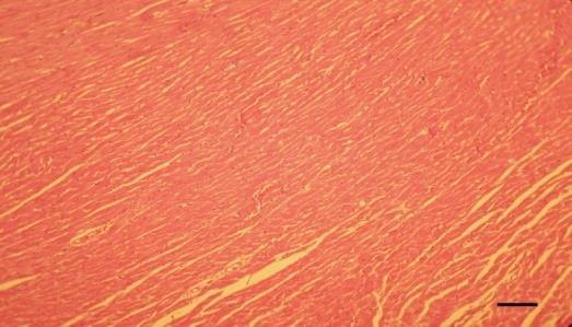
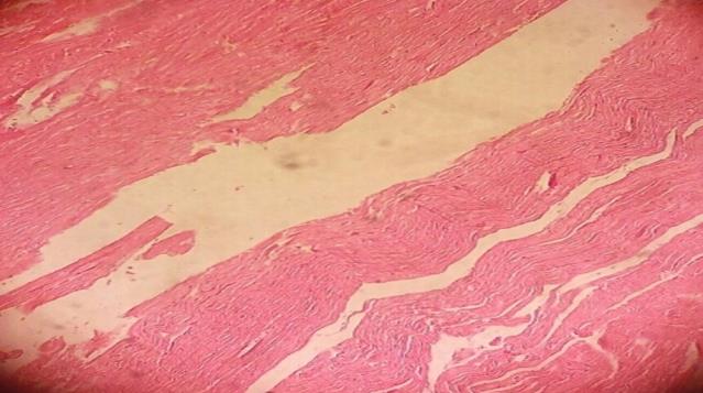
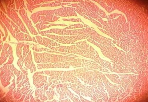
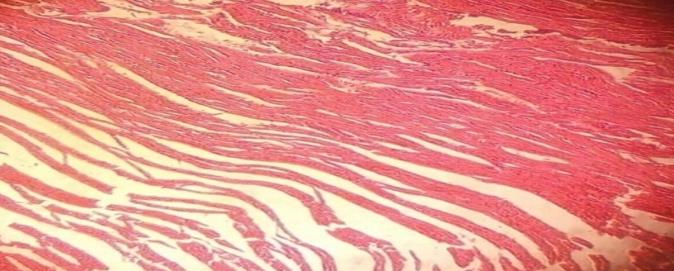
Figure 1: represents control (not treated with monosodium glutamate. There is no change in heart tissues of Wistar rats. Figure 2 showed slightly change in heart myofibers of Wister rat treated with low dose (0.5g). Figure 3 indicated degenerations change on heart tissues of Wister rat treated with medium dose (1.0g). Figure 4 indicated congestions change and hemorrhage on heart tissues of Wister rat treated with high dose (1.5g).
Conclusion
The present study concluded that MSG effect on lipid profile (Free Cholesterol, triglyceride, LDL and HDL-C) is significance increase in low, mid and high dose compared with control. Monosodium glutamate also effect on heart histology of Wister rat as in figure 1,2,3,4 respectively.
References
-
Adjene JO, Iteire KA, Cynthia I (2017) Effects of long- term consumption of indomie noodles on the body and brain weights of adult Wistar rats. World J Pharmacy Pharma Sci 6(12): 152-158.
-
Zailani HA, Umaru HA, Samuel G (2016) Effects of Instant Noodles Formulated Diet on Weanling Albino Rats. Direct Research Journal of Agriculture and Food Science 4(7): 161-168.
-
Sanni ME, Daniel E, Friday TB, Karachi C, Oglala E (2013) Effects of chronic administration of indomie noodles on the activity of alanine aminotransferases of rat kidney. J Pharm Biomed Sci 30(30): S65-S71.
-
Okediran BS, Olurotimi AE, Rahman SA, Michael OG, Olukunle JO (2014) Alterations in the lipid profile and liver enzymes of rats treated with monosodium glutamate. Sokoto Journal of Veterinary Sciences 12(3): 42-46.
-
Saeed AA (2016) Adverse Effects of Monosodium Glutamate on Serum Lipid Profile. Research Journal of Pharmaceutical, Biological and Chemical.
-
Madiha SAM, Muhammad SK, Qurratul-ain RK (2017) Instant Noodles: Are they Really Good for Health? A review. Electronic J Biol 13(3): 222-227.
-
SANZ (Standards Australia New Zealand) (2003) Monosodium Glutamate. A safety assessment. Technical Report. Food Standards Australia New Zealand. Series No: 20.
-
Hongbao MA, Kuan-Jiunn (2006) Cholesterol and Human Health. J Ame Sci (517) 432-0623.
-
Huizen (2018) What is cholesterol? Medical News Today.
-
HEART UK- the Cholesterol Charity 7 North Road Maidenhead Berkshire SL6 1PE, Email: ask@heartuk.org.uk. www.heartuk.org.uk.
-
Colpo A (2005) LDL Cholesterol: “Bad” Cholesterol, or Bad Science? J Ame Phy Surgeons 10(3).
-
Meiattini F, Prenipe L, Bardelli F, Giannini G, Tarli P (1978) The 4-hydroxybenzoate / 4- aminophenazone chromogenic system used in enzymatic determination of serum cholesterol. Clin Chem 24(12): 2161-2165.
-
Allian CC, Poon LS, Chan GSC, Richmond W, Fu PC (1974) Enzymatic determination of total serum cholesterol. J Clin Chem 20: 470-475.
-
Friedman A, Young G (1997) Effect of diseases on clinical laboratory tests, 3rd (Edn.), AACC Press.
-
Fossati P, Prencips L (1982) Serum triacylglycerol determined calorimetrically with an enzyme that producer hydrogen peroxide. Clin Che 28(10): 2077- 2080.
-
Burstein M, Scholnick HR, Morfin R (1980) Rapid method for isolation of lipoprotein from human serum by precipitation with polyanions. J Lipid Res 11(6): 583-595.
-
Grove TH (1979) Effect of reagent pH on determination of high density lipoprotein 560-564.
-
Salah E, Sabahelkhier MK, Shama IYA (2015) Effects of Aluminum Sulphate Treated in Deionizable and Tap Water on Lipid Profile of Wister Rats. ARPN Journal of Science and Technology 5(5): 268-270.
-
Saeed AA (YEAR) Cholesterol Status and Blood Glucose in Adult Rats. Research Journal of Pharmaceutical, Biological and Chemical Sciences.
-
Saeed AA (2016) Adverse Effects of Monosodium Glutamate on Serum Lipid Profile, Cholesterol Status and Blood Glucose in Adult Rats. RJPBCS 7(1): 732- 739.
-
Albert KGMM (1998) Diabetic Medicine; Triglyceride. HEART UK–The Cholesterol Charity 5: 275/281.
-
Husarova V, Ostatnikova D (2013) Monosodium glutamate toxic effects and their implications for human intake: a review. J Med Res 1-12.
-
Kingsley OA, Jacks PTW, Amaza DS, Peters TM, Otong ES (2013) The effect of monosodium glutamate (MSG) on the gross weight of the heart of Albino rats. Scholars J App Med Sci 2320-6691.
-
Magda ME, Nassr HAA, Marwa AEA, Eman MS, Mussab MR (2018) Effect of some food additives on lipid profile, kidney function and liver function of adult male albino rats. J Basic Environ Sci 5(2018): 52-59.
-
Tushar Kanti Bera SK, Prem KY, Prithwiraj M, ShankarY, Bishal J (2017) Effects of monosodium glutamate on human health: A systematic review. World Journal of Pharmaceutical Sciences 5(5): 139- 144.
- Superposition of Cryo-EM and AlphaFold Predictions of Dengue Antigen-Antibody Complexes
- Jugular-Applied Coherent Low-Level Laser Therapy Enhances Systemic Mitochondrial Metabolic Function and Antioxidant Response
- Role of OMC32 Polypeptide in Acrosin-Mediated Exocytosis during the Bovine Sperm Acrosome Reaction
- Association of Galectin-3 but not Laminin in Tamoxifen-Induced Growth Suppression in Breast Cancer MCF-7 Cells
- Effect of Different Wavelengths of Light on the Rate of Photosynthesis
- Nutritional, Therapeutic, and Environmental Effect of Oyster Mushrooms: An Editorial