Identification of Shooter using Scanning Electron Microscope Coupled with Energy Dispersive X-Ray (Sem-Edxa) Technique: A Case Study
In a shooting incident on 26.11.2012, the gunshot residue (GSR) sample were collected by the police department using double edged stubs from both the hands of the suspected shooter. These two stubs were sent to the laboratory for the presence of gunshot residue analysis. The search was carried out for firearm and ammunition later. The exhibits including stubs were sent to the laboratory on 21.04.2017. The analysis of the stubs was conducted using Scanning Electron Microscope Coupled with Energy Dispersive X-Ray (Sem-Edxa– Model Leo1430vp/1430vp-06-56) Technique. The GSR particles viz., Lead (Pb), Barium (Ba) and Antimony (Sb) were detected indicating his presence in the shooting incidence [1,2]. The specific case examination focuses on the detection of these particles even after five years after collection of these stubs and their careful preservation.
Introduction
The priming composition, the propellant, and the projectile are the three chief ingredients responsible for the development of the gunshot residues. The major portion of the residues is blown out from the muzzle along with the powder gases. The minor portion of the residue goes out from the openings of the firearm and most prominently the space between the front end of the cylinder and the rear end of the barrel. The purpose is to obtain gunshot residue samples from the persons suspected of recently discharging a firearm. The residue consists of sub microscopic particles and can be deposited on the shooter’s hand, face or clothing, and inside nostrils in varying amounts depending on the type calibre, and condition of the weapon or ammunition used as well as the environmental conditions at the time of shooting. Washing of hands and other activities on the part of the shooter can remove substantial amount of residues. Therefore, it is imperative to obtain samples as soon as possible.
When a gun fires, the firing discharge residues travels through the barrel and deposits on the target. Due to the space, especially in the case of country made firearm/improvised firearm and the revolvers, the GSR deposits on the shooter’s hand, face etc. and on the bystanders those who are on the proximity [3]. To identify the shooter, the gunshot residue analysis plays a very crucial/vital role in criminal investigation. The police Sergeant collected the GSR samples with the help of the GSR stubs from both the hands of the accused on the same day of the crime occurred. The two GSR stubs were sealed and sent to the laboratory on 21.04.2017.
Materials and Methods
The stubs, made up of aluminum, are placed inside the chamber and electron beam is allowed to fall on the same. Cobalt is used as a standard in the chamber. For detection of the gun shot residues, back scattered electrons are used. Back scattered electrons are those scattered backward and emitted out of the specimen, when incident electron scatter from the specimen. Since scattered electron possesses high energy than secondary electrons, information from a relatively deep region is contained in the back scattered electrons. The back scattered electrons are sensitive to the composition of the specimen. If the atomic number of the constituent atom is larger, the back scattered electron yield is larger i.e., an area consists of a heavy item appears bright in the back scattered electron image. The working distance is fixed at 15mm, EHT at 20 kV and with a probe current at about 100 micro Amps. The EDXA is used for the detection of elemental profile of the elements present in the sample. The specimens as a whole and at various points were analyzed and their respective spectrum are studied [4, 5].
Results and Discussion
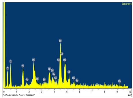
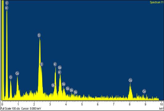
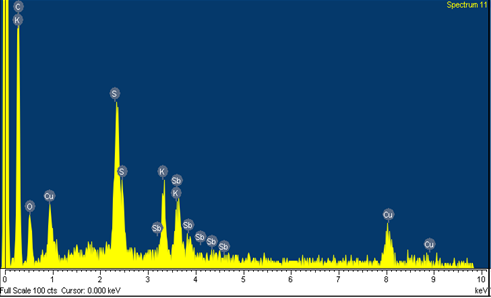
The spectrum shown in Figures 1 & 2 are of right hand and Figures 3 & 4 are of left hand of the shooter. Their weight percent and the atomic percent of the elements present therein are shown in Tables 1-4 respectively. Table 2 reveals the presence of the GSR particles viz., Lead (Pb), Barium (Ba) and Antimony (Sb) in weight percent of 12.07 %, 45.38% & 8.67% and atomic percent of 2.11%, 11.95%, and 2.57% respectively. Table 1, Table 3 and Table 4 reveals the presence of one of the element or two elements of the GSR particles viz., Lead (Pb), Barium (Ba) and Antimony (Sb). The peaks of the elements of respective spectrum can be corroborated with the weight percent and the atomic percent as provided in the table.
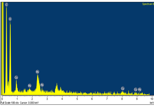
| Element | Weight % | Atomic % | ||||||
|---|---|---|---|---|---|---|---|---|
| C K | 41.72 | 51.83 | ||||||
| O K | 50.79 | 47.37 | ||||||
| Cu K | 1.6 | 0.38 | ||||||
| Pb M | 5.89 | 0.42 | ||||||
| Totals | 100 |
Table 2: Elements of the Right Hand.
| Element | Weight % | Atomic % | ||||||
|---|---|---|---|---|---|---|---|---|
| C K | 14.83 | 44.66 | ||||||
| O K | 14.30 | 32.34 | ||||||
| Al K | 4.76 | 6.38 | ||||||
| Sb L | 8.67 | 2.57 | ||||||
| Ba L | 45.38 | 11.95 | ||||||
| Pb M | 12.07 | 2.11 | ||||||
| Totals | 100.00 |
Table 3: Elements of the Right Hand.
| Element | Weight % | Atomic % | ||||||
|---|---|---|---|---|---|---|---|---|
| C K | 38.18 | 48.24 | ||||||
| O K | 52.84 | 50.12 | ||||||
| Al K | 1.72 | 0.97 | ||||||
| Ba L | 3.95 | 0.44 | ||||||
| Pb M | 3.31 | 0.24 | ||||||
| Totals | 100 |
Table 4: Elements of the Left Hand.
| Element | Weight % | Atomic % | ||||||
|---|---|---|---|---|---|---|---|---|
| C K | 61.4 | 78.84 | ||||||
| O K | 15.35 | 14.8 | ||||||
| S K | 3.99 | 1.92 | ||||||
| K K | 3.64 | 1.43 | ||||||
| Cu K | 8.86 | 2.15 | ||||||
| Sb L | 6.76 | 0.86 | ||||||
| Totals | 100 |
Table 1: Elements of the Left Hand.
Conclusion
The stubs, which were collected and preserved carefully, were analyzed after five years under SEM-EDXA. Even after five years of time span, the samples on the stubs responded to the electron beam and the GSR particles viz., Lead (Pb), Barium (Ba) and Antimony (Sb) were found to be present. Later, the seized firearm and ammunition were examined and found that the fired cartridge case could be linked with the suspected firearm because every firearm has its own individual characteristic marks. This information acts as a vital piece of evidence in the Hon’ble Courts.
References
-
Basu S (1982) Formation of gunshot residues. J Forensic Sci 27(1): 72-91.
-
Romolo F, Margot P (2001) Identification of gunshot residue: a critical review. Forensic Sci Int 119 (2): 195-211.
-
Lindsay E, McVicar M, Gerard R, Randall E, Pearson J (2011) Passive exposure and persistence of gunshot residue (GSR) on bystanders to a shooting: comparison of shooter and bystander exposure to GSR. Can Soc Sci J 44(3): 89-96.
-
Andrasko J, Maehly A (1977) Detection of gunshot residues on hands by scanning electron microscopy. J Forensic Sci 22(2): 279-287.
-
American Society for Testing and Materials, ASTM. ASTM Standard Guide for Gunshot Residue Analysis by Scanning Electron Microscopy/Energy Dispersive X-Ray Spectrometry, ASTM International, West Conshohocken,PA,201.
- Narcotics and Digital Forensics: Bridging Crimes in the Digital Age
- Ethics in Forensic Psychiatry: Principles, Dilemmas, and Human Rights
- Impact of Acute Stress on Attentional Orienting to Social Cues
- Head Injury and Intracranial Hemorrhage in Western Region of Libya
- A Forensic Study on Handedness: Examination of Handwriting Features in Right and Left Handed Writers
- Techniques for Latent Fingerprint Development Using Natural and Synthetic Powders: A Review