The Alternative Science of the Robert Blake Criminal Trial
Robert Blake, former television and movie star, was accused of shooting his wife, Bonnie Bakley, twice while she was sitting in the front passenger seat of their car. The shots came from outside the car while Blake alleged he was not present. There were no witnesses to the shooting. Blake stated to the police that upon returning to his car, he had sat on the driver’s seat before realizing his wife had been shot. Blake was taken into custody by the police, his hands GSR sampled, and released. There were no measures taken by the police to protect Blake’s hands or clothing from gunshot residue (GSR) contamination while he was in the police environment (police car and station). Blake had on his person a .38 caliber pistol at the time of the homicide that was not the murder weapon, but also a potential source of GSR contamination to his hands and clothing. The clothing Blake allegedly wore when the shooting occurred, was later collected by the police from Blake at his home (i.e., there was a break in the evidence chain). The clothing had been placed in an open cardboard box in the trunk of a police car for 48 h before proper packaging but was sampled and analyzed for GSR anyway. The evidence of Blake’s hands and clothing was presented in court by the prosecution, despite all the samplers being negative for GSR, alleging that they were inculpatory. Even if the GSR analyses found significant concentrations of GSR on any of these samples, it would have had no probative value. Errors were made interpreting the spectra from the scanning electron microscopy/energy dispersive X-ray analyses.
Abbreviations
GSR: Gun Shot Residue; SOP: Standard Operating Procedure.
Introduction
The shooting
Robert Blake, a former popular television and movie star [1], was accused of shooting his wife, Bonnie Bakley, after they dined at a restaurant. The shooting occurred on May 4, 2001 at approximately 2130 while Bakley was sitting in the passenger seat of their 1991 Dodge Stealth. The vehicle was parked. Bakley was shot while waiting in the car for her husband, Blake, when he had allegedly returned to the restaurant to retrieve his forgotten .38 caliber revolver. The murder weapon, a World War II vintage 9 mm Walther P38 Luger pistol, was found in a large construction debris dumpster behind which Blake had parked his car. The pistol was covered with motor oil and dirt. Two Remington 9 mm casings were found near the passenger side of the car.
The Author’s Retention
The author, a certified gunshot residue (GSR) expert Burnett BR [2], was retained by Los Angeles attorney Thomas Mesereau, who subsequently withdrew from the case. Mesereau was replaced by attorney Gerald Schwartzbach. No discovery was transmitted to the author until Schwartzbach took over the case. Schwartzbach retained another GSR “expert,” Celia Hartnett of Forensic Analytical, Hayward, California, but agreed to transmit the discovery to the author upon learning that a retainer had been paid. The following is mainly based on the discovery from attorney Schwartzbach.
The Autopsy
The autopsy report stated that Bakley was shot twice. One bullet entered her right cheek area, went through the skin of the cheek, through the bones at the base of the skull, and into the left temporal muscle (the bullet core) and the left temporal lobe of the brain (the bullet jacket). The bullet’s direction of travel was right to left and slightly upward. No soot or powder stippling was noted around the entrance wound; thus, the weapon was fired at a distant/indeterminate range. This was a fatal gunshot wound. The other bullet entered the right shoulder, passed through the soft tissue of the neck, through the right carotid artery, and ended in the area of the right cervical spine of C-7. The projectile core and jacket were recovered at this location. Again, no soot or stippling was noted around the wound. This shot was also at a distant/ indeterminate range and was “potentially” fatal.
Gunshot Residue Testing
The GSR analyst for the police who did most of the analyses by SEM/EDS was criminalist Steve Dowell of the Los Angeles County Coroner’s Office. Criminalist Yamauchi of the Los Angeles Police Department analyzed the GSR samples from the victim’s car.
The author’s evaluation of the GSR evidence, described below, were presented to attorney Schwartzbach, which he chose to ignore.
Results and Discussion
The 9mm Pistol
The 9 mm pistol, which was found in the dumpster, could not be traced to Blake [3]. The vintage Walter 9 mm pistol was cleaned and test fired by a criminalist with the Los Angeles Police Crime Lab. The objective was to determine whether the pistol deposited gunshot residue (GSR) on the shooter’s hand. The police and prosecutor decided that the results of scanning electron microscopy/energy dispersive spectroscopy (SEM/EDS) of Blake’s hand and clothing samples (analyses conducted during the latter part of May, 2001) linked Blake to the homicide.
Before test firing, the weapon was rinsed in isopropyl alcohol “as a precaution.” Two CCI Blazer Pb-free cartridges were fired prior to the two test shots of Remington 9 mm hollow point (147 gr bullet). There was no explanation as to why the pistol was rinsed with isopropyl alcohol (the exterior of the gun apparently had already been cleaned of the oil and dirt), nor was a reason provided as to why Pb- free ammunition preceded the test shots. The analysis data of this test was not included in the discovery reviewed for this paper only that the pistol was capable of depositing GSR on the shooter. The test firing of the pistol prevented the determination of the composition of the particles (e.g., by a dry wipe sample of the bore) that were produced from the pistol before the test firing. However, the oil may have entered the bore of the pistol, making such sampling impossible. Standard 9 mm Remington cartridges use a three-metal primer composed of PbSbBa [4]. Remington cartridges, as most cartridges from other companies, produce GSR particles composed of PbSbBa, SbBa, PbBa, PbSb and Pb [5].
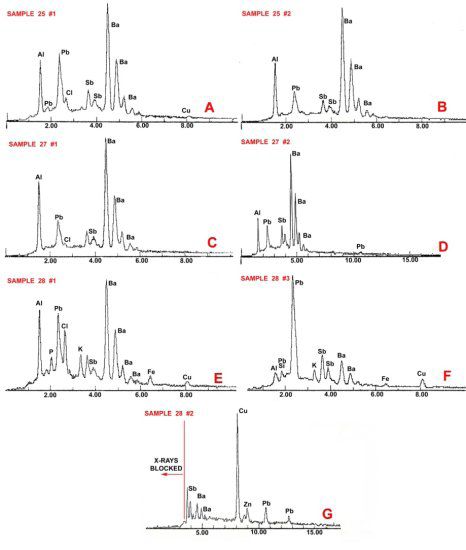
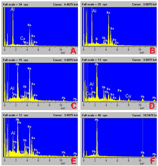
![Figure 3: A-G. Spectra of particles found in sample 25 (A and B), sample 27 (C and D) and sample 28 (E, F and G); sample 26 had no particles that could be attributed to GSR. Analyses were performed by criminalist Yamauchi of the Los Angeles Police Department. The lack of peaks below 1.0 keV indicate that a beryllium window detector was used for these analyses. Gunshot Residue Samples of victim Bakley Hands and the Vehicle Front Passenger Area The SEM/EDS analysis of the GSR samples from Bakley’s hands revealed only five characteristic (PbSbBa) [6] GSR particles on Bakley’s left hand and only one Pb particle on her right hand. Spectra of these particles are shown in Figure 1. Bakley’s left hand was exposed to at least one of the two shots; the car door shielded her right hand from GSR deposition. The vehicle in which Bakley was sitting was extensively sampled by standard adhesive SEM stubs. Ten samples were taken (Figure 2), of which four (samples 25, 26, 27 and 28) were examined by scanning electron microscopy. Of these four samplers, three were positive for characteristic GSR (samples 25, 27 and 28). The GSR data from Bakley’s hands (Figure 1) and the car Figure 3 show that aluminum is a feature of this characteristic GSR. However, aluminum is not a declared ingredient in Remington cartridges [4], and this may have been a component of previously fired ammunition (e.g., CCI makes ammunition with aluminum casings). An alternative explanation may be that the Remington cartridges were old and had a primer composition that included aluminum.](/fulltextimages/12902/fig_3.png)
Figure 3. A-G. Spectra of particles found in sample 25 (A and B), sample 27 (C and D) and sample 28 (E, F and G); sample 26 had no particles that could be attributed to GSR. Analyses were performed by criminalist Yamauchi of the Los Angeles Police Department. The lack of peaks below 1.0 keV indicate that a beryllium window detector was used for these analyses. Gunshot Residue Samples of victim Bakley Hands and the Vehicle Front Passenger Area The SEM/EDS analysis of the GSR samples from Bakley’s hands revealed only five characteristic (PbSbBa) [6] GSR particles on Bakley’s left hand and only one Pb particle on her right hand. Spectra of these particles are shown in Figure 1. Bakley’s left hand was exposed to at least one of the two shots; the car door shielded her right hand from GSR deposition. The vehicle in which Bakley was sitting was extensively sampled by standard adhesive SEM stubs. Ten samples were taken (Figure 2), of which four (samples 25, 26, 27 and 28) were examined by scanning electron microscopy. Of these four samplers, three were positive for characteristic GSR (samples 25, 27 and 28). The GSR data from Bakley’s hands (Figure 1) and the car Figure 3 show that aluminum is a feature of this characteristic GSR. However, aluminum is not a declared ingredient in Remington cartridges [4], and this may have been a component of previously fired ammunition (e.g., CCI makes ammunition with aluminum casings). An alternative explanation may be that the Remington cartridges were old and had a primer composition that included aluminum.
The analysis of the GSR samples of victim Bakley’s hands and the vehicle shows that the 9 mm pistol deposited GSR both on her and the interior of the car.
Blake in the Police Environment
Blake was taken to the police station in a police car. His hands were sampled for GSR at approximately 2355 on May 4, 2001. The time between the shooting (2130) and GSR sampling falls within the usual protocol range of 4 h [7, 8, 9, 10].
However, there was no attempt to isolate Blake from possible GSR contamination from sources other than the shooting while he was in the police environment. Gunshot residue contamination can come from the police handling of Blake [11, 12], the police car [13], or the police station [14] before GSR sampling.
Blake’s .38 Caliber Revolver
Blake carried a .38 caliber Smith and Wesson revolver during his visit to the restaurant. He claimed that he took the revolver out of his belt holster and placed it beside him on the bench under a sweatshirt. The revolver was alleged to have fallen to the floor under the booth table unnoticed when Blake and Bakley left the restaurant. Victim Bakley was shot during Blake’s alleged return to the restaurant to recover his revolver. His contact with the revolver could have contaminated him with GSR.
The GSR sampling of Blake’s revolver included a holster, which was collected by the police the night of the shooting. The revolver, like the Walther 9 mm Pistol, was also test- fired prior to sampling for GSR. Because this revolver was test-fired before sampling, any GSR information regarding the .38 caliber revolver’s contribution to the crime scene was compromised. If the exterior surface of the revolver had been sampled prior to test firing, those samples could have shown whether Blake had the potential to be contaminated by GSR particles that were originally associated with the revolver.
The GSR EDS spectra from the sampling (after the test firing) of the .38 caliber revolver are shown in Figure 4.
Although there are aluminum peaks in some of these spectra, the aluminum is not as prominent as the GSR particles from the samples from Bakley’s hands (Figure 1) or Blake’s car (Figure 3). It is likely that the aluminum contribution to many of these spectra comes from aluminosilicate particles near the analyte particles.
The Gunshot Residue Samples of Blake’s Hands
The Standard Operating Procedure (SOP) used by Criminalist Steve Dowell and the Los Angeles Department of the Coroner in their GSR examinations [15]:
- The gunshot residue samplers are placed within the SEM and the samplers are automatically scanned for gunshot residue candidate particles.
- The SEM-associated computer records the position of particles of interest. When the automated run is completed, the analyst relocates the particles of interest and performs a confirmation elemental analysis for each.
- The analyst summarizes the data and records by hand- written notes as well as acquiring spectra and particle images.
- A report is then written that summarizes the analysis results as well as provides a conclusion.
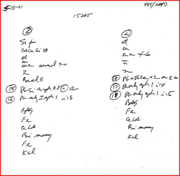
Figure 5. Handwritten notes written by analyst Steve Dowell summarizing the EDS results of the Blake hand sample analyses; the left column shows the results from the right-hand sampler. The right column shows the results from the left hand. The circled numbers are the particles where he returned to particles in the SEM for confirmation analyses by EDS. Figure 6 provides the interpretation of these notes.
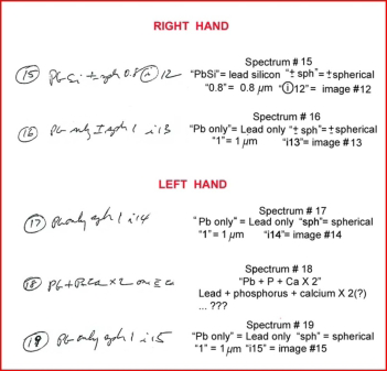
Figure 6. Interpretation of Dowell’s handwritten notes (from Figure 5), where spectra and images were recorded from the analyses of the gunshot residue hand samples of Blake. On the left, the circled number is the file number followed by the elemental composition interpretation of the particle. The “sph” means the particle is spherical; “+/- sph” means it is somewhat spherical; following the particle shape is the size (e.g., for particle 15, the size is 0.8 mm). The “i” followed by a number is the image number.
It has never been clear what is summarized in 3 of the crime lab’s SOP [15]. Is it from the computer-generated list, or did the analyst actually go to each of the particles noted in the handwritten document and confirm the elemental compositions?
Criminalist Dowell’s handwritten notes of the analysis of the Blake hand samples are shown in Figure 5. He did not provide a key to his notes in this discovery or in the author’s experience, anywhere else. After working on this case and previous cases that Dowell had reports, the author gained an understanding of his notes (e.g., Figure 6 left) to provide a translation (Figure 6, right). The circled numbers (Figure 5) are the particles that Dowell saw fit to return the particles of potential GSR composition in the SEM and the record spectra and images. The recorded spectra and particle images Figures 7 & 8 were matched to Dowell’s handwritten notes (top left of each spectrum, from Figures 5 & 6).
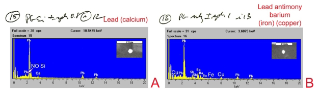
Figure 7. Handwritten notes with associated spectra referenced and particle images of the two particles for which the data were recorded from the right hand; the actual elemental identity is provided in the upper right of each spectrum. Elements noted in parentheses may be from surrounding particles (background). A. Particle 15, image 12; the note “NO Si” added by the author. B. Particle 16, image 13; elements Sb, Ba, copper Cu, and Fe were not noted by Dowell, but are in spectrum. This spectrum is of a characteristic GSR particle.
Analysis of the right-hand sampler from Blake’s hand revealed two possible consistent GSR particles and two additional possible consistent particles (Figure 5, left column). The “BaCaSiS” and “BaAlS” may be particles of interest in this case, but spectra and images were not recorded for these particles. For the other two particles of interest in this sample, spectra and images were recorded (Figure 7). Curiously, the spectra and size values for both of these particles do not match the written description. For spectrum 15 (Figure 7A), no Si is apparent in the spectrum despite the handwritten note stating otherwise. (The small peak at about 1.8 keV is consistent in size with only a Pb M-line peak at this location.) The size of this particle is approximately 3.5 microns (according to the scale bar in the image), not 0.8 microns. A small amount of calcium may be from a nearby particle. For spectrum 16 (Figure 7B), Dowell notes “Pb only.” However, Ba, Sb, Cu, and a small Fe peak are present making this a characteristic GSR particle. The reported size of this particle, 1 micron, is also incorrect. The particle measures approximately 2 microns according to the scale bar provided in the image.
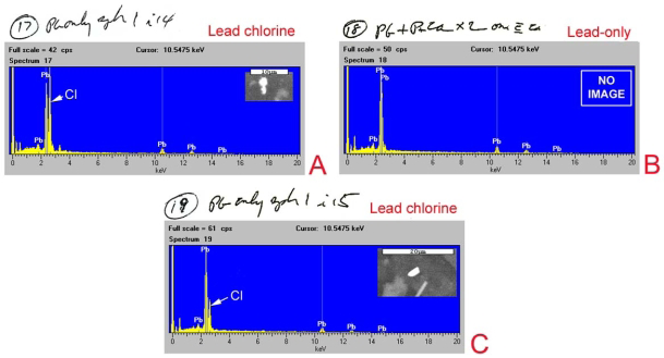
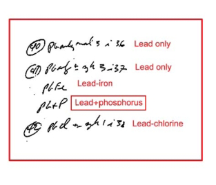
Figure 8. Handwritten notes, spectra, and particle images of the three particles for which these data were recorded from the left hand; the actual elemental identity is provided in the upper right of each spectrum. A. Particle 17, image 14; Cl in the spectrum, but not noted. B. Particle 18, no particle image; P and Ca are not represented in the spectra but are claimed to be present in the handwritten summary. C. Particle 19, image 15; again, Cl is in the spectrum, but not noted. Three possible consistent GSR particles were found in the analysis of the sampler from Blake’s left hand (Figure 5, right column). For the particle that spectrum 17 Figure 8A represents, Dowell claims “Pb only,” but he has neglected a prominent Cl peak in the spectrum. The particle is reported as spherical and 1 micron, but it is irregular and more than twice as large (2.4 x 5.0 microns). In spectrum 18 (Figure 8B), the handwritten notes “P” (phosphorus) and “Ca” (calcium) are present. There are no P or Ca peaks in this spectrum, but the handwritten note is confusing. (Such a particle would not be considered GSR, anyway.) Particle 19 spectrum Figure 8C does not match the written composition; a Cl peak is present in the spectrum. The particle is reported to be 1 micron, but is more than twice as large (2.2 x3.6 microns). It is also irregular and not “sph” (spherical). None of the spectra reported for Blake’s hand samples (Figures 7 & 8) match the reported composition.
The GSR Samples of Blake’s Clothing: The Storage Box Control
The clothing allegedly worn by Blake the night of the shooting was collected from his home the following day by a police officer. There was a break in the chain for this evidence. The officer stated during the trial that he “hoped and assumed it was the same clothing.” Despite this break in the chain of custody, the police proceeded with an analysis of Blake’s clothing, the results of which were part of the discovery. It is unknown to the author whether the results of the analyses of the clothing for GSR were presented in trial although Dowell in his 2005 presentation talked about these samples [16].
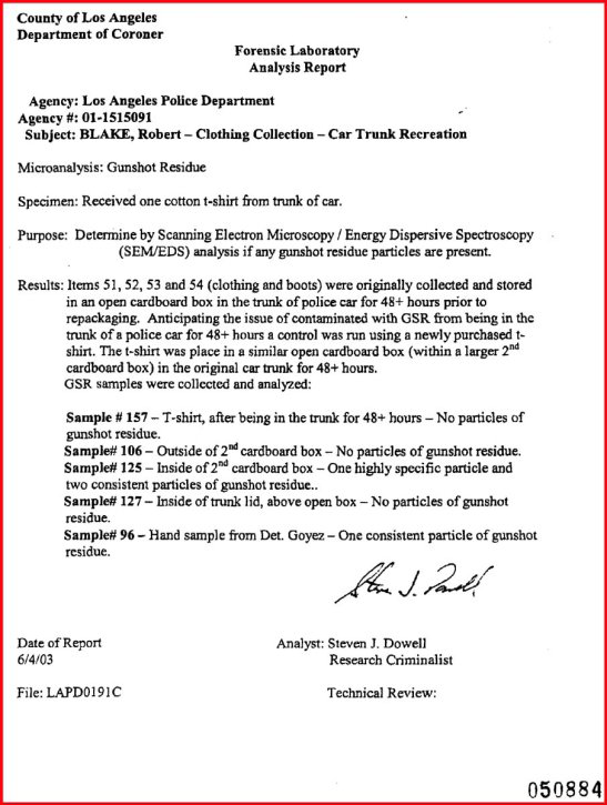
Sampling and analyses of the alleged clothing worn by Blake during the shooting were again performed by Steve Dowell of the Los Angeles County Department of the Coroner. Blake’s clothing after collection was not packaged in paper bags but was bulk stored in an open cardboard box in the trunk of a police car for more than 48 h. “Anticipating the issue of contamination with GSR from being in the trunk of a police car for 48+ hours, a control was run using a newly purchased shirt. The t-shirt was placed in a similar open card board box …” (Report from Dowell, June 4, 2003). This report indicated that there were only four of these control samples (Figure 9). However, the presentation by Dowell [16] suggested that additional samples were taken at that time. There were two boxes in the experiment, apparently one within the other. As to which was the “2 nd box” (Figure 9) is unclear. It is also unclear whether there was a box-within-a- box for the storage of the evidence in the back of the police car that this experiment attempts to simulate. The “control” samples:
The inner trunk door. This is a tape lift from the interior of the hood directly above the box. The report, dated 6/4/03, states “No particles of GSR” were found (Figure 9). Dowell’s later account in 2005 of this sampling notes, “we collected a number of samples from … different areas on the inside of the trunk.” He goes on to say, “… there were some particles on the trunk lid, not directly over the area where the t-shirt was.” Perhaps additional samples that were collected at that time were analyzed later although his report (Figure 9) does not mention this. The outside of the box. Dowell concluded that there were “no particles of GSR” (Figure 9) from the sampler taken from the outside of the test open cardboard box. This conclusion contradicted his handwritten notes (Figure 10) for this sampler. He noted three particles of PbBa and “Pb only many.” PbBa particles are common for.22 caliber rimfire cartridges [17]. PbBa particles from an environmental source are rare (fireworks [18] and nail guns [19] are the reported sources). There were no confirmation spectra. If the PbBa composition of these particles is real, a firearm source is likely.
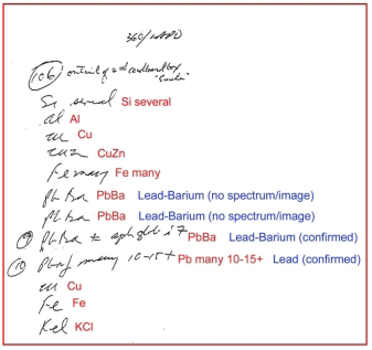
Figure 10. Dowell’s handwritten notes summarizing the results of the analysis of the SEM sample from outside of the control box; the PbBa particles, if real, likely have a firearms origin. The red notes: translations of the Dowell notes. The blue notes: identification of the elemental symbols and comments; (confirmed) = spectrum reflects note designation.
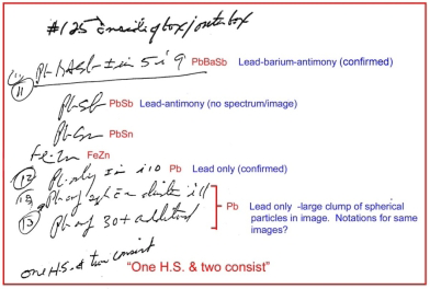
Figure 11. Dowell’s handwritten notes that summarize the results of the analysis of the SEM sample from inside the control box; in the interest of saving space, the particles of little or no interest (e.g. KCl, FeCl, CuZn, etc.) were redacted from these notes. The PbSbBa particle is a characteristic GSR particle. The red notes: translation of the Dowell notes. The blue notes: identification of the elemental symbols and comments; (confirmed) = spectrum reflects Dowell’s designation.
The inside of the box. Dowell found “One H.S. & two consist” (Figure 11), which means that he identified one particle that he is calling “highly specific” (i.e., a characteristic PbSbBa particle, Figure 12A) and two that are consistent with GSR. Indeed, Dowell’s report dated 6/4/03 Figure 9 states, “Inside of the 2nd cardboard box – One highly specific [characteristic] particle and two consistent particles of gunshot residue.” The spectra correspond to Dowell’s handwritten notes. However, for the particles that he calls “consistent,” there are not two but three: the “PbSb” particle (no spectrum/image), the single Pb particle (Figure 12B), and the clump of Pb particles (Figure 12C). Curiously, Dowell noted in his oral professional presentation Dowell S [16] that this sample did not exist: “They [defense counsel] criticized me for not taking a sample from the inside of the cardboard box. And at the time I said, ‘Well, I did not have a really good reason why I did not take a sample from the inside of the cardboard box.” Did he lie in his presentation or perjurer himself on the witness stand? The new t-shirt that was placed in the box had no detectable particles with GSR-like compositions. He apparently did not take control samples prior to the experimental exposure to the interior of the police car. Although the t-shirt was not contaminated by GSR, the presence of GSR particles on both surfaces of the box indicated that items present in the trunk of the police car were subject to GSR contamination. The account of these results given by Dowell [16] differs markedly from his case report (Figure 9), which, in turn, differs from his notes and spectra (Figures 10-12).
Figure 12. A–C. Notes, spectra, and images from the analysis of the sampler from the interior of the control box (Figure 11); the actual elemental identity is provided in the upper right of each spectrum. In this sample, the elemental compositions match reflects Dowell’s designation, except that the lower spectrum shows Cl is present in this spectrum.
The GSR Samples of Blake’s Clothing
A number of GSR samples were taken from the various items of Blake’s clothing, despite the potential contamination from Blake’s contact with the police car, the police station, his sitting in his car after victim Bakely was shot [16], and while the clothing was stored in an open box in the trunk of a police car [16].
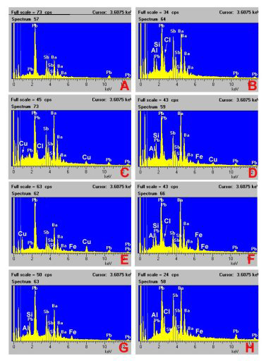
Figure 13. Spectra, images, and handwritten notes of particles associated with the boot sample; the actual elemental identity is provided in the upper right of each spectrum. Elements in parentheses may be from surrounding particles. A. Particle 5, image 4. B. Particle 7, image 6. C. Particle 8, image 7. D. Compilation of Dowell’s handwritten notes of other PbSb-bearing particles in the sample; there is no guidance provided as to how many particles were counted in, “Pb+P several.”
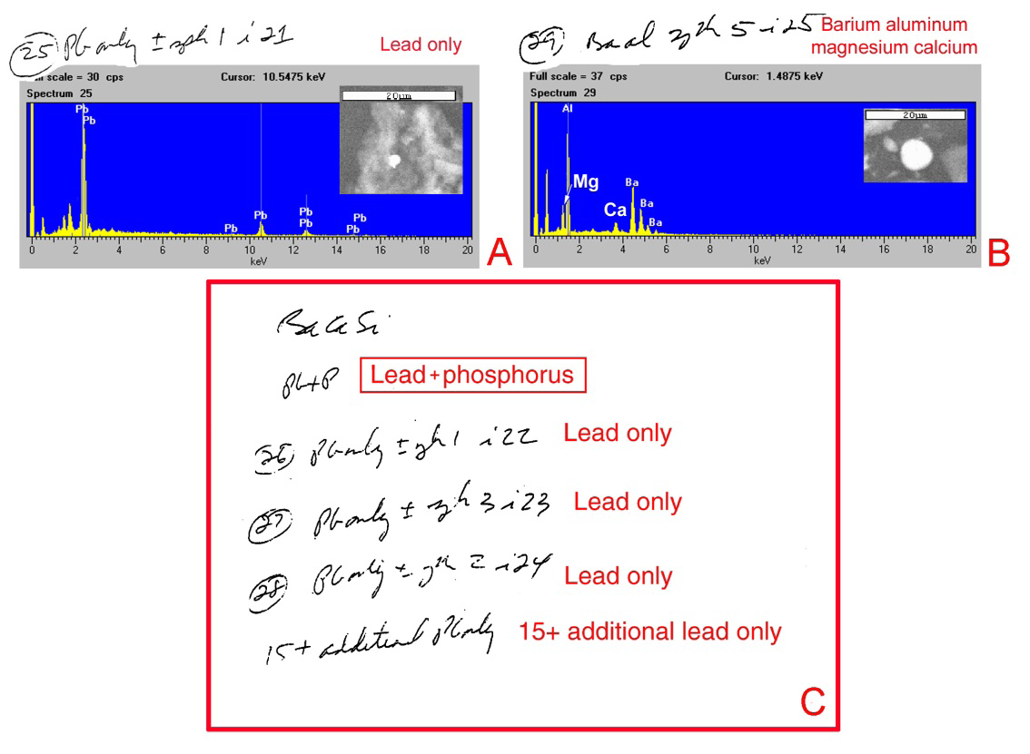
Figure 14. Spectra, images, and handwritten notes of particles associated with the jeans (2nd sample); the actual elemental identity is provided in the upper right of each spectrum. A. Particle 25, image 21. B. Particle 29, image 25; Mg and Ca are part of this particle but were not noted by Dowell. C. Selected list of particles of interest in the analysis.
The scenario offered by the prosecution is that if Blake shot his wife, then the clothing he was wearing would be subject to GSR contamination from the 9 mm pistol. However, the following excerpt from an email authored by Dowell, dated May 11, 2001, in the discovery identified another problem: “The Levi jeans appear to be ‘dirty’, that is they appear to have a history of use as do the black leather boots. In understanding and correctly interpreting the finding of GSR on items such as pants and boots, it is important to understand the history of the item(s) use – might the item been in contact at some other time, GSR may persist on such items for long periods of time and therefore a finding of GSR on such an item may not directly relate to the event that you are trying to understand. The presence of consistent particles of GSR on the samples collected from the hands, t-shirt and socks may have been transferred from the pants and/or boots. …” “Survey samples” were taken by Dowell [16] of the t-shirt worn by Blake (item #156) and the blue jeans + belt/belt buckle (item #152). Neither handwritten notes nor spectra of the “survey samples” were provided in the discovery. These “samples” were likely a non-automated examination of the samples taken from these items prior to automated SEM/EDS analysis. Boots. Dowell reports “one highly specific particle of gunshot residue and several consistent particles of gunshot residue.” Particle 5 (Figure 13A), Dowell claims calcium as a part, although this particle and the small amounts of Si and Al are likely from nearby particles. The “highly specific” (characteristic) particle appears to be the particle analyzed in spectrum 7 (Figure 13B). Dowell failed to note in particle 7 that Al and S (plus small amounts of sodium & magnesium) are likely from surrounding particles. Particle 8 BaAl, (Figure 13C) the Na and a small contribution of Al and Si are also likely from nearby particles.
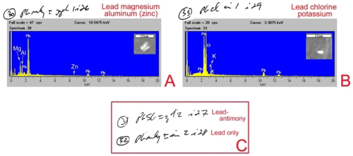
Figure 15. Spectra, images, and handwritten notes of particles associated with the black belt and buckle; the actual elemental identity is provided in the upper right of each spectrum. Elements in parentheses may be from surrounding particles. A. Particle 30, image 26; the presence of Mg, Al, and Zn) in the particles, but they were not noted in the spectrum. B. Particle 33, image 29. K was missing in note. C. Selected listing of particles of interest in this analysis.
Jeans (2nd sample). Particle 25 Figure 14A is likely correctly reported. The Al and Si are probably from nearby particles. Particle 29 Figure 14B also contains Mg and Ca. These elements are part of the particle.
The Mg would exclude this particle as GSR [6]. Dowell reports “several consistent particle [sic] of gunshot residue.” There are 19 Pb only particles in this sample (Figure 14C).
Black belt and buckle. Particle 30 (Figure 15A), which is mistakenly reported as having “Pb only,” also has Al, Mg, and zinc (Zn). The Mg would exclude this particle as GSR (Wolten et al, 1979). Particle 33 (Figure 15B), reported as having “Pb only,” also has Cl and K. A PbSb and a Pb-only particle were also reported Figure 15C.
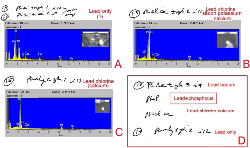
Figure 16. Spectra of interest and particle note summary from the analysis of the SEM sampler from the pair of black socks; the actual elemental identity is provided in the upper right of each spectrum. Elements in parentheses may be from surrounding particles. A. Particles 11 and 12, image 10; particles are probably Pb-only, (?): Si, Al. K and Ca X-rays are likely from the surrounding debris. B. Particle 13, image 11; Si and K are present but not noted. C. Particle 15, image 13; Cl and Ca are likely part of this particle. D. Selected list of other particles of interest in this analysis.
Pair of black socks. Particles 11 and 12 (Figure 16A) contain Al, K, Ca, and Fe. The “PbSi” assignment by Dowell is not correct. These two particles are probably Pb-only. The elements Si, Al, K, Ca, and Fe are likely from the particles are closely associated with these Pb particles. Particle 13 (Figure
16B) also contains Si and K, which appear to be part of this particle. With particle 15 (Figure 16C), Dowell failed to note that spectral peaks of Cl and Ca are also associated with the particle.
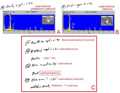
Figure 17. Spectra, images, and handwritten notes of particles associated with the black t-shirt (2nd sample); the actual elemental identity is provided in the upper right of each spectrum. Elements noted in parentheses may be from surrounding particles. A. Particle 36, image 32; Cl, K, and Ca are in the spectrum but not noted. B. Particle 37, image 33. C. Selected list of other particles of interest in this analysis; the actual elemental identity is provided in the upper right of each spectrum.
Black T-shirt (2nd sample). Particle 34 also contains a small amount of zinc (Zn). Particle 36 (Figure 17A), reported as having “Pb only,” also contains Cl, K, and Ca. Particle 37 Figure 17B is reported as having “PbCl” also has Ca) An example of the inconsistency of element assignments shown by Dowell: the proportion of Cl to Pb in particle 36 (Figure
17A) equals that of particle 37 (Figure 17B); however, in the former, Cl is not reported.
Boots (2nd sample). All particle compositions were confirmed. Dowell’s notes on Pb-bearing particles are presented in Figure 18.
| Elements | # |
|---|---|
| Pb only | 38 |
| PbCl (Ca, K) | 7 |
| PbP | 8 |
| PbCa +/- | 3 |
| PbFe | 2 |
| PbAlMg(Zn) | 1 |
| PbSb* | 1 |
| PbBaCa(Zn) | 1 |
| PbSbSn | 1 |
| BaAlMg(Ca) | 1 |
| PbBa* | 1 |
| BaAlSi* | 1 |
| BaSbAISiS* | 1 |
| Sb* | 1 |
Table 1: Summary of the Pb-bearing and other possible GSR particles associated with the clothing belonging to Blake; this list co
Table 1. Summary of the Pb-bearing and other possible GSR particles associated with the clothing belonging to Blake; this list comprised the corrected element compositions where spectra were available. Elements in parentheses are in trace amounts. The particle spectra with asterisk are consistent with GSR (without characteristic GSR other sources are probable).
The number of Pb-, Sb-, and Ba-containing particles for the six clothing samples is estimated to be 67 and is summarized in Table 1. Noteworthy is the lack of “highly specific” (characteristic) GSR particles (i.e., those composed of PbSbBa) in this combined listing. However, Dowell apparently calls the one particle (from the boots) with an elemental composition of “BaSb” (Figure 13B—actually BaSbAlS (Si?) as “highly specific” (characteristic) without Pb. At the time of this analysis, BaSb particles, without Pb were considered as “unique” [7]. But Torre C, et al. [24] found BaSb particles associated with friction brake dust. A confirmed environment source resulted in these particles are now considered consistent GSR. The 9 mm pistol used in this homicide could generate these particles (based on the GSR deposition in the car and on victim Bakley) with these elements. The particles with an asterisk in the list Table 1 could have been produced by a firearm (i.e., consistent GSR).
Conclusions
The Samples from the Victim and Car
Characteristic GSR particles were found on Bakley’s left hand and the interior of the car, most of which had strong aluminum peaks. The 9 mm pistol generated these particles at the shooting. Blake’s Hand GSR samples?
Dowell attached incorrect images of the spectra and particles to the report on Blake’s hand GSR burdens. Particle size determinations for all these particles are incorrect. Dowell likely mixed the Blake samples with another case. There was a lack of oversight (i.e., peer review) for Dowell’s work on this case. The potential for GSR contamination of Blake when he entered the police environment was not controlled [10]. Some other jurisdictions put paper bags over the hands of a suspect before being placed in the police environment if that suspect cannot be sampled at the place of arrest [20]. Even if characteristic GSR particles were found on Blake, the police car where he was placed, the police station, his .38 caliber revolver, sitting in his car after Bakley was shot (gunshot residue was deposited within the car at the shooting of Bakley) origins cannot be ruled out. The Walther 9 mm Pistol The handling of the 9 mm pistol and casings can be viewed as incompetent. It would have been appropriate to have obtained a bore wipe prior to the test firing of the pistol. If it was not possible to sample the bore of the pistol, then samples from the two casings may have provided information as to the nature (characteristic and consistent) of the GSR produced by the Walther 9 mm pistol. There was no additional mention of the casings after their collection at the scene and examination by the firearm expert.
Sweatshirts in the Back Seat of Blake’s Car
The flawed handling of this case extends to the proposed GSR analysis of two sweatshirts found in the backseat of Blake’s car. Detective Ronald Ito had requested GSR tests on the two sweatshirts to “determine whether either one may have been used as a silencer.” Criminalist Yamauchi of the Los Angeles Police Department’s Scientific Investigation Division noted in a memo, “Explained to Detective Ito that gunshot residue particle analysis cannot prove or answer his question. Any interpretation of the presence or absence of gunshot residue on surfaces other than bare hands is unfounded and possibly misleading.” If one of the sweatshirts was used as a silencer, SEM/EDS analysis would not be necessary because bullet holes and soot would be found by gross examination if one of the sweatshirts was used as a silencer.
The sampling of one or more inanimate objects associated with a shooting or at a shooting scene can have probative value (e.g., [21, 22]) and contrary to Yamauchi’s opinion, sampling and analysis of Blake’s clothing for GSR was conducted by Dowell and Yamauchi did analyze the samplers for Blake’s car (Figure 2). Both Yamauchi and Dowell failed to understand that for sampling and analysis of inanimate objects for GSR, an understanding of the shooting scene is necessary and whether such sampling has potential probative value. Blake’s Clothing Samples Most of the Pb particles found on Blake’s clothing were without Sb or Ba. Sources of Pb with Cl, Ca, Fe are leaded paint, gasoline, and pesticides [23]. Lead-rich particles persist in the environment despite their discontinued commercial use for a number of years [23]. The “lead only” particles noted by Dowell, if they are likely composed of Pb carbonate (the carbon of the particle cannot be distinguished from sampler adhesive). Phosphorus (actually PO4) associated with Pb is an insoluble compound [23].
Fertilizers with P-bearing compounds are added to soils to remediate the toxic forms of Pb (e.g., PbCl, PbCa, etc) to the nontoxic form, PbPO4 [23]. In addition, compounds of Pb in soil convert naturally to PbPO4 when free phosphate is available [23]. Dowell’s report on May 11, 2001 as noted above, “The Levi jeans appear to be ‘dirty’, that is, they appear to have a history of use…” The point is that the population of Pb particles found on Blake’s clothes came from soil, not from a firearm.
Additional control samples (i.e., samples from Blake’s home and the soils around) are necessary to make Dowell’s results meaningful for the analysis goal (e.g., particles found in the clothing samples have a statistically probable origin other than from Blake’s home), but not for probative value of these samples (i.e., Blake was the shooter). Indeed, Dowell’s answer to question 4 [16] admitted that the control samples to the clothing samples would have been appropriate. Without control samples, the results from the samples taken from Blake’s clothing are meaningless. It is misleading to call these particles “consistent” GSR without the detection of characteristic GSR particles [6].
The potential for GSR contamination from sources other than the shooting of victim Bakley, means that any positive GSR (> 3 characteristic plus consistent GSR particles [6]) for the clothing would be meaningless. Contamination of Blake’s clothing from the police car and station, from the trunk of the police car where the Blake clothing was stored, from the car where Blake apparently momentarily sat beside his dying wife, and from Blake’s .38 caliber revolver are all possible GSR sources for the few particles that could be attributed to a firearm.
There is a discrepancy between the reported composition and the submitted spectra for many of the alleged GSR particles. The predominance of Pb without Sb or Ba suggests that the Pb-bearing particles are from a source other than a centerfire cartridge firearm. The presence of PbP particles (Figures 13D, 14A, 16D, 17C, and 18) in this particle population indicates a soil origin [23].
Populations of GSR Particles
On page 58 of the pioneering work of GSR analysis by SEM/EDS by the Aerospace Group is the rule, “The presence of substantial numbers of inconsistent particles overrules the evidentiary significance of particles consistent with gunshot residue” [7]. The US Federal Bureau of Investigation Crime Lab criteria for a positive (significant ) GSR sample is 3 or greater characteristic accompanied by consistent GSR particles [6]. Torre C, et al. [24] reported that automotive friction products (i.e., brake pads) produced by different manufacturers generate GSR-like particles with combinations of PbSbBa [24]. There is a diversity of these particles that are made up of Pb, Sb, and Ba, usually with a significant Fe contribution to the spectra. When found on a sample, these brake-origin particles could be mistaken for GSR. Torre, et al. proposed an addition to Wolten’s Rule: “… before judging a sample as positive, the type of ammunition fired in the investigated crime must always be taken into consideration: only by comparison between the sample and the ammunition’s particles is it possible to attain a decisive answer” [24]. Giacalone JR [25] expressed the same position. However, the rule requires an additional modification due to residual GSR from previous discharges (the memory effect [17] and references therein) which may contribute GSR particles or combinations of a different composition from the most recent primer discharge. A SEM/EDS analysis of a dry bore wipe before testing firing of a firearm would be the best option if a comparison of suspect hand/crime scene GSR to a suspect’s firearm.
Kowal and Dowell stated, “…we report what we see on a submitted GSR sample including consistent P-GSR [primer-gunshot residue] of both spherical and irregular morphologies. In our report we include statements about the possible origins of those consistent particles” [26]. Many of the reports by Dowell (e.g., Figure 9), contrary to their crime lab’s policy, do not state the “possible origins of those consistent particles.” Alternative Science Counselor to the US President, Kellyanne Conway, in a 2017 interview described that she relied on “alternative facts” in advising the President [27]. In the People v. Robert Blake case, the “science” presented was junk, or in deference to Ms. Conway, “alternative” science and like the misinformed President, so was the Blake court. If this evidence was properly reviewed by a competent, expert in SEM/EDS and presented in pretrial motion, Steve Dowell’s junk science would never have been allowed to be presented to the court:
- Mixing of the Blake’s hand GSR SEM/EDS evidence with another case.
- Many instances of mistaken interpretation of EDS spectra.
- A break in the chain of custody of Blake’s clothing; was the clothing tested for GSR the same clothing worn by Blake at the time of the shooting?
- A waste of resources in the GSR analysis of Blake’s clothing even without the chain break; the GSR sampling and testing of only Blake’s shirt would have sufficed for determining GSR burden, if any, but the sampling and testing of the socks, belt, etc.? The GSR burden of Blake’s clothing was negative; there was no point in running the GSR control samples of the box in the trunk of the police car.
- Even if Blake’s hand and/or clothing samples were positive for GSR, the many potential sources of contamination would make any positive GSR result meaningless.
References
-
Robert Blake (actor) https://en.wikipedia.org/wiki/ Robert_Blake_(actor)
-
Curriculum vitae of Burnett B www.meixatech.com/ cv.pdf
-
(2023) Robert Blake, actor acquitted in wife’s murder, dies at 89. www.cbsnews.com/news/RobertBlake
-
Remington MSDS (2001) Material Safety Data Sheet. Centerfire/rimfire ammunition, Remington Arms Company.
-
Charles S, Geusens N, Nys B (2022) Interpol review of gunshot residue 2019 -2021. Forensic Science International: Synergy.
-
Wright DM, Trimpe MA (2006) Summary of the FBI Laboratory’s Gunshot residue sysposium, May 31-June 3, 2005. Forensic Science Communications 8(3): 1-19.
-
Wolten GM, Nesbitt RS, Calloway AR, Loper GL, Jones PF (1977) Final Report on Particle Analysis for Gunshot Residue Detection. The Aerospace Corporation pp: 1-197.
-
Zeichner A, Levin N (1995) Casework experience of GSR detection in Israel on samples from hands, hair and clothing using autosearch SEM/EDX system. J Forensic Sci 40(6): 1082-1085.
-
Mastruko V (2002) Forensic applications of an ESEM. International Association of MicroAnalysis 3(1): 4-7.
-
Jaeger L (2004) The value of primer gunshot residue (p-GSR) evidence: A pilot study regarding p_GSR contamination. Scanning Proceedings 26(2): 67-68.
-
French JC, Morgan R, Davy J (2014) The secondary transfer of gunshot residue: An experimental investigation carried out with SEM-EDX. X-ray Spectrum 43(1): 56-61.
-
Cook M (2016) Gunshot residue contamination of the hands of police officers following start of shift handling their firearms. Forensic Sci Inter 269: 56-62.
-
Kowal D, Sandberg K, Dowell S (2000) Gunshot residue in the law enforcement environment. California Association of Criminalists Seminar Presentation. pp: 12-14.
-
Crowson CA, Cullum HE, Hiley RW, Lowe AM (1996) A survey of high explosive traces in public places. J Forensic Sci 41(6): 980-989.
-
Anon (1997) Procedure: GSR Interpretation. Los Angeles County, Department of the Coroner. Microanalysis Section (A SOP document).
-
Dowell S (2005) Gunshot residue in the Robert Blake case-Lessons learned. Transcript of Presentation to Scanning, Monterey, California.www.meixatech.com/ BlakeGSR-Dowell.pdf.
-
Burnett BR, Nunziata F (2023) Divergent gunshot residues and characterization of the memory effect in a .22 caliber revolver and pistol. Egyptian Journal of Forensic Sciences 13(13): 1-20.
-
Trimpe MA (2003) Analysis of fireworks for particles of the type found in primer residue. International Association for MicroAnalysis 4(1): 1-8.
-
Lindsay E, Ballantyne D (2009) Regarding nail guns. Forensic_SEM@Yahoogroups.com; www.meixatech. com/NAILGUNS.pdf.
-
Kimmett MJ (2001) Incidence of gunshot residue transfer to paper bag hand covers. International Association for MicroAnalysis 1(3): 2-4.
-
Burnett BR, Lebiedzik J (2017) Discharge of a pistol out a car window with the breech within the interior of the car: Analysis of gunshot residue on a car’s interior surface. J Forensic Sci 62(3): 768-772.
-
Burnett BR (2018) A case of alleged discharge of a firearm within a vehicle. Forensic Sci Int 289: e1-e8.
-
Cao X, Ma LQ, Singh SP, Chen M, Harris GW, et al. (2003) Field demonstration of metal immobilization contaminated soils using phosphate amendments. Final Report, Florida Institute of Phosphate Research 97-01- 148R, pp: 1-98.
-
Torre C, Mattutino G, Vasino V, Robino C (2002) Brake linings: A source of non-GSR particles containing lead, barium and antimony. J Forensic Sci 47(3): 494-504.
-
Giacalone JR (2002) Distinguishing characteristics for the determination of forensic microtrace origination. International Association for MicroAnalysis 3(2): 8-10.
-
Kowal D, Dowell S (2001) How do you weigh morphology in the interpretation of your P-GSR results?. International Association for MicroAnalysis 2(2): 10.
-
Alternative facts. https:/en.wikipedia.org/wiki/ alternative_facts
- Narcotics and Digital Forensics: Bridging Crimes in the Digital Age
- Ethics in Forensic Psychiatry: Principles, Dilemmas, and Human Rights
- Impact of Acute Stress on Attentional Orienting to Social Cues
- Head Injury and Intracranial Hemorrhage in Western Region of Libya
- A Forensic Study on Handedness: Examination of Handwriting Features in Right and Left Handed Writers
- Techniques for Latent Fingerprint Development Using Natural and Synthetic Powders: A Review