Protein Profile of Biopolymers from Marine Lobster Panulirushomarus
Panulirushomarus is an economically important spiny lobster that is widespread through the Indian Ocean in the southern part of Tamilnadu, India. The aim of the study is to analyze the presence of proteins in chitin and chitosan obtained from the shells of crustacean Panulirushomarus. The Quantification of the proteins was determined using the Bradford method and the associated proteins were analyzed by using the SDS PAGE analysis. The Biopolymers chitin and chitosan were studied by using XRD.
Isaac Dhinakaran D* and Gomathi M
Tamilnadu, India
626124, Tamilnadu, India, Email: isaacdhina@yahoo.co.in
chitosan were studied by using XRD.
Keywords: Panulirushomarus; Chitin; Chitosan; XRD; SDS Page
Introduction
Biopolymers are natural polymers that are abundantly available and extractable from natural sources. Some of them such as cellulose, starch, chitin and chitosan possess high potential that can be biologically and chemically synthesized into wide range of material applications [1]. Capture based aquaculture showed that Lobsters (both spiny and slipper varieties) act as the most important natural resources of Gujarat. The development of sea cage culture was adopted for the development of mud spiny lobster, Panulirus polyphagus [2]. The anticancer property of microbial pigment prodigiosin, isolated from Serratia marcescens, a marine crustacean, found to be against human cervix carcinoma cells. They showed dose dependent inhibition of cell proliferation and apoptosis activity against the Hela cells [3]. Chitin and chitosan are valuable marine biopolymers, recovered from the shrimp wastes, in the shrimp processing industry of Vietnam. It is estimated that 200,000 metric tons are produced per year. The obtained chitin and chitosan are characterized by their purity and functional properties. The polymers show good quality with low residual ash and protein content (<1%) [4]. Scygonadin is an anionic antimicrobial peptide recently identified from the seminal plasma of crab Scylla serrata. It inhibited the growth of Micrococcus luteus, E. coli, P. aeruginosa, S. aureus and Streptococcus pyogenes. The peptide was approximately a 43-kDa fusion protein and was highly stable and active [5]. Crustacean shell wastes are a rich source of astaxanthin, which is the pigment responsible for their orange-pink coloration. Crustaceans are able to modify some carotenoids such as beta carotene and transform them into astaxanthin. Crustacean haemocytes play important roles in the host immune response including recognition, phagocytosis, melanization, cytotoxicity and cell to cell communication [6]. Spiny lobsters (Palinuridae) are one of the most commercially important groups of decapod crustaceans that are usually inhabitants of hard substrates associated with coral reefs, rocky shores and boulder-strewn bottoms. There are eleven extant genera of spiny lobsters. Panulirushomarus which has a wide distribution in the Indo-West Pacific region is the most dominant species along the southwest and southeast coasts of India. P. homarus_is having three recognized sub-species. They are _P.-homarus-homarus, P. homarusmegasculptus and P. Homarusrubellus [7]. The objectives of the present study are to detect the presence of proteins in chitin and chitosan from the shells of P. homarus.
Materials and Methods
Sample Collection
The Spiny lobsters were collected from a local fish landing Centre at Chinnamuttam, Kanyakumari and South India and brought to the laboratory. The spiny lobsters were identified as Panulirushomarus. The samples were weighed and packed into the airtight containers.
Extraction of Chitin and Chitosan
The obtained lobster shells were washed thoroughly with distilled water and dried in an oven to constant weight at a temperature of 35°C. Then a 100g shell of Panulirushomarus was taken for the extraction process. The extraction method was based on the standardized protocol [8]. Demineralization: Demineralization 20gm of sample powder was demineralised with 300ml of 2N HCl or 24 hours with constant stirring and thus filtered. The filtrate was again washed with distilled water and filtered till the liquid showed neutral pH. The filtrate was then dried in a vacuum dryer and weighed. Deprotenization: The sample was then deprotenized with 300ml of 1N NaOH at 80°C for 24 hour with constant stirring. The NaOH was exchanged intermittently and the sample was washed with distilled water every time before adding fresh NaOH. After 24 hour the sample was filtered. The sample filtrate was washed as before and dried. Deacetylation: Chitosan was extracted from Panulirushomarus lobster. Chitin through deacetylated following the method of (Taga et al., 1984). Briefly, chitin was deacetylated with 40% NaOH, heated for 6hrs at 110°C in constant stirring then 10% acetic acid was added to the sample and stored for 12hrs at room temperature with constant stirring. The dissolved sample was re precipitated by adding 40% NaOH to pH 10. The sample was then dialyzed by deionized water to a pH of 6.5 and centrifuged at 10,000 rpm for 10minutes and freeze dried
Estimation of Proteins using Bradford Method
Bradford method (9) was used in this study to estimate the protein in chitin and chitosan. This technique was based on the binding between the protein in the sample solution and the Coomassie Brilliant blue G-250 dye in the Bradford reagent. The standard bovine serum albumin (BSA) solution (stock solution= 1mg/mL) of various series of concentration (20µg to 100µg) was taken and made up to 1mLwith distilled water in test tubes. The unknown samples, 1- 5µl, were taken and made up to 1mLdistilled water. 2.5ml of Bradford reagent was added to each tube. After 2min optical density at 595nm was read using an ultraviolet- visible (UV-Vis) spectrophotometer (Which one?). The standard graph was plotted as X axis representing BSA standard protein concentrations and Y axis represents the OD at 595nm of the standard. From this graph the protein concentration of unknown sample was calculated at different aliquots of samples (20µl to 100µl).
SDS-PAGE Analysis
In this study, SDS-PAGE was used to analyze the presence of protein. The gels, which had the thickness of 0.75 mm, had two parts: the stacking gel (5 % polyacrylamide) and the separating gel (12 % polyacrylamide). This kind of gel had the separating resolution range between 15 and 60 kDa. The purpose of the stacking gel was to concentrate proteins before passing through the separating gel, therefore resulting in shaper protein bands.
Preparing the Working Solutions
Solution A (Acrylamide stock solution), 100 ml: 29.2 g acrylamide, 0.8 g bis-acrylamide and add water to make up 100 ml. Solution B (4 x Separating gel buffer), 100 ml: 75 ml 2M Tris-HCl (pH 8.8), 4 ml 10 % SDS and 21ml water. Solution C (4 x Stacking Gel Buffer), 100mL: 50 ml 1M Tris-HCl (pH 6.8), 4 ml 10 % SDS and 46 ml water.
Preparation of the Gel
The formulation of gels used in this study was described in Table 1.
| 12% separating | 5% stacking | |||||||
|---|---|---|---|---|---|---|---|---|
| Reagents | ||||||||
| gel | gel | |||||||
| Solution A | 4ml | 2.3ml | ||||||
| Solution B | 2.5 ml | 0.67ml | ||||||
| Solution C | - | 1ml | ||||||
| Milli Q water | 3.5ml | 4.8ml | ||||||
| 10% APS | 50 µl | 30 µl | ||||||
| TEMED | 5 µl | 5 µl |
Table 1: [INLINE_TABLE:1:0]
Preparing Samples and Operation of SDS-PAGE
The prepared protein extracts were diluted to make a final concentration of 1 mg/ml, and then mixed with the loading dye. An amount of 15 or 20 µg of each protein mixture was introduced into the wells. The Precision plus dual stain protein marker 10 – 250 kDa (Bio-Rad, USA) was also added to one of the wells as a reference to estimate the molecular weight of the separated protein bands. The apparatus was run into two stages: stage 1 with 80 voltages in 20 minutes to have the proteins settled in the stacking gel, and stage 2 with 180 voltages in 50 minutes to separating the protein solution. After the run was completed, the gels were removed from the glass plate assembly and put in the Coomassie blue stain solution for 10 minutes with shaking and then washed in the destain solution for 3 hours till the protein bands were visible as dark blue bands against a transparent background. The gels were then scanned their images and saved in computer for later analysis.
XRD
The prepared samples was characterized by X-ray diffraction (XRD) technique using an X-ray diffractometer (Bruker Germany, D8 Advance, 2.2 KW Cu Anode, Ceramic X-ray) with CuKa radiation (k = 1.5406 A ° ´). The measurement was in the scanning range of 5–70 at a scanning speed of 50s-1.
Results
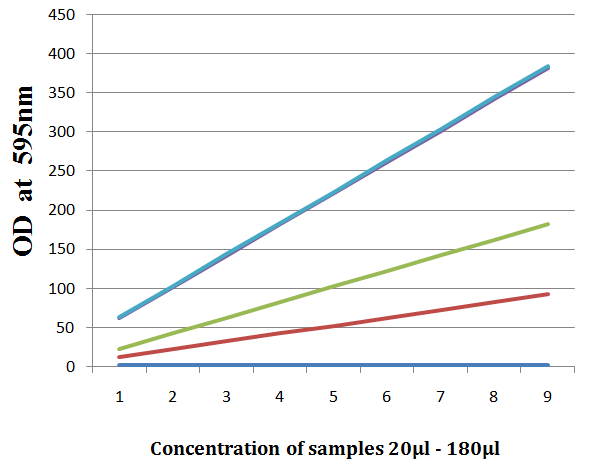
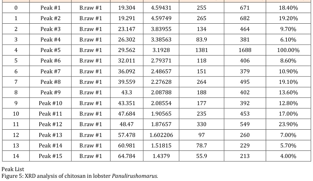
In the present investigation the chitin and chitosan extracts were prepared from marine spiny lobster Panulirushomarus. From the Figure 1 and Figure 2 the quantity of proteins present in chitin and chitosan of marine lobster Panulirushomarus using Bradford method was observed In Figure 3 the protein profile of the spiny lobster chitin and chitosan is associated proteins were identified using SDS-PAGE analysis. The molecular eight of the protein such as 205 KDa and 29 KDa were analyzed. The Figure 4 explains the XRD pattern of chitin prepared from lobster shells illustrates 8 characteristic broad diffraction peaks and highest peak observed at second peak shows the high intensity. The Figure 5 denotes the XRD pattern of chitosan prepared from lobster shells waste illustrates 15 characteristic broad diffraction peaks and the highest peak was observed at fifth peak shows the high intensity.
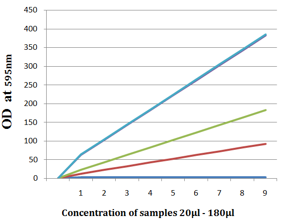
Figure1: Estimation of proteins in chitin using Bradford method.
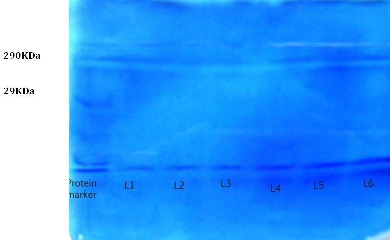
Lane1, Lane2, Lane3 Chitin Lane4, Lane5, Lane6 Chitosan Figure 3: SDS-PAGE analysis of Chitin and chitosan associated proteins in Panulirushomarus.
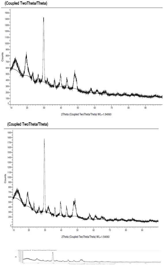
| Index | Name | Scan | Angle | d value | Net Intensity | Gross Intensity | Rel. Intensity | ||||||||||||||||
| 0 | Peak#1 | A.raw#1 | 26.491 | 3.36188 | 117 | 488 | 11.40% | ||||||||||||||||
| 1 | Peak#2 | A.raw#1 | 29.732 | 3.00245 | 1025 | 1397 | 100.00% | ||||||||||||||||
| 2 | Peak#3 | A.raw#1 | 31.89 | 2.80403 | 70.8 | 412 | 6.90% | ||||||||||||||||
| 3 | Peak#4 | A.raw#1 | 36.23 | 2.47744 | 107 | 399 | 10.40% | ||||||||||||||||
| 4 | Peak#5 | A.raw#1 | 39.955 | 2.26751 | 188 | 483 | 18.40% | ||||||||||||||||
| 5 | Peak#6 | A.raw#1 | 43.4489 | 2.25465 | 163 | 457 | 15.90% | ||||||||||||||||
| 6 | Peak#7 | A.raw#1 | 49.489 | 2.07924 | 150 | 426 | 14.60% | ||||||||||||||||
| 7 | Peak#8 | A.raw#1 | 49.341 | 1.84549 | 120 | 373 | 11.70% |
Index Name Scan Angle d Value Net Intensity Gross Intensity Rel. Intensity
Crustaceans compose a large, ancient and diverse animal group that includes many well-known, commercially exploited members, such as shrimp, crab, crayfish, and lobster. AMPs or proteins are one of the major components of the innate immune defence and are ubiquitously found in crustaceans [9]. Wild and cultured lobsters harbour a diverse bacterial flora which includes the dominant generas like Aeromonas, Pseudomonas, Bacillus, E.coli, Salmonella and Vibrio. These organisms are having the ability to produce enzymes. These enzymes are produced during the utilization of certain nutrients such as proteins; lipid and carbohydrate extracellular lipases [10]. In Panulirusjaponicus, digestive proteinase was highly active at pH 7.5. But in Norwegian Lobster Nephropsnorvegicus, three proteases were identified in SDS-PAGE including digestive cathepsin D1 [11]. In this present study zymogram showed the presence of chitin and chitosan in the shells of marine Lobster Panulirushomarus based on the bands on SDS-PAGE with the molecular weight of 29kDa. The crystal structures of chitin and chitosan are characterized by XRD measurements. The XRD is a valuable tool to calculate the chiin and chitosan size. Previous studies in the crab the range of chitin and chitosan was from 10 mm. These observation with the results of other studies that the diffracted intensities were recorded from 20° to 40°C at the spherical structure. It reported that XRD pattern obtained for chitin and chitosan showed number of bragg’s reflection [12]. The present study shows that degree of deacetylation, bulk density, and water binding capacity of chitin and chitosan in the shell extracts of Panulirushomarus.
Conclusion
The marine spiny lobster Panulirushomarus have biopolymers like chitin and chitosan with associated proteins. The highest amounts of proteins are found in the chitin and chitosan. The biopolymers could be used as the sources for the drug discovery.
References
-
Alvarenga ESD (2011) Characterization and Properties of Chitosan. In: Elnashar PM (ed.) Biotechnology of Biopolymers. In Tech 34(1): 45-89.
-
Mohammed G, Rao GS, Ghosh J (2010) Aquaculture of spiny lobsters in sea cages in Gujarat, India. J Mar Biol Ass India 52 (2): 316-319.
-
Kavitha R, Aiswariya S, Chandana R, Ratnavali MG (2010) Anticancer activity of red pigment from Serratia marcescens in Human cervix carcinoma. Int J Chem Tech Res 2(1): 784-787.
-
Trung TS, Bao HND (2015) Physicochemical Properties and Antioxidant Activity of Chitin and Chitosan Prepared from Pacific White Shrimp Waste. International Journal of Carbohydrate Chemistry 2: 59- 64.
-
Wang KJ, Huang WS, Yang M, Chen HY, Bo J, et al. (2007) A male specific expression gene, encodes a novel anionic antimicrobial peptide, scygonadin, in Scylla serrata. Mol Immunol 44(8): 1961-1968.
-
Yederi R, Reddy K (2009) Purification and characterization of antibacterial proteins from granular hemocytes of Indian mud crab, Scylla serrata. Acta Biochim Pol 56(1): 71-82.
-
Wang SL, Shih IL, Liang TW, Wang CH (2002) Purification and characterization of two antifungal chitinases extracellularly produced by Bacillus amyloliquefaciensV656 in a shrimp and crab shell powder medium. J Agric Food Chem 50: 2241-2248.
-
Yamaguchi T, Takamura H, Matoba T, Terao J (1998) HPLC method for the evaluation of the free radical- scavenging activity of foods by using 1, 1-Diphenyl-2- picrylhydrazyl. Biosci Biotechnol Biochem 62(6): 1201-1204.
-
Paul S, Jayan A, Sasikumar CS, Cherian S (2014) Extraction and purification of chitosan from chitin isolated from sea prawn enneropenaeusindicus. Asian J Pharm Clin Res 7(4): 1-4.
-
Aider M (2010) Chitosan application for active bio- based films production and potential in the food industry. Food Science and Technology 43(6): 837- 842.
-
Tayel AA, Moussa S, Opwis K, Knittel D, Schollmeyer E (2010) Inhibition of microbial pathogens by fungal chitosan. Int J Biol Macromol 47(1): 10-14.
-
Jiravanichpaisal P, Lee BL, Soderhall K (2006) Cell- mediated immunity in arthropods: Hematopoiesis, coagulation, melanization and opsonization. Immunobiology 211(4): 213-36.
- Genetic Improvement of Nile Tilapia (Oreochromis niloticus): Advances in Selective Breeding and Genomic Approaches for Sustainable Aquaculture
- Microplastics, Contaminants, and Waste Hotspots: Divergences and Faults in Prioritizing Control Efforts
- Creating a Healthier, More Vibrant Open and Closed Aquatic Environment. A Submersible, Centrifugal Magnetically Affixed Current Changing Aquarium Pump
- An Attempt to Assess Alpha Diversity and Sample Size: Using the Ostracod Assemblages off Kumamoto Port, Japan
- Assessment of the Efficiency of Common Fishing Gears and Crafts Used at Mohananda River of Chapai Nawabganj, Bangladesh
- Fish Productivity and Biodiversity Status of Sundarban Mangrove in Bangladesh