Antimicrobial, Antioxidant and Anticoagulant Properties of Chitosan Extracted From Mole Crab, Emerita emeritus (Linnaeus, 1767)
The present study evidences the immunological properties of chitosan extracted from the shells of Emerita emeritus which includes antimicrobial, antioxidant, and anticoagulant activity. The extraction proceeded by subjecting the shells to demineralization, deproteinization, and deacetylation reactions. The presence of chitosan was analyzed using Fourier transform infrared (FTIR) spectroscopy. Antibacterial activity of chitosan displayed maximum inhibition for Bacillus subtilis and Pseudomonas aeruginosa when compared to Escherichia coli and Staphylococcus aureus. Similarly, the extracted chitosan highly inhibited Candida albicans than Aspergillus fumigatus. The minimum inhibitory concentration (MIC) of chitosan for different bacterial and fungal strains was also documented. The free radical scavenging activity of chitosan using 1,1-diphenyl- 2-picrylhydrazyl (DPPH), increased by 60.40%. The anticoagulant activity of chitosan was discovered when mixed with chicken blood. Overall, the study emphasizes the conversion of aquaculture waste products like crustacean exoskeleton into a beneficial chitosan and signifies the effectiveness of chitosan against invading microbes in aquaculture.
Introduction
The Indian economy’s fisheries and marine sectors have enormous potential for development as a source of addressing community nutritional needs. A group of crustacean species such as the marine mole crab, which belongs to the Hippidae family, are medium-sized crustacean that occurs in the intertidal zone and upper subtidal sandy marine environment is one fisheries item with high nutritional value [1]. Crustaceans are a globally distributed faunal group from the equator to the poles and they are found across all habitats [2]. Decapods are the most recognizable of all crustaceans and comprise a dominant group of benthic invertebrates, including many species of economic importance [3]. Decapod crustaceans such as penaeid shrimp, lobsters, mud crabs, and blue swimming crabs are considered as high-value resources and become protein sources for human [4]. In India shrimp (Penaeus vannamei) farming and prawn (Macrobrachium rosenbergii) culture brought a considerable socio-economic benefit [5, 6].
Likewise, crab fishery is also considered one of the important economic beneficiaries of India especially mud crabs [7]. However, apart from the economic importance the anomuran and brachyuran crabs are proved to be ecologically important as they play an important role in the estuary and marine ecosystems and any changes in the species will lead to biodiversity loss [8].
In crustaceans, the exoskeleton or cuticle is a salient feature with numerous functions among which is to provide protection against pathogens. Despite being a simple physical barrier, the immune protection of the cuticle is slightly more complex [9]. In response to a mechanical scratch or microbial invasion, melanization occurs in the cuticle. This defense response results from the presence of phenoloxidase zymogens, which are produced by hemocytes and transported to the cuticle across the epidermis [10].
This process is often used for wound healing and results in the formation of dark melanized plugs around the damage zone of the cuticle [11]. The exoskeleton or shells of crustaceans such as crabs, shrimps, and crayfish consist of 30-40% protein [12], 30-50% calcium carbonate, and calcium phosphate and 20-30% chitin [13].
Chitin is the second most common polysaccharide found in nature and present in exoskeleton of crustaceans and insects, parasitic nematode eggs, gut linings, and in the cell wall of fungi [14]. It is a linear, neutrally charged polymer of β-(1,4)-linked N-acetyl glucosamine (GlcNAc) [15, 16].
Chitosan, a cationic polymer of glucosamine (GlcN), is primarily produced from chitin by exhaustive alkaline deacetylation; this process involves boiling chitin in concentrated alkali for several hours. Since this N-deacetylation is never complete, chitosan is considered a partially N-deacetylated derivative of chitin [16, 17]. The first investigation of chitosan using X-ray diffractometry was carried out on fibers prepared from lobster tendon chitin by a solid-state N-deacetylation [18]. A subsequent X-ray pattern was obtained and fully analyzed with tendon chitosan prepared from a crab tendon chitin by a similar deacetylation method [19].
Chitosan was quoted as a bio-molecule that is biodegradable and biocompatible in nature [20]. The biological properties of chitosan are non-toxicity, cytocompatibility, anticholesterolemic, antioxidant, anti- inflammatory activity, analgestic action, hemostatic action, angiogenesis stimulation, macrophage activation, granulation and scar formation, adsorption enhancer [21], mucoadhesive [22], antimicrobial and wound healing [23]. It has been recognized as a valuable material for potential applications in drug and gene delivery systems [24].
The increasing economic and social concern to decrease the use of antibiotics and other therapeutic chemicals has encouraged controlling diseases [25]. Numerous bacteria and fungi are highly pathogenic causing various infectious diseases [26]. Interestingly, chitin and chitosan have been investigated as antimicrobial agents against a wide range of target organisms like algae, bacteria, yeasts, and fungi in experiments involving in vivo and in vitro [27].
Chitosan and its derivatives possess antibacterial properties against various spoilage and pathogenic microorganisms. Its derivatives generally exhibit bacteriostatic rather than bactericidal activities [27]. It also exhibited antifungal activity against molds and yeasts, such as Fusarium oxysporum, Botrytis cinerea, Rhizoctonia solani, Candida lambica and Phomopsis asparagi [28]. These properties could be owned to be fungistatic to a greater extent than fungicidal [29]. Chitosan is also expected to function more rapidly on fungi than bacteria [27]. Some chitosan derivatives have been reported to have potent antifungal activities than that of original chitosan [30].
Furthermore, antioxidant properties of fungal chitosan from shiitake stipes have also been reported Yen MT, et al. [31]. But, the antioxidant properties of chitosan derived from crab shells are less available. Accordingly, the objective of this study was to assess the antioxidant properties of chitosan prepared from crab chitin by N-deacetylation using a concentrated sodium hydroxide solution [31]. Excluding hemostatic effect sulfonated derivatives of chitosan found to possess blood anticoagulant activity [32]. In the present work, an attempt was made to extract chitosan from the shells of mole crab E. emeritus and to analyze its antimicrobial, antioxidant and anticoagulant properties as an effective immune molecule of crustacean exoskeleton.
Materials and Methods
Collection and Maintenance of Experimental Mole Crabs
The animals with approximate body weight (7 - 9 ± 2.32 g) were collected from the intertidal zone along the coast of Elliots Beach, Besant Nagar, Chennai, Tamil Nadu, India. The specimen was transported to the laboratory in plastic troughs containing seawater. The animals were maintained in glass tanks containing sand and sea water with conventional aeration. The animal being a filter feeder does not require any supplementary feed. The seawater was changed twice on a daily basis. These animals were kept for acclimatization to the laboratory condition for a few days prior to the experiment.
Chitosan Extraction
Pretreatment of Mole Crab Shells
The E. emeritus shell waste was collected and washed three times thoroughly using tap water to remove the salt on its surface followed by washing with distilled water and then spreading on blotting paper to remove excess water. They were dried in a hot air oven at 70 °C overnight. All dried shells were milled using a mixer in order to obtain a particle size less than 0.5 mm following Shimahara, et al. [33]. In order to obtain purified chitin, it must be separated from the proteins, minerals, and other components. This separation was achieved by the following three steps.
Demineralization With Hydrochloric Acid
The dried mole crab shell chips (5 g) were immersed in 250 ml of 2N hydrochloric acid (HCl). The mixture is kept for 2 days at room temperature with occasional stirring using a glass rod. In the initial stage of the reaction, frequent stirring is required to prevent the floating of the shell chips caused by the generation of carbon dioxide gas. In the middle of the treatment, the exhausted hydrochloric acid is exchanged. After 2 days of immersing, de-mineralized shell chips are collected and washed with deionized water until neutral [33].
Deproteinization with Aqueous Sodium Hydroxide
The demineralized shell chips (5 g) were added to 250 ml of 1 N aqueous sodium hydroxide (NaOH). The mixture was boiled until the vaporization proceeded. The exhausted alkaline solution was exchanged for a fresh one every 6 hrs after the beginning of the reaction. After 21 hrs, the demineralized shell chips or crude chitin chips were collected and washed with deionized water until neutral followed by Foster and Hackman [34].
Deacetylation of Chitin with Aqueous Sodium Hydroxide
The deproteinized shells were subjected to deacetylation under 50% 1N NaOH for 36 hrs at 45 ºC. After this process, the NaOH solution was decanted and the chitosan powder was obtained. It was washed with distilled water until neutral and left to dry overnight at 60-65 ºC [33].
Confirmation of the Chitosan
The quality of the chitosan produced was checked by a solubility test with 1% acetic acid. Chitosan dissolves completely in 1% acetic acid. For the estimation of chitosan produced we took the sample out of the storage and weighed a few flakes of the shells. Then the sample was placed inside a clean beaker and 10 to 20 ml of 1% acetic acid was added to it. The solution was kept in BOD shaker for 30 to 40 min. Then the sample was taken out and weighed, carefully.
Fourier Transforms Infrared (FTIR) Spectroscopy Analysis of Chitosan
The chitosan extracted sample was dried and ground to a fine powder. About 0.5 g of the sample was then given for FTIR spectroscopy analysis [35].
Culturing of Microbes
All the microbial strains used in this assay were obtained from the Unit of Biocontrol and Metabolites laboratory, Centre for Advance Studies in Botany, University of Madras, Guindy Campus, and Chennai-600 025, India. The bacterial species E. coli, P. aeruginosa (gram-negative), and S. aureus, B. subtilis (gram-positive) were grown overnight in nutrient broth and kept in rotary shaker for 12 hrs and used for antibacterial activity.
Fungal strains such as C. albicans and A. fumigatus (Pathological strains) were sub-cultured on potato dextrose agar and allowed to grow well for 5 days under sterile conditions and subsequently stored at 10 ºC until used.
Antibacterial Activity of Chitosan
The antibacterial activity of chitosan was done by following agar well diffusion method using Muller Hinton agar (MHA) [36]. Briefly, the MHA medium was and poured onto the sterilized Petri plates placed in the biohazard chamber. After solidification, four bacterial strains such us E. coli, P. aeruginosa, B. subtilis and S. aureus (1-2 million cells per CFU, calibrated with McFarland reagent and absorbance measured) were spread on the agar with sterile swabs. Amphotericin was used as a positive control. Then, the chitosan (10 mg.ml-1) samples of different concentrations (50, 75, 100 µl/well) were added to each well. The plates were incubated at 37 ºC for 24 hrs and the antibacterial activity was determined by measuring the zone of inhibition. Triplicates were used for each test and their mean values were calculated.
Antifungal Activity of Chitosan
Two fungal strains such C. albicans and A. fumigatus were used and ketoconazole was used as a positive control. The antifungal activity was done by following Magaldi S, et al. [36] as same as described for antibacterial activity.
Minimum Inhibitory Concentration
To evaluate the minimum inhibitory concentration (MIC), the serial dilution technique was used following Qi LF, et al. [37]. Initially, chitosan solution was prepared for 10 mg.ml-1 in 1% acetic acid at pH 6.0. Then serial dilutions were performed from 1: 1 to 1: 512 and decreasing concentrations ranging from 1 - 0.001%. 10 µl of 1.5 × 10⁸ CFU/ml bacterial suspensions were transferred to each one in the series of tubes and incubated at 37 ºC for 24 hrs and OD measured at 450 nm.
For fungal strains, 10 µl of 1.5 × 10⁸ CFU/ml of fungal suspension was added to each well having different dilutions and kept for 48 hrs at 37ºC and OD measured at 450 nm. The microbial growth inhibition by serum was calculated using the formula.
$$ \text{Inhibition}\% = \frac{\text{OD of Control} - \text{OD of Sample}}{\text{OD of Control}}\times 100 $$
Scavenging Ability on 1, 1-Diphenyl-2- Picrylhydrazyl Radicals
Each chitosan sample (0.10 - 10 mg.ml-1) in 1% acetic acid solution was mixed with 100 µl of methanolic solution containing DPPH radicals, resulting in a final concentration of 0.1 mM DPPH. The mixture was shaken vigorously and left to stand for 30 min in the dark, and the absorbance was then measured at 517 nm against a blank [38]. IC50 value (mg.ml-
1) is the effective concentration at which DPPH radicals were scavenged by 50% and ascorbic acid was used as a standard. The free radical scavenging activity (RSA) was calculated by the following formula.
Absorbanceof DPPH Absorbanceof Sample RSA 100 Absorbanceof DPPH NC − = ×
( )
Anticoagulant Activity
For anticoagulant activity, different volumes (3, 5 and 7 ml) of 10 mg.ml-1 of chitosan were taken in sterile test tubes with cotton plugs. The control did not contain chitosan. Then fresh chicken blood was added to each test tube and made to a final volume of 10 ml. The test tubes were plugged properly and kept at room temperature for observation.
Results
Fourier Transform Infrared (FTIR) Spectrum of Chitosan
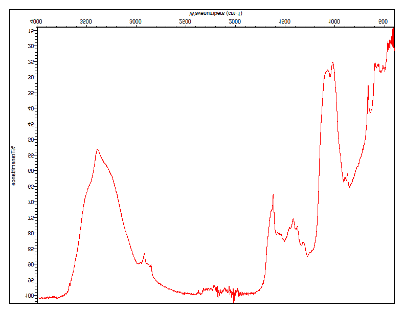
The chitosan extracted was analyzed using FTIR and the following graph was obtained (Figure 1). Characteristic peaks obtained were correlated with the chitosan standard graph cited in the literature. A strong band in the region 3400–400 cm-1 corresponds to N-H and O-H stretching, as well as the intramolecular hydrogen bonds. The absorption bands at around 3291 cm-1 can be attributed to C-H symmetric stretching. The presence of residual N-acetyl groups was confirmed by the bands at around 1645 cm-1 C=O stretching of amide I) and 1350 cm-1 (C-N stretching of amide III), respectively. The CH2 bending and CH3 symmetrical deformations were confirmed by the presence of bands at around 1423 and 1375 cm-1, respectively. The absorption band at 1153 cm-1 can be attributed to the asymmetric stretching of the C-O-C Bridge. The bands at 1066 correspond to C-O stretching.
Antibacterial Activity of Chitosan
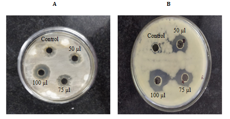
The results depicted that 50 µl of 10 mg ml-1of chitosan highly inhibited P. aeruginosa (9 ± 0.06) followed by E. coli (7 ± 0.01), B. subtilis and S. aureus. At 75 µl concentration highest inhibition was displayed by B. subtilis (13 ± 0.03) followed by P. aeruginosa (12 ± 0.03), E. coli (9 ± 0.02), and S. aureus (8 ± 0.01). Similarly, at 100 µl concentration, the inhibition was highest against B. subtilis (17 ± 0.01) followed by P. aeruginosa (15 ± 0.01), E. coli (11 ± 0.02) and S. aureus (10 ± 0.05) respectively (Figure 2A-D).

Antifungal Activity of Chitosan
The maximum inhibition at 50 µl of chitosan was higher for C. albicans (7 ± 0.03) than A. fumigatus (5 ± 0.02). This expression was followed at higher concentrations like 75 µl and 100 µl respectively. In brief, C. albicans showed more inhibitory potential than A. fumigatus (Figures 3A & 3B).
Minimum Inhibitory Concentration (MIC) of Chitosan
Antibacterial activity
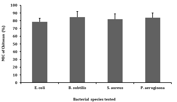
Minimum inhibitory concentration (MIC) of chitosan for different bacterial strains was determined (Figure 4) by assessing their turbid growth after 24 hrs respectively. It was observed that decreasing concentration of the chitosan increased the growth of the microbes. The MIC of chitosan for different bacterial strains were B. subtilis (84.50%) followed by P. aeruginosa (83.90%), S. aureus (81.90%) and for E. coli (78.60%).
Antifungal Activity of Chitosan
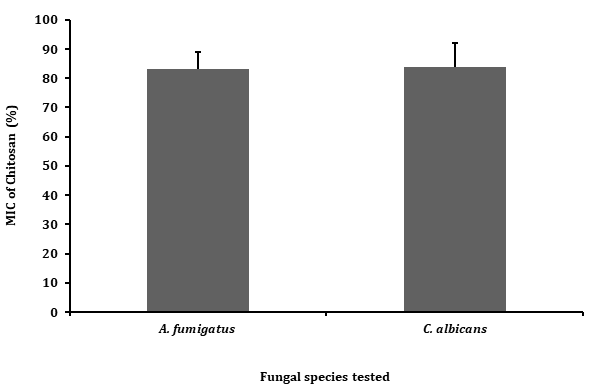
Similarly, MIC of chitosan for two fungal strains was determined (Figure 5) by assessing their turbid growth after 48 hrs respectively. The MIC of chitosan for C. albicans is 83.80% and for A. fumigatus 83%.
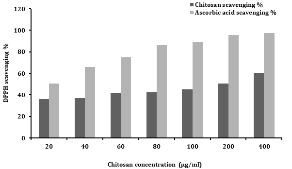
Antioxidant Activity of Chitosan
It is proved that antioxidant activity of chitosan was concentration-dependent. The change in colour of the solution (sample + DPPH reagent) was immediately observed which indicates the chitosan has potent quenching effect towards free radicals in the range of 36 to 60.40%. The
maximum radical scavenging of chitosan occurred at 400 µg/ ml while the 50% scavenging occurred at 200 µg/ml (Figure 6).
Anticoagulant Activity of Chitosan
A preliminary study to test the anticoagulant properties of chitosan was established. It was found that increasing concentration of chitosan increased the ability to prevent the coagulation of chick blood (Table 1).
| Chitosan | 10 min | 30 min | 60 min | |
|---|---|---|---|---|
| 1 | 3 ml | No clot | Minor clot | Semi clot |
| 2 | 5 ml | No clot | No clot | Minor clot |
| 3 | 7 ml | No clot | No clot | No clot |
Table 1: ** Different time intervals of chick blood anticoagulant properties in chitosan from mole crab Emerita emeritus.
Data represents triplicate repeats of five determinations. Table 1: Different time intervals of chick blood anticoagulant properties in chitosan from mole crab Emerita emeritus.
Discussion
The present study expresses the extraction of chitosan from mole crab E. emeritus shell waste and the subjecting the extracted chitosan to potential biochemical assays such as antibacterial, antifungal, antioxidant, and anticoagulant activities. The extracted chitosan showed a strong band in the region 3400-4000 cm-1 corresponding to N-H and O-H stretching, as well as the intramolecular hydrogen bonds.
The absorption bands at around 2921 can be attributed to C-H symmetric stretching. This band is a characteristic typical polysaccharide and is found in other polysaccharide spectra, such as xylan, glucans and carrageenans [39]. The presence of residual N-acetyl groups was confirmed by the bands at around 1645 cm-1 (C=O stretching of amide I) and 1350 cm-1 (C-N stretching of amide III), respectively. We did not find the small band at 1550 cm-1 that corresponding to N-H bending of amide II. This is the third band characteristic of typical N-acetyl groups, and it was probably overlapped by other bands. The CH2 bending and CH3 symmetrical deformations were confirmed by the presence of bands at around 1423 and 1375 cm-1, respectively. The absorption band at 1153 cm-1 can be attributed to the asymmetric stretching of the C-O-C Bridge. The bands at 1066 correspond to C-O stretching. All bands found in the spectra of E. emeritus chitosan were similar to reported by others [40, 41].
Assessing the antibacterial activity of extracted chitosan gave significant results against P. aeruginosa followed by E. coli, B. subtilis and S. aureus. As expected, the antimicrobial activity of chitosan was increased by increasing concentration [42]. The antibacterial activity of E. emeritus chitosan against B. subtilis showed highest inhibition while no activity or mild activity of chitosan from prawn shell waste against B. subtilis was reported by Tarafdar A, et al. [43]. However, Gram-positive bacteria (S. aureus and S. pneumonia), and gram-negative bacteria (E. coli) were inhibited markedly by Schiff base modified chitosan (acrylonitrile) as compared to unmodified chitosan [30]. The present study displayed the minimum inhibitory percentage of chitosan against B. subtilis, P. aeruginosa, S. aureus and E. coli were 84.50%, 83.90%, 81.90% and 78.60% similar to Parapenaeus longirostris chitin which exhibited lowest MIC against E. coli. In this study, both gram-positive and gram-negative strains showed moreover similar inhibitory percentage. But chitosan has profound antagonistic activity against gram- negative bacteria as compared to gram-positive [44].
The most prevalent proposed antibacterial activity of chitosan is by binding to the negatively charged bacterial cell wall causing disruption of the cell, thus altering the membrane permeability, followed by attachment to DNA causing inhibition of DNA replication and subsequently cell death [45]. Another possible mechanism is that chitosan acts as a chelating agent that selectively binds to trace metal elements causing toxin production and inhibiting microbial growth [46].
Interestingly, chitosan of E. emeritus depicted a dose- dependent inhibition against both the fungal strains C. albicans and A. fumigatus. But comparing the inhibition against two strains, C. albicans was higher than that of A. fumigatus. Previous studies have denoted chitosan as fungistatic rather than fungicidal [47, 48, 49] and proved to inhibit the growth of several fungi [50]. The antifungal activity of chitosan is known to be dependent on pH, molecular weight, degree of acetylation, temperature, ionic strength, and the presence of metal salts [49, 51]. Although various theories have been proposed to explain the mechanism of the antifungal activity of chitosan, the intracellular leakage hypothesis is widely accepted [52, 53]. The positive charge of chitosan binds to the negative charge on the surface of the fungal cell causing altered membrane permeability which leads to leakage of intracellular constituents causing cell death. Therefore, the antifungal activity of chitosan is suggested to be limited to the acidic condition as the positive charge of the chitosan amino group is lost at neutral pH [37, 54].
Antioxidant activity is a fundamental property and much important for life. Many biological functions such as anti- aging properties, anti-mutagenicity, and anti-carcinogenicity originate from antioxidant activity [55]. The DPPH is a stable nitrogen-centered free radical which can be effectively scavenged by antioxidants [56]. The ability of the protein to scavenge DPPH radical was determined by the decrease in it is absorbance in a spectrophotometer. In this study, the scavenging ability of chitosan increased from 20-400 µg/ml concentrations in comparison with the standard ascorbic acid. It has been shown that coupling chitosan with carbohydrate or oligosaccharide groups increases the antioxidant capabilities tremendously. The free radical quenching activity of chitosan extracted from shrimp wastes proved a dose dependent action [57]. A correlation between the antioxidant activity of chitosan and the degree of acetylation was reported by Hajji S, et al. [58]. Chitosan with a higher deacetylation degree because of more amino groups [31] and with low molecular weight exhibits improved antioxidant activity [59]. The scavenging activity of chitosan may be due to the reaction between the free radicals and the residual free amino group to form stable macromolecule radicals or the amino groups can form ammonium groups by absorbing hydrogen ions from the solution and then reacting with radicals through an additional reaction [60].
Heparin, one of the most widely used blood anticoagulants, is an expensive product. Attempts have been made to prepare a number of synthetic coagulants, but none are as non-toxic as heparin [61]. But our results about the dose-dependent increase in the anticoagulation ability of chitosan with fresh chicken blood may have use in the future as natural and less expensive anticoagulant. Several studies confirmed the anticoagulant property of chitosan extracted from crab shells [62, 63] and lobster shells [64]. Literature signifies that sulfonated derivatives of chitosan possess blood anticoagulant activity [65] and can be used as an effective anticoagulant with less toxicity.
Conclusion
In the present study, we conclude that chitosan extracted from shells of E. emeritus has antimicrobial, antioxidant, and anticoagulant properties. Because of its non-toxic and edible nature, it serves as a novel byproduct to enhance the quality of seafood and can be added in seafood products of aquaculture. Further, the antimicrobial and antioxidant properties depict the role of chitosan in fighting against microbes in the marine environment rather than just acting as a protective exoskeleton.
References
-
Mahapatro D, Karna SK, Mohanty SK, Mohanty B, Muduli PR, et al. (2018) First record of a burrowing mole crab Emerita emeritus (Decapoda: Anomura: Hippodae) from Chilika LAke, East Coast of India. Indian Journal of Geo Marine Sciences 47(01): 109-113.
-
Buhay JE (2011) Population dynamics of crustaceans: introduction to the symposium. Integrative and Comparative Biology 51(4): 577-579.
-
Silva JM, Creer S, Santos AD, Costa AC, Cunha MR, et al. (2011) Systematic and evolutionary insights derived from mtDNA COI barcode diversity in the Decapoda (Crustacea: Malacostraca). PLoS ONE 6(5): e19449.
-
Hamid A, Wardiatno Y (2018) Diversity of decapod crustaceans in Lasongko Bay, Southeast Sulawesi, Indonesia. Biodiversity Journal 9(3): 303-311.
-
Kutty MN (2005) Towards sustainable freshwater prawn aquaculture lessons from shrimp farming, with special reference to India. Aquaculture Research 36(3): 255- 263.
-
Kumaran M, Anand PR, Ashok Kumar J, Ravisankar T, Paul J, et al. (2016) Is Pacific white shrimp (Penaeus vannamei) farming in India is technically efficient? - A comprehensive study. Aquaculture 486(1): 262-270.
-
Sathiadhas R, Najmudeen TM (2004) Economic evaluation of mud crab farming under different production systems in India. Aquaculture, Economics and Management 8(1-2): 99-110.
-
Kunsook C, Dumrongrojwatthana P (2017) Species diversity and abundance of marine crabs (Portunidae: Decapoda) from a collapsible crab trap fishery at Kung Krabaen Bay, Chanthaburi Province, Thailand. Tropical Life Sciences Research 28(1): 45-67.
-
Moret Y, Moreau J (2012) The immune role of the arthropod exoskeleton. International Surgery Journal 9(2): 200-206.
-
Ashida M, Brey PT (1995) Role of the integument in insect defense: pro-phenol oxidase cascade in the cuticular matrix. Proceedings of the National Academic Sciences USA 92(23): 10698-10702.
-
Plaistow SJ, Outreman Y, Moret Y, Rigaud T (2003) Variation in the risk of being wounded: an overlooked factor in studies of invertebrate immune function?. Ecology Letters 6(6): 489-494.
-
Allan CR, Hadwiger LA (1979) The fungicidal effect of chitosan on fungi of varying cell wall composition. Experimental Mycology 3(3): 285-287.
-
Muzzarelli RAA (1977) Chitin. Pergamon Press, Oxford, USA, pp: 83-252.
-
Bueter CL, Specht CA, Levitz SM (2013) Innate sensing of Chitin and Chitosan. PLoS Pathogens 9(1): e1003080.
-
Merzendorfer H (2011) The cellular basis of chitin synthesis in fungi and insects: Common principles and differences. European Journal of Cell Biology 90(9): 759- 769.
-
Victor RDS, Santos AMDC, Sousa BVD, Neves GDA, Santana LNDL et al (2020) A review on chitosan’s uses as biomaterial: Tissue engineering, drug delivery systems and cancer treatment. Materials 13(21): 4995.
-
Dina Raafat GF (2008) Chitosan as an antimicrobial compound: Modes of action and resistance mechanisms. PhD thesis, University of Bonn, Germany, pp: 1-195.
-
Clark GL, Smith AF (1936) X-ray diffraction studies of chitin, chitosan, and derivatives. Journal of Physiological Chemistry 40(7): 863-879.
-
Ogawa K (1991) Effect of heating an aqueous suspension of chitosan on the crystallinity and polymorphs. Agricultural & Biological Chemistry 55(9): 2375-2379.
-
Chandy T, Sharma CP (1990) Chitosan-as a biomaterial. Biomaterials, Artificial cells and Artificial Organs 18(1): 1-24.
-
Kumirska J, Weinhold MX, Sauvageau JCM, Thöming J, Kaczynski Z, et al. (2009) Determination of the pattern of acetylation of low-molecular-weight chitosan used in biomedical applications. Journal of Pharmaceutical and Biomedical Analysis 50(4): 587-590.
-
Sogias IA, Williams AC, Khutoryanskiy VV (2008) Why is Chitosan Mucoadhesive?. Biomacromolecules 9(7): 1837-1842.
-
Rabea EI, Badawy MET, Stevens CV, Smagghe G, Steurbaut W (2003) Chitosan as antimicrobial agent: Applications and mode of action. Biomacromolecules 4(6): 1457- 1465.
-
Lin QK, Ren KF, Ji J (2009) Hyaluronic acid and chitosan- DNA complex multilayered thin film as surface-mediated nonviral gene delivery system. Colloids and Surface B Biointerfaces 74(1): 298-303.
-
Torrecilla D, Lozano MV, Lallana E, Neissa JI, Carballal RN, et al. (2013) Anti-tumor efficacy of chitosan- g-poly(ethylene glycol) nanocapsules containing docetaxel: Anti-TMEFF-2 functionalized nanocapsules vs. non-functionalized nanocapsules. European Journal of Pharmaceutics and Biopharmaceutics 83(3): 330-337.
-
Mousavi SM, Ghotaslou R, Kordi S, Khoramdel A, Aeenfar A, et al. (2018) Antibacterial and antifungal effects of chitosan nanoparticles on tissue conditioners of complete dentures. International Journal of Biological Macromolecules 118(A): 881-885.
-
Goy RC, de Britto D, Assis OBG (2009) A review of the antimicrobial activity of chitosan. Polimeros 19(3): 241- 247.
-
El-Hack MEA, El-Saadony MT, Shafi ME, Zabermawi NM, Arif M, et al. (2020) Antimicrobial and antioxidant properties of chitosan and its derivatives and their applications: A review. International Journal of Biological Macromolecules 164: 2726-2744.
-
Qin Y, Li PC, Guo ZY (2020) Cationic chitosan derivatives as potential antifungals: A review of structural optimization and applications. Carbohydrate Polymers 236: 116002.
-
Sabaa MW, Elzanaty AM, Abdel-Gawad OF, Arafa EG (2018) Synthesis, characterization and antimicrobial activity of Schiff bases modified chitosan-graft- poly(acrylonitrile). International Journal of Biological Macromolecules 109: 1280-1291.
-
Yen MT, Yang JH, Mau JL (2008) Antioxidant properties of chitosan from crab shells. Carbohydrate Polymer 74(4): 840-844.
-
Horton D, Just EK (1973) Preparation from chitin of (1→4)-2-amino-2-deoxy-β-D-glucopyranuronan and its 2-sulfoamino analog having blood-anticoagulant properties. Carbohydrate Research 29(1): 173-179.
-
Shimahara K, Takiguchi Y, Ohkouchi K, Kitamura K, Okada O (1984) Chitin, chitosan, and related enzymes. In: Zikakis JP (Ed.), Academic Press, New York, USA, pp: 239.
-
Foster AB, Hackman RH (1957) Application of ethylenediaminetetra-acetic acid in the isolation of crustacean chitin. Nature 180(4575): 40-41.
-
Queiroz, MF, Melo KRT, Sabry DA, Sassaki GL, Rocha HAO (2015) Does the use of chitosan contribute to oxalate kidney stone formation?. Marine Drugs 13: 141-158.
-
Magaldi S, Mata-Essayag S, De Capriles CH, Perez C, Colella MT, et al. (2004) Well diffusion for antifungal susceptibility testing. International Journal of Infectious Diseases 8(1): 39-45.
-
Qi LF, Xu ZR, Jiang X, Hu CH, Zou XF (2004) Preparation and antibacterial activity of chitosan nanoparticles. Carbohydrate Research 339: 2693-2700.
-
Shimada K, Fujikawa K, Yahara K, Nakamura T (1992) Antioxidative properties of xanthone on the auto oxidation of soybean in cyclcodextrin emulsion. Journal of Agriculture and Food Chemistry 40: 945-948.
-
Wolkers WF, Oliver AE, Tablin F, Crowe JH (2004) A Fourier-transform infrared spectroscopy study of sugar glasses. Carbohydrate Research 339(6): 1077-1085.
-
Vino AB, Ramasamy P, Shanmugam V, Shanmugam A (2012) Extraction, characterization and in vitro antioxidative potential of chitosan and sulfated chitosan from Cuttlebone of Sepia aculeata Orbigny. Asian Pacific Journal of Tropical Biomedicine 2(1): 5334-5341.
-
Song C, Yu H, Zhang M, Yang Y, Zhang G (2013) Physicochemical properties and antioxidant activity of chitosan from the blowfly Chrysomya megacephala larvae. International Journal of Biological Macromolecules 60: 347-354.
-
Lim SH, Hudson SM (2003) Review of chitosan and its derivatives as antimicrobial agents and their uses as textile chemicals. Journal of Macromolecular Science Part C 43(2): 223-269.
-
Tarafdar A, Biswas G (2013) Extraction of chitosan from prawn shell wastes and examination of its viable commercial applications. International Journal on Theoretical and Applied Research in Mechanical Engineering 2(3): 17-24.
-
Limam Z, Selmi S, Sadok S, El Abed A (2011) Extraction and characterization of chitin and chitosan from crustacean by-products: biological and physicochemical properties. African Journal of Biotechnology 10(4): 640- 647.
-
Nagy A, Harrison A, Sabbani S, Munson RS, Dutta PK, et al. (2011) Silver nanoparticles embedded in zeolite membranes: release of silver ions and mechanism of antibacterial action. International Journal of Nanomedicine 6: 1833-1852.
-
Divya K, Smitha V, George TK, Jisha MS (2017) Antimicrobial properties of chitosan nanoparticles: Mode of action and factors affecting activity. Fibers and Polymers 18(2): 221-230.
-
Coma V, Martial-Gros A, Garreau S, Copinet A, Deschamps A (2002) Edible antimicrobial films based on chitosan matrix. Journal of Food Science 67(3): 1162-1169.
-
Toan NV, Hanh TT, Thien PVM (2013) Antibacterial activity of chitosan on some common food contaminating microbes. The Open Biomaterials Journal 4: 1-5.
-
Ke CL, Deng FS, Chuang CY, Lin CH (2021) Antimicrobial actions and applications of chitosan. Polymers 13(6): 904.
-
Islam M, Masum S, Mahbub KR (2011) In vitro antibacterial activity of shrimp chitosan against Salmonella paratyphi and Staphylococcus aureus. Journal of the Bangladesh Chemical Society 24(2): 185-190.
-
Velasquez CL, Avelizapa LR (2020) A review on the physicochemical and biological aspects of the chitosan antifungal activity in agricultural applications. Journal of Research Updates in Polymer Science 9: 70-79.
-
Badawy MEI, Rabea EI, Rogge TM, Stevens CV, Smagghe G, et al. (2004) Synthesis and fungicidal activity of new N, O-Acyl chitosan derivatives. Biomacromolecules 5: 589-595.
-
Kong M, Chen XG, Xing K, Park HJ (2010) Antimicrobial properties of chitosan and mode of action: A state of the art review. International Journal of Food Microbiology 144(1): 51-63.
-
Liu H, Du Y, Wang X, Sun L (2004) Chitosan kills bacteria through cell membrane damage. International Journal of Food Microbiology 95: 147-155.
-
Cook NC, Samman S (1996) Flavonoids-Chemistry, metabolism, cardioprotective effects, and dietary sources. Journal of Nutritional Biochemistry 7(2): 66-76.
-
Vilano D, Fernandez-Pachon MS, Moya ML, Troncoso AM, Garcia-Parrilla MC (2007) Radical scavenging ability of polyphenolic compounds towards DPPH free radical. Talanta 71(1): 230-235.
-
Samar MM, El-Kalyoubi MH, Khalaf MM, Abd El-Razik MM (2013) Physicochemical, functional, antioxidant and antibacterial properties of chitosan extracted from shrimp wastes by microwave technique. Annals of Agricultural Sciences 58(1): 33-41.
-
Hajji S, Chaker A, Jridi M, Maalej H, Jellouli K, et al. (2016) Structural analysis, and antioxidant and antibacterial properties of chitosan-poly (vinyl alcohol) biodegradable films. Environmental Science and Pollution Research 23: 15310-15320.
-
Chien PJ, Sheu F, Huang WT, Su MS (2007) Effect of molecular weight of chitosans on their antioxidative activities in apple juice. Food Chemistry 102(4): 1192- 1198.
-
Kim KW, Thomas RL (2007) Antioxidative activity of chitosans with varying molecular weights. Food Chemistry 101(1): 308-313.
-
Jayakumar R, New N, Tokura S, Tamura H (2007) Sulfated chitin and chitosan as novel biomaterials. International Journal of Biological Macromolecules 40: 175-181.
-
Vongchan P, Sajomsang W, Subyen D, Kongtawelert P (2002) Anticoagulant activity of a sulfated chitosan. Carbohydrate Research 337(13): 1239-1242.
-
Vikhoreva G, Bannikova G, Stolbushkina P, Panov A, Drozd N, et al. (2005) Preparation and anticoagulant activity of a low-molecular-weight sulfated chitosan. Carbohydrate Polymer 62(4): 327-332.
-
Arasukumar B, Prabakaran G, Gunalan B, Moovendhan M (2019) Chemical composition, structural features, surface morphology and bioactivities of chitosan derivatives from lobster (Thenus unimaculatus) shells. International Journal of Biological Macromolecules 135: 1237-1245.
-
Heise L, Hobisch M, Sacarescu L, Maver U, Hobisch J, et al. (2018) Low-molecular-weight sulfonated chitosan as template for anticoagulant nanoparticles. International Journal of Nanomedicine 13: 4881-4894.
- Genetic Improvement of Nile Tilapia (Oreochromis niloticus): Advances in Selective Breeding and Genomic Approaches for Sustainable Aquaculture
- Microplastics, Contaminants, and Waste Hotspots: Divergences and Faults in Prioritizing Control Efforts
- Creating a Healthier, More Vibrant Open and Closed Aquatic Environment. A Submersible, Centrifugal Magnetically Affixed Current Changing Aquarium Pump
- An Attempt to Assess Alpha Diversity and Sample Size: Using the Ostracod Assemblages off Kumamoto Port, Japan
- Assessment of the Efficiency of Common Fishing Gears and Crafts Used at Mohananda River of Chapai Nawabganj, Bangladesh
- Fish Productivity and Biodiversity Status of Sundarban Mangrove in Bangladesh