A Retroperitoneal Peritoneal Cyst, a Rare Cystic Lesion, Case Report
Background: Retroperitoneal cystic masses are benign fluid filled lesions arising within the retroperitoneal space surrounding the organs of the compartment. Peritoneal cysts are defined as tumors of the abdomen and pelvic peritoneum. Both are rare conditions that commonly assume an asymptomatic presentation, posing a challenge in diagnosis. Case presentation: A 42-year-old male was admitted to the surgical ward for right lower abdominal pain which had been present for one year. Ultrasound revealed that the mass was cystic in consistency and localized to the right side of the urinary bladder having debris and multiple calcifications. Intraoperatively, a retroperitoneal mass (cyst) about (9×9cm) in size was found adherent to the surrounding tissue, and when opened, fluid and mucus gelatinous material with debris and two staghorn stones (3×3cm) were found inside it. The mass extended downward through the right femoral canal to the thigh. An incision below the right inguinal ligament was done, and another stag horn of (3×3cm) size was found inside it. Total excision of the whole cyst was performed. Fluid was sent for cytopathology for microscopic examination, and the result was negative for malignancy. Conclusion: A rare incidence of retroperitoneal peritoneal cyst grows to massive measures and thus leads to nonspecific abdominal symptoms. Total excision is the treatment of choice for this case.
Introduction
As the name denotes, retroperitoneal cystic masses are benign fluid filled lesions arising within the retroperitoneal space surrounding the organs of the compartment. It is a rare condition with an incidence of 1/5750 to 1/250,000 [1]. According to many reports, there are no clinical signs which are pathognomonic of retroperitoneal cyst. it is an asymptomatic condition, yet half of the patients present with nonspecific symptoms such as vague abdominal pain and bloating [1, 2]. For this reason, the cyst is discovered incidentally. However, due to the massive space the retroperitoneum occupies, these sacs tend to augment in size eventually causing significant manifestations and complications [3]. The diagnosis is made by using radiographic investigations with computed tomography (CT) scan being the most useful modality for narrowing down the potential list of differentials. Moreover, the treatment of choice is complete surgical excision. One case report suggests that incomplete excision is associated with a tremendous rate of local recurrence [4].
Similarly, cystic lesions arising from the peritoneum are less commonly met. Peritoneal cysts are defined as tumors of the abdomen and pelvic peritoneum [5]. These sacs usually present in women in the childbearing age as a complication of extremely rare disease entities. The most common clinical presentation caused by these lesions is often nonspecific with progressive pelvic pain and palpable abdominal mass [6]. Less than half of the cases are diagnosed accidentally underlining the frequency of asymptomatic patients [6].
This case report aims to share a rare case of retroperitoneal peritoneal cyst. Despite the continuous advancement in numerous imaging modalities, this condition still poses diagnostic as well as therapeutic challenges. To this day, various case reports presented the diverse types of retroperitoneal cystic masses, but to our knowledge only two cases of this specific kind are found in literature.
Case Presentation
A 42-year-old male was admitted to the surgical ward for right lower abdominal pain which had been present for one year. Pain was aching in nature. Its intensity increased with time, and it was worsened by walking. In the last three months, pain started to radiate to the right lower limb, to the anterior and medial aspects of the right thigh. Later, the patient had started limping in the right lower limb. At this point, pain was not relieved by analgesics. There was no history of change in bowel habits, anorexia, or loss of weight. The patient had a history of newly discovered hypertension, controlled by treatment. He also has an earlier history of hiatus hernia and is now on treatment for duodenal ulcer. Additionally, the patient was a heavy smoker. On physical examination, the abdomen was soft and lax, and there was a palpable mass in the right lower abdomen. On deep palpation, the mass was not tender, oval, and about eight*10 cm in size. It also had a smooth surface, and the edges were well defined in the upper part but ill-defined in the lower part. The mass was not pulsatile, not compressible, and not mobile. Furthermore, there was multiple right inguinal lymph nodes enlargement.
Investigations
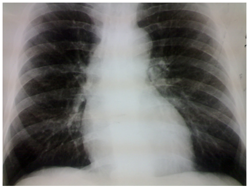
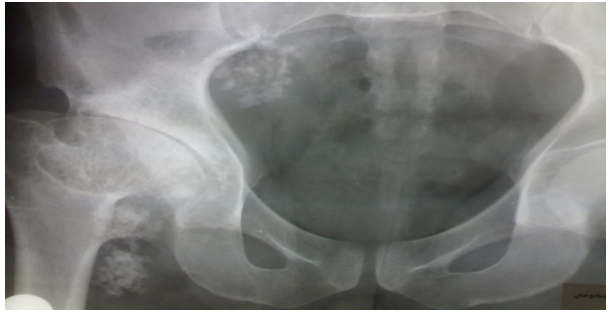
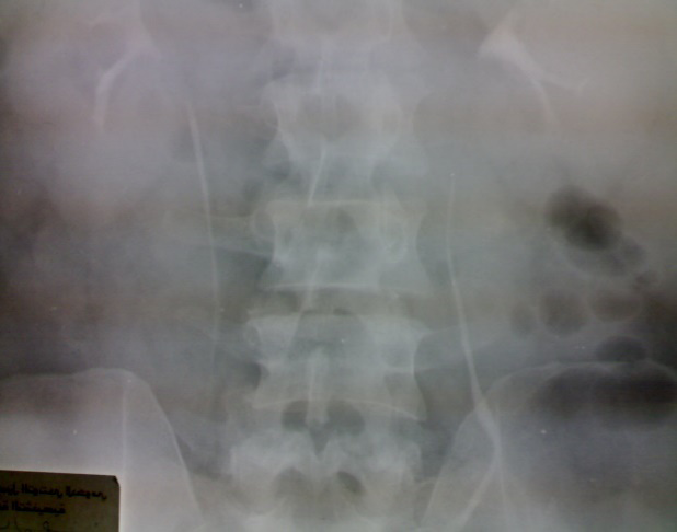
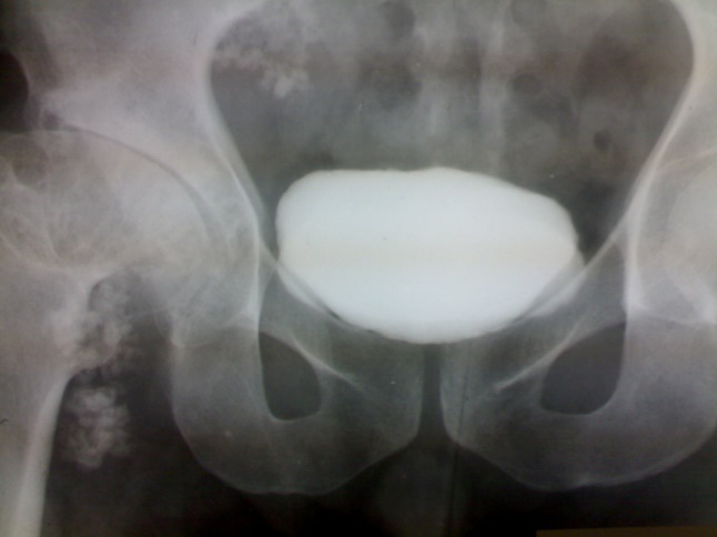
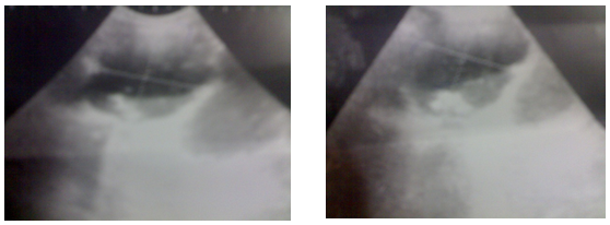
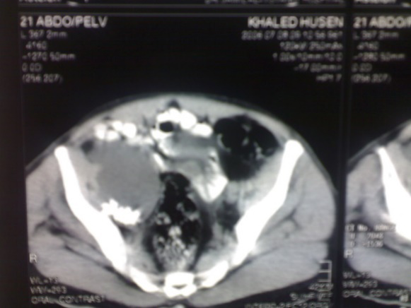
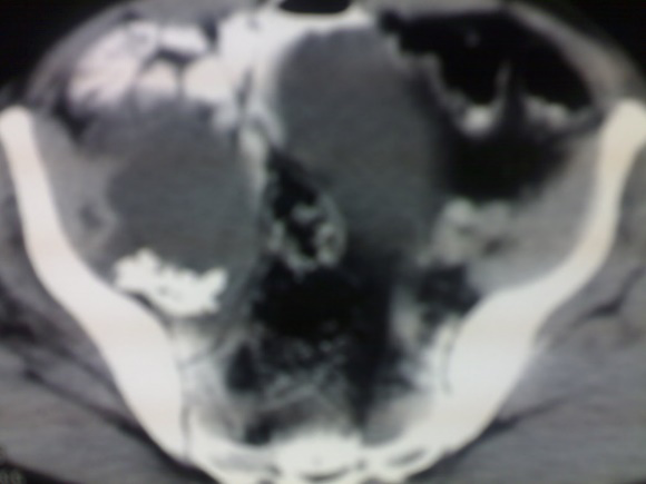
Blood tests including CBC, KFT, LFT, and URINALYSIS were performed and were normal. Radio-Imaging Regarding imaging, a chest x-ray was done, and it was normal (figure1). A Plain abdomen showed that there were clusters of calcifications on the right side of the pelvis and clusters of calcifications medial to the right head of the femur (Figure 2). After giving the IV contrast, kidneys, ureters, and urinary bladder are normal with symmetrical excretion, and no residual urine (Figure 3). On abdominal ultrasound and abdominal CT scan the mass size was (8.5×8.5cm).in addition, ultrasound reviles that the mass was cystic in consistency and localized to the right side of the urinary bladder having debris and multiple calcifications (Figure 4).
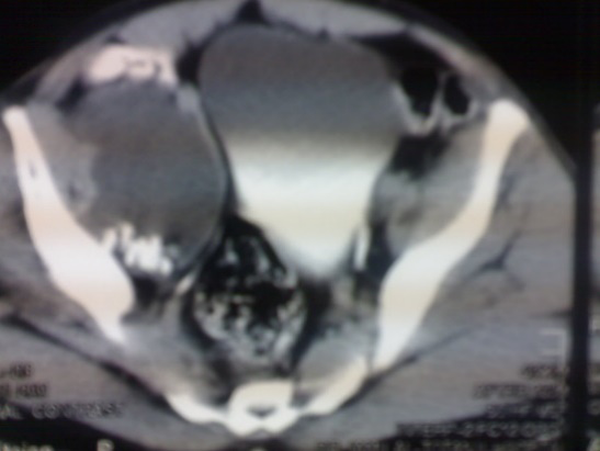
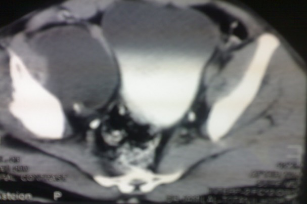
Abdominal and Pelvic C.T scans with and without contrast were done and they reviled that the liver, spleen, pancreas, and both suprarenal glands were normal. There was cystic structure to the right side of the urinary bladder having calcification not connected to it, suspected that the mass originated from pelvic muscle (Figure 5).
Intraoperative, after preparing the patient and under general anesthesia a lower midline incision was done. The findings were retroperitoneal mass (cyst) about (9×9cm) in size adherent with the surrounding tissue. When the cyst had been opened, serous fluid and mucus gelatinous material with debris and 2 stag horn stones (3×3cm) each holding inside it were shown. The mass extended downward through the right femoral canal to the thigh and an incision below the right inguinal ligament was done. The skin was opened in layers, the size of this part is about (4×4×6 cm) and another stag horn of (3×3cm) size was found inside it. Total excision of the whole cyst was done. Fluid was sent for cytopathology for microscopic examination and the result was Negative for malignancy, benign mesothelial cells. Regarding the cyst and Inguinal lymph node samples that were sent for histopathology; mesothelium lined peritoneal cyst containing 3 nodular white calcified bony masses {3×3cm} inside it and was Negative for malignancy. Inguinal lymph node histopathology showed reactive lymphoid hyperplasia.
Discussion
Benign multicystic peritoneal mesothelioma is a rare benign tumor originating from the peritoneum, affecting mostly young, fertile women, and it is exceedingly rare in men [7]. The incidence of the disease is 0.15/per 100,000
annually, which makes it challenging to diagnose and treat [8].
Handfield-Jones; Texas. University. USA: defined retroperitoneal-peritoneal cysts as those cysts lying in the retroperitoneal fatty tissue which have no clear connection with any adult anatomical structure save by areolar tissue.” This studied case is unique because the combination of retroperitoneal and peritoneal cyst in the same situation is rarely reported. Retroperitoneal peritoneal cyst in the literature reported just two cases. Also, the gender of the patient, in this case, makes it rare as the disease does not occur in males routinely. Moreover, the three staghorn stones that were found after excision increase the uniqueness of this case.
The uncertainty about their nature is reflected in the terminology used for these lesions in the literature; they have been called benign multicystic mesothelioma, cystic mesothelioma, multilocular peritoneal inclusion cysts, inflammatory cysts of the peritoneum, and postoperative peritoneal cyst. Associated risk factors include endometriosis and pelvic inflammatory disease in women, and prior abdominal surgery in all genders [9].
Moreover, the pathogenesis of this condition is still controversial until now. However, according to a study done by Khurram, et al. [8]. There are three hypotheses have been proposed, First, the disease is secondary to a reactive process, and second, some authors believe the origin to be neoplastic. The third theory may be a hormonal hypothesis, within which the event and progression of BMPM (Benign Multicystic Peritoneal Mesothelioma) are tightly linked to its sensitivity to sex hormones. This theory is supported by the evidence of upper incidence in women during their reproductive age and the responsiveness of BMPM to certain endocrine agents like tamoxifen and gonadotropin-releasing hormone analogs. Some genetic and familial associations have also been documented.
Khurram, et al. [8] said in their study “Two of our cases had a family history of colon, breast, and lung cancer and leukemia, respectively”. The potential for malignant transformation in patients with benign multicystic peritoneal mesothelioma is low [7]. On the other hand, there were only two cases reported in the literature.
The diagnosis of benign cystic peritoneal mesothelioma is difficult [10]. In most cases, the diagnosis is made incidentally during routine investigation or surgery for another purpose.
Radiological modalities including ultrasonography, CT, and MRI may detect the lesions but cannot differentiate them from other cystic lesions. Exploratory laparoscopy is the most exact diagnostic method because it allows local biopsy of the suspected tissue.
As the recurrence rate is high when incomplete resection of the cyst is done. As a result, complete resection is recommended. An aggressive surgical approach including cytoreductive surgery with peritonectomy is also recommended to avoid recurrence. Because malignant transformation is reported in two cases, some choose aggressive surgery followed by heated intraperitoneal chemotherapy (HIPEC).
Conclusion
Benign multicystic peritoneal mesothelioma is a rare benign tumor originating from the peritoneum, affecting mostly pre-menopausal women. The diagnosis of this case is difficult. Surgery stays the mainstay of treatment.
Acknowledgments
This research was mentored and supervised by the Mutah Research and Audit Society (MRAS).
References
-
Vilos AG, Vilos GA, Marks J, Pollett A (2013) Retroperitoneal pelvic cyst: a diagnostic and therapeutic challenge. J Obstet Gynaecol Can 35(2): 164-167.
-
Abedini L, Hosseinpour R, Mehrabi S, Hejazinia S, Barhaghtalab MJY (2022) An asymptomatic huge primary retroperitoneal pseudocyst: A case report and review of the literature. BMC surgery 22: 58.
-
Matthew GW, Matthew F, Alexander S, Mark E (2007) Retroperitoneal cyst of Müllerian origin: A case report and Review of the Literature. Journal of Pelvic Medicine and Surgery 13(3): 149-152.
-
Maurya SK, Bhot FB, Ghosh DK, Nayak VM (2003) Retroperitoneal cyst. Med J Armed Forces India 59(1): 73-74.
-
Uchikova E, Milchev N, Dimitrova E, Anavi B, Uchikov A (2003) Peritoneal cyst--a case report. Akusherstvo i ginekologiia 42(3): 27-28.
-
Natkanska A, Bizon-Szpernalowska MA, Milek T, Sawicki W (2021) Peritoneal inclusion cysts as a diagnostic and treatment challenge. Ginekologia polska 92(8): 583-586.
-
György A, Schmal F, Szabó H, Tóth LB, Lukovich P (2019) Benignus multicysticus peritonealis mesothelioma. Orvosi hetilap 160(21): 839-843.
-
Khurram MS, Shaikh H, Khan U, Edens J, Ibrar W, et al. (2017) Benign Multicystic Peritoneal Mesothelioma: A Rare Condition in an Uncommon Gender. Case reports in pathology 2017: 9752908.
-
Chand MT, Edens J, Lin T, Anderson I, Berri R (2020) Benign multicystic peritoneal mesothelioma: literature review and update. Autopsy & case reports 10(3): e2020159.
-
Khuri S, Gilshtein H, Abboud W, Assalia A, Kluger Y (2012) Benign cystic mesothelioma of the peritoneum: a rare case and review of the literature. Case reports in oncology 5(3): 667-670.
- Reconstruction of Nasal Defect Using a Local Flap Based in the Nanoperforants Concept: A Case Report
- Transplant Tourism in Japan: Insights from Nationwide Surveys and Emerging Ethical Challenges
- The Challenge of Peritoneal Vaginoplasty in Transgender Women: Review Literature and Personal Perspectives
- Algorithmic Paternalism: Autonomy Versus Automation
- Dermatoscopy in Gorlin Syndrome: Avoiding the Disfigurement in Patients
- How Artificial Intelligence is Ushering in a New Era of Innovation in General and Plastic Surgery: A Short Communication