Ovicidal Efficacy of Silver Nanoparticle against Vectors MOSQUITOS
Vector control is a critical requirement in epidemic disease situations, as is an urgent need to develop new and improved mosquito control methods that are economical and the environment. Mosquitoes transmit serious human diseases, causing millions of deaths every year. Use of synthetic insecticides to control vector mosquitoes has caused physiological resistance and adverse environmental effects in addition to high operational cost. Insecticides of botanical origin may serve as suitable alternative biocontrol techniques in the future. In view of the recently increased interest in developing plant origin insecticides as an alternative to chemical insecticide, in the present study, the Ovicidal activity of silver nanoparticles (AgNPs) synthesized using Sida acuta plant leaf extract against eggs of Anopheles stephensi, Aedes aegypti, and Culex quinquefasciatus was determined. The range of concentrations of synthesized AgNPs (20, 40, 60, 80, and 120 μg mL−1) and aqueous leaf extract (75,150,225,300,375 and 450 μg mL−1) were tested against the adults of A. stephensi, A. aegypti, and C. quinquefasciatus. Eggs were exposed to varying concentrations of aqueous leaf extract and synthesized AgNPs for 24 h. Considerable mortality was evident after the treatment of S. acuta for all three important vector mosquitoes. The synthesized AgNPs from S. acuta were highly toxic than aqueous leaf extract to three important vector mosquito species. In this recorded from UV–visible spectroscopy, Fourier transform infrared spectroscopy (FTIR), Scanning electron microscopy (SEM) with energy-dispersive X-ray spectroscopy analysis (EDX), and Transmission electron microscopy (TEM). These results suggest that synthesized silver nanoparticles are a rapid, eco-friendly, and single-step approach; the AgNPs formed can be potential mosquito Ovicidal agents.
Introduction
Mosquito-borne diseases are endemic in more than over 100 countries, causing mortality of nearly two million people every year, and at least one million children die of such diseases each year, leaving as many as 2100 million people at risk around the world. Mosquitoes constitute a major public health problem as vectors of serious human diseases like malaria, filariasis, Japanese encephalitis, dengue fever, chikungunya, and yellow fever. Mosquitoes alone transmit disease to more than 700 million people annually [1, 2]. Malaria is one of the serious scourges inflicted upon humanity. It causes human mortality and morbidity along with great financial loss. In general, transmission of malaria occurs between 64°N and 32°S of the Earth in more than 100 countries throughout the Africa, Asia, and Latin America along with certain Caribbean and Pacific islands where there are favorable conditions for completion of life cycle of malaria parasite [3]. Anopheles stephensi are major malaria vectors in India. With an annual incidence of 300–500 million clinically manifested cases and a death toll of 1.1– 2.7 million. Currently, about 40% of the world’s population lives in areas where malaria is endemic [4].
Culex quinquefasciatus is a vector of lymphatic filariasis affecting 120 million people worldwide, and approximately 400 million people are at risk of contracting filariasis worldwide, resulting into the annual economic loss of 1.5 billion dollars [5]. This attempts to develop novel materials as mosquito larvicides are still necessary. With the progress of nano-technology, many laboratories around the world have investigated silver nanoparticles (AgNPs) production as the nanoparticle possesses more surface atoms than a micro particle, which greatly improves the particle's physical and chemical characteristics. Some physical or chemical methods that are currently available for silver nanoparticle production include mechanical smashing, a solid-phase reaction, freeze-drying, spread drying, and precipitation (co- and homo-precipitation). In general, these methods consume a lot of energy in order to maintain the high pressures and temperatures that are needed for them to work. In contrast, many bioprocesses occur under normal air pressure and temperature, resulting in vast energy savings. As a consequence, this type of procedure attracted the attention of microbiologists and chemists [6]. Nanoparticles form a link between bulk materials and molecular structures, thus developing research interest for their utility in various fields. Due to their unique properties, metal nanoparticles have potential applications in catalysis, biological tagging, drug delivery, diagnostics, imaging, sensing, gene delivery, artificial implants, and tissue engineering [7]. Nowadays, synthetic insecticides/larvicides have created many ecological problems due to their long-term residual accumulation in the environment, development of resistance in target vectors, and chronic effects in non-target organisms, thereby ecological imbalance through food chain and harm cattle and human beings. In recent years plant mediated biological synthesis of nano particles is gaining importance due to its simplicity and eco-friendliness [8].
The crude methanol leaf extracts of Ficus benghalensis showed good larvicidal activity against the early second, third, and fourth instar larvae of C. quinquefasciatus, A. aegypti, and A. stephensi [9]. The Low-cost and eco- friendly green synthesis of silver nanoparticles using Feronia elephantum (Rutaceae) against C. quinquefasciatus, A. stephensi, and A. aegypti [10]. However, the silica nanoparticles have been tested against the larvae and pupae of A. stephensi, C. quinquefasciatus, and A. aegypti [11]. The pediculocidal and larvicidal activities of synthesized silver nanoparticles using aqueous leaf extract of Tinospora cordifolia have been reported against the human capitis and fourth instar larvae of A. subpictus and C. quinquefasciatus [12].
The larvicidal and repellent properties of essential oils from various parts of four plant species Cymbopogon citrates, C. zeylanicum, R. officinalis, and Z. officinale against C. tritaeniorhynchus and A. subpictus [13]. The mosquito larvicidal properties of silver nanoparticles synthesized using Heliotropium indicum (Boraginaceae) against A. aegypti, A. stephensi, and C. quinquefasciatus [14]. In the present study, we reported the Silver nanoparticle (Ag NPs) would be useful in promoting research aiming at the development of new agent for mosquito Ovicidal activity. So far, there are no reports on aqueous leaf extract by S. acuta or synthesized AgNPs on mosquito Ovicidal activity.
Materials and Methods
Collection of Materials
Fresh leaves of S. acuta (Malvaceae) Figure 1 were collected from in and around Valayamadevi, Chidambaram area, and Tamil Nadu, and the taxonomic identification was made by the Department of Botany, Annamalai University, Annamalai Nagar, Tamil Nadu, India. The voucher specimen was numbered and kept in our research laboratory for further reference. Silver
nitrate was obtained from Qualigens Fine Chemicals, Mumbai, India.
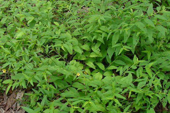
Figure1: S. acuta plant.
Preparation of Plant Extracts
The leaves of S. acuta were dried in the shade and ground to fine powder in an electric grinder. Aqueous extract was prepared by mixing 50 g of dried leaf powder with 500 mL of water (boiled and cooled distilled water) with constant stirring on a magnetic stirrer [15]. The suspension of dried leaf powder in water was left for 3 h and filtered through Whatman no. 1 filter paper, and the filtrate was stored in an amber-colored airtight bottle at 10 °C temperature till use.
Synthesis of Silver Nanoparticles S. Acuta Leaf Extract
Leaves were washed with distilled water and dried for 5days at room temperature. A plant leaf Broth was prepared by placing 10 g of the leaves (finely cut) in a 300mL flask with 100mL of Sterile distilled water. This mixture was boiled for 20min, decanted, stored at - 4°C, and used in our tests within 1 week. The filtrate was treated with aqueous 1 mM AgNO3 solution in an Erlenmeyer flask and incubated at room temperature. Formation of AgNPs was indicated by the Brown- yellow coloration of the solution suggesting that aqueous silver ions can be reduced by Aqueous extract of plant parts to generate extremely stable silver nanoparticles in water.
Characterization of Silver Nanoparticles
Synthesized silver nanoparticles were confirmed by sampling the reaction mixture at regular Intervals and the absorption maxima was scanned by UV-vis spectra, at the wavelength of 300–800 nm in UV-3600 Shimadzu spectrophotometer at 1 nm resolution. Further, the reaction Mixture was subjected to centrifugation at 15,000 rpm for 20 min; resulting pellet was dissolved in deionized water and filtered through Millipore filter (0.45Dm). An aliquot of this filtrate containing silver nanoparticles was used for SEM, EDS and FTIR studies. The structure and Composition of freeze-dried purified silver particles was analyzed by using a 10 kV ultra high Resolution scanning electron microscope with 25Dl of sample was sputter coated on copper stub and the images of nanoparticles were studied using (FEI QUANTA- 200SEM). The surface Groups of the nanoparticles were qualitatively confirmed by using Fourier transform infrared (FTIR) spectroscopy, with spectra recorded by a Perkin-Elmer Spectrum 2000 FTIR Spectrophotometer. An aliquot of this filtrate containing silver nanoparticles was used for X-ray diffraction (XRD) analysis. In addition presence of metals in the sample was analyzed.
Ovicidal Activity
Ovicidal activity: For Ovicidal activity, slightly modified method of Su, et al. [16] was performed. A. stephensi, A. aegypti and C. quinquefasciatus eggs were collected from vector control laboratory, Department of Zoology, Annamalai University. The leaf aqueous extracts and silver nanoparticle were to achieve various concentrations ranging from 75 to 450 µg/ml and 20 to 120 µg/mL. The freshly laid egg raft containing 100 eggs of was exposed to each dose of leaf aqueous extract and silver nanoparticle until they hatched or died. Each concentration was replicated six times. After 24 h treatment, the eggs from each concentration were individually transferred to distilled water cups for hatching assessment after counting the eggs under microscope. Each experiment was replicated six times along with appropriate control. The hatch rates were assessed 48 h post treatment by following formula:
Number of hatched larvae
% hatchability = ∈100 Total number of eggs raft/ eggs
Results
Ovicidal activity of Aqueous Crude Extract and Synthesized AgNps
In the laboratory test, the oviposition cups treated with different concentrations of S. acuta leaf aqueous extract in 100 ml of distilled water received different number of egg rafts/ eggs at different concentrations. The
Silver nanoparticle containing served as a control received only a small amount of egg rafts/eggs. The different age of egg rafts/eggs of A. stephensi, A. aegypti and C. quinquefasciatus treated with different concentrations of leaf aqueous extract caused Ovicidal activity resulting in failure to hatch the egg rafts/eggs Table 1. The table clearly indicates that the higher level of Ovicidal activity by the extract was observed in the early stage of egg development. The Silver nanoparticle containing water that served as a control showed 94% hatchability in 0–18-h-old egg rafts/eggs, but the 100% hatchability was noted in egg rafts/eggs beyond the age of 0–18 h old Table 2. From the above results, it is quite clear that younger age groups of egg rafts/eggs showed a poor hatchability rate when exposed to higher concentrations of the extract, and older age groups of egg rafts/eggs showed a high hatchability rate when exposed to lower concentrations of the extract.
| Mosquitoes | Age of the egg raft/eggs (h) | Percentage of egg hatchability | ||||||
|---|---|---|---|---|---|---|---|---|
| Concentration (µg/mL) | ||||||||
| Control | 75 | 150 | 225 | 300 | 375 | 450 | ||
| An. stephensi | 0-6 | 100±0.0 | 27.4±0.8 | 15.2±1.2 | NH | NH | NH | NH |
| 12-Jun | 100±0.0 | 37.3±1.6 | 24.1±1.0 | 17.4±0.8 | NH | NH | NH | |
| 18-Dec | 100±0.0 | 51.1±1.8 | 37.4±1.4 | 23.0±0.8 | 16.3±1.3 | NH | NH | |
| Ae. aegypti | 0-6 | 100±0.0 | 52.3±0.8 | 34.1±1.2 | 18.2±1.7 | NH | NH | NH |
| 12-Jun | 100±0.0 | 68.7±0.9 | 56.4±1.3 | 39.3±1.8 | 19.5±1.0 | NH | NH | |
| 18-Dec | 100±0.0 | 74.0±1.0 | 62.1±1.6 | 54.7±0.9 | 33.2±1.3 | 16.4±1.5 | NH | |
| Cx.quinquefasciatus | 0-6 | 100±0.0 | 67.2±1.4 | 53.4±1.7 | 36.3±0.9 | 19.3±1.3 | NH | NH |
| 12-Jun | 100±0.0 | 86.1±1.8 | 75.3±0.8 | 56.4±1.2 | 30.3±1.5 | 16.3±1.8 | NH | |
| 18-Dec | 100±0.0 | 95.4±0.8 | 86.2±1.2 | 69.7±1.5 | 55.4±1.8 | 34.3±0.9 | 18.2±1.2 |
Table 1: Ovicidal activity of Sida acuta aqueous leaf extract against egg raft of Anopheles stephensi, Aedes aegypti and Culex qu
| Mosquitoes | Age of the egg raft/eggs (h) | Percentage of egg hatchability | ||||||
|---|---|---|---|---|---|---|---|---|
| Concentration (µg/mL) | ||||||||
| Control | 20 | 40 | 60 | 80 | 100 | 120 | ||
| An. stephensi | 0-6 | 100±0.0 | 27.4±0.9 | 15.2±1.3 | NH | NH | NH | NH |
| 12-Jun | 100±0.0 | 39.1±1.3 | 27.5±1.6 | 16.3±1.8 | NH | NH | NH | |
| 18-Dec | 100±0.0 | 49.2±0.8 | 36.3±1.4 | 29.1±1.6 | 19.4±0.9 | NH | NH | |
| Ae. aegypti | 0-6 | 100±0.0 | 45.5±1.4 | 32.1±1.8 | 18.3±1.1 | NH | NH | NH |
| 12-Jun | 100±0.0 | 61.3±1.8 | 49.2±0.8 | 31.4±1.5 | 16.3±1.0 | NH | NH | |
| 18-Dec | 100±0.0 | 71.4±1.0 | 58.6±1.5 | 45.1±1.8 | 36.4±0.9 | 18.2±1.1 | NH | |
| Cx.quinquefasciatus | 0-6 | 100±0.0 | 52.2±1.4 | 39.4±1.6 | 27.1±1.8 | 16.2±0.8 | NH | NH |
| 12-Jun | 100±0.0 | 84.3±0.8 | 69.2±1.0 | 51.5±1.3 | 36.1±1.7 | 18.3±0.9 | NH | |
| 18-Dec | 100±0.0 | 95.7±1.4 | 83.4±1.8 | 62.5±1.2 | 48.0±0.9 | 33.1±1.7 | 19.3±1.1 |
Table 2: Ovicidal activity of Silver nanoparticle against egg raft of Anopheles stephensi, Aedes aegypti and Culex quinquefasciat
Characterization of Silver Nanoparticles
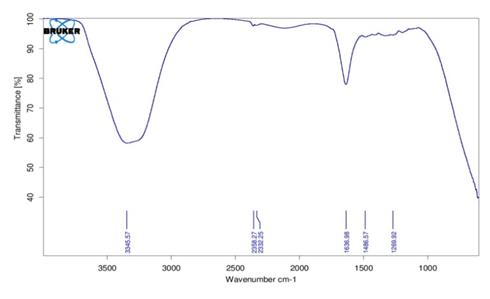
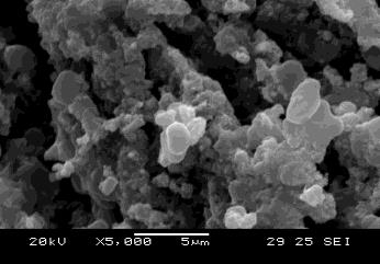
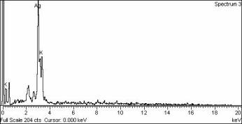
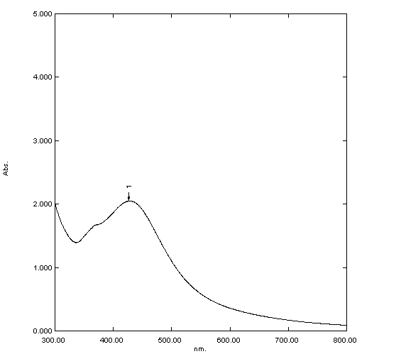
The change in color was noted by visual observation in the S. acuta extract when it was incubated with AgNO3 solution. S. acuta extract without AgNO3 did not show any change in color Figures 2a-2c. The color of the extract changed to light brown within an hour and then later changed to dark brown during the 30 min incubation period. No significant change occurred after 30 min. The absorption spectrum of S. acuta extract at different wavelengths ranging from 300 to 800 nm revealed a peak at 420 nm Figure 3. FTIR analysis of the purified nanoparticles showed the presence of bands due to O-H group (1269.92 cm−1), C=N stretch (1486.57), -NH2 (1636.98), =NH (2332.25), -H stretch (2358.27), and O-H stretch (3345.57) Figure 4. SEM micrographs of the synthesized AgNPs of S. acuta magnified at ×1,000 and ×5,000, times its size was measured at 20 to 60 nm are shown in Figures 4a & b. It is clear that the triangles, pentagons, and hexagons structures. EDX proves the chemical purity of the synthesized AgNPs Figure 5b. The
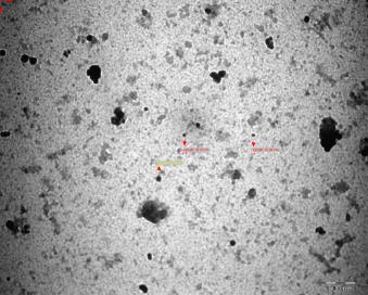
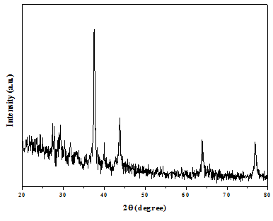
electron microscopic study of the nanoparticles using TEM revealed that the nano- Ag predominates with spherical, triangle, truncated triangles, and decahedral morphologies ranging from 18 to 35 nm with an average size of 25 nm Figure 5. The control thin films of the leaf extract as well as the AgNO3 did not show the characteristic peaks. The XRD pattern shows four intense peaks in the whole spectrum of 2θ values ranging from 25 to 60. The XRD spectrum compared with the standard confirmed spectrum of silver particles formed in the present experiments were in the form of nano crystals, as evidenced by the peaks at 2θ values of 27.33°, 31.69°, 39.89°,43.74°,63.91,and 76.85° corresponding to 29,31, 46, ,42, 152,and 146 planes for silver, respectively. The XRD pattern clearly shows that the silver nanoparticles formed by the reduction of AgNO3 ions by S. acuta are crystalline in nature Figure 6.
a b $$ y = \frac {1}{2} x - 1 $$
c Figure 2: a) Photographs showing change in color after adding AgNO3 before reaction. B) After reaction time of (6 h). c. UV–Vis spectra of aqueous silver nitrate with S. acuta leaf extract.
Discussion
The broad spectrum antimicrobial properties of silver nanoparticles have attracted researchers to evaluate their potential against the parasites of world's most threatening diseases, malaria, and dengue. Synthesis of AgNps using bio organisms like bacteria, fungi, actionmycetes, and extracts of various plant parts have advantage over other processes, as they are environment friendly. Fungi are beneficial than other biological agents for AgNp synthesis as bearing high metal uptake and tolerance and better wall binding capability (Chen et al., 2003). Fungi secrete copious amount of proteins, fungal biomass can be produced on large scale, and easy recovery of silver nanoparticles attracted the researchers. In addition, myco synthesized silver nanoparticles have good monodispersity, controlled size, and shape with hydrophilic nature [17].
The maximum mortality, 40, 36, 32, 30, and 26 %, was observed at 1 % concentration level in all instars and pupa, respectively, and their LC50 values are 1.19, 1.39, 1.77, 1.46, and 1.97 % on treatment with aqueous leaf extract alone. Similar findings have been reported for insecticidal activity in ethanolic leaf extract of Mimosa pudica at 2.5 ppm level against the instar stages and pupae of malarial vector A. stephensi [18]. Earlier authors reported that the methanol extract of Cassia fistula exhibited LC50 values of 17.97 and 20.57 mg/L, A. stephensi and C. quinquefasciatus, respectively [19]. The bioactivity of latex-producing plant Pergularia daemia as well as AgNPs against the larval instars of A. aegypti and A. stephensi mosquito larvae was determined, the results AgNPs shows excellent larvicidal activity of first, second and third instar larvae and fourth-instar larvae did not exhibit any noticeable effects after either 24 or 48 h of exposure at their LC50 and LC90values [20]. Several larvicidal investigations against mosquitoes have been carried out with various plant extracts. It has been reported that leaf extracts of Ocimum canum, Ocimum sanctum, and Rhinacanthus nasutus have been found to be only moderately toxic against the larvae of A. aegypti with LC50 values ranging between 99.42 and 81.56 ppm [21].
The benzene, hexane, ethyl acetate, methanol, and chloroform leaf extract of A. paniculata was found to be more effective against C. quinquefasciatus than A. aegypti. The LC50 values were 112.19, 137.48, 118.67, 102.05, and 91.20 and 119.58, 146.34, 124.24, 110.12, and 99.54 ppm, respectively [22]. In the adulticidal activity of Synthesized AgNPs against the vector mosquitoes A. stephensi, A. aegypti, and C. quinquefasciatus had the following LD50 and LD90 values: A. stephensi had LD50 and LD90 values of 18.041 and 32.575 μg mL−1; A. aegypti had LD50 and LD90 values of 20.399 and 37.534 μg mL−1; and C. quinquefasciatus had LD50 and LD90 values of 21.798 and 39.596 μg mL−1. and The LD50 and LD90 values of the F. elephantum aqueous leaf extract appeared to be effective against A. stephensi (LD50 88.866 μg mL− 1 and LD90 161.368 μg mL−1) followed by A. aegypti (LD50 101.166 μg mL− 1 and LD90 183.296 μg mL−1) and C. quinquefasciatus (LD50 108.420 and LD90 194.650 μg mL−1) respectively [23, 24, 25, 26, 27, 28, 29, 30, 31, 32, 33].
In conclusion, an attempt has been made to evaluate the role of S. acuta extracts and synthesized AgNPs ovicidal bioassay against A, stephensi, A. aegypti, and C. quinquefasciatus activity. The synthesized AgNPs with aqueous extract and the isolation and purification of aqueous leaf extract of S. acuta are in progress. The silver nanoparticles have also been tested for their Ovicidal agent against mosquito A. stephensi, A. aegypti, and C. quinquefasciatus .The plant-mediated silver nanoparticles can have an immediate impact on mosquito control. These nano Ovicidal are environmentally safer, greener, and rapidly effective against mosquito vectors. We can, therefore, develop that the aqueous leaf extract- synthesized silver nanoparticles could be a better, environmentally safer, and greener approach for the vector control.
References
-
Jang, Young Su, Kim, Moo-Key, Ahn, et al, (2002) Larvicidal activity of Brazilian plant against Aedes aegypti and Culex pipiens (Diptera: Culicidae). Journal of applied Biological Chemistry 45(3): 131- 134.
-
WHO (2006) Chikungunya and dengue in the south west Indian Ocean. Epidemic and Pandemic Alert and Response, EPR.
-
Zarchi AKA, Mohmoodzadeh AH, Vatani HA (2006) A survey on malaria and some related factors in south east of Caspian Sea. Pak J Med Sci 22(4): 489-492.
-
Wernsdorfer G, Wernsdorfer WH (2003) Malaria at the turn from the 2nd to the 3rd millennium. Wien Klin Wochenschr 115(3): 2-9.
-
WHO (2002) Lymphatic filariasis the disease and its control. Technical report 71, WHO, Geneva.
-
Chen JC, Lin ZH, Ma XX (2003) Evidence of the production of silver nanoparticles via pretreatment of Phoma sp.3.2883 with silver nitrate. Lett Appl Microbiol 37(2): 105-108.
-
Thakkar KN, Mhatre SS, Parikh RY (2010) Biological synthesis of metallic nanoparticles. Nanomedicine 6(2): 257-262.
-
Farooqui, Arshad, Chauhan, Prakash Singh; Krishnamoorthy, et al. (2010) Extraction of silver nano-particles from the leaf extracts of Clerodendrum inerme. DJNB 5(1): 43-49.
-
Govindarajan M (2010) Larvicidal efficacy of Ficus benghalensis L. plant leaf extracts against Culex quinquefasciatus Say, Aedes aegypti Linn. Parasitol Res 108: 693-702.
-
Veerakumar K, Govindarajan M, Rajeswary M, Muthukumaran U (2014) Low-cost and eco-friendly green synthesis of silver nanoparticles using Feronia elephantum (Rutaceae) against Culex quinquefasciatus, Anopheles stephensi, and Aedes aegypti (Diptera: Culicidae) Parasitol Res 113(5): 1775-1785.
-
Barik TK, Kamaraju R, Gowswami A (2012) Silica nanoparticles: a potential new insecticide for mosquito vector control. Parasitol Res 111(3): 1075- 1083.
-
Jayaseelan C, Rahuman AA, Rajakumar G, Vishnu Kirthi A, Santhoshkumar T, et al. (2011) Synthesis of pediculocidal and larvicidal silver nanoparticles by leaf extract from heartleaf moonseed plant, Tinospora cordifilia Miers. Parasitol Res. 109(1): 185-194.
-
Govindarajan M (2011) Larvicidal and repellent properties of some essential oils against Culex tritaeniorhynchus Giles and Anopheles subpictus Grassi (Diptera: Culicidae). Asian Pac J Trop Med 4(2): 106-111.
-
Veerakumar K, Govindarajan M, Rajeswary M, Muthukumaran U (2014) Mosquito larvicidal properties of silver nanoparticles synthesized using Heliotropium indicum (Boraginaceae) against Aedes aegypti, Anopheles stephensi, and Culex quinquefasciatus (Diptera: Culicidae). Parasitol Res 113(6): 2363-2373.
-
Minjas JN, Sarda RK (1986) Laboratory observations on the toxicity of Swartzia madagascariens (Leguminaceae) extract to mosquito larvae. Trans R Soc Trop Med Hyg 80(3): 460-461.
-
Su T, Mulla MS (1998) Ovicidal activity of neem products (Azadirachtin) against Culex tarsalis and Culex quinquefasciatus (Diptera: Culicidae). J Am Mosq Control Assoc 14(2): 204-209.
-
Govindarajan M, Sivakumar R (2012) Adulticidal and repellent properties of indigenous plant extracts against Culex quinquefasciatus and Aedes aegypti (Diptera: Culicidae). Parasitol Res 110(5): 1607-1620.
-
Mohanpuria P, Rana NK, Yadav SK, (2008) Biosynthesis of nanoparticles: technological concepts and future applications. J Nanopart Res 10(3): 507- 517.
-
Aarthi N, Vasuki C, Panneerselvam C, Prasanakumar K, Madhiyazhagan, et al. (2011) Toxicity and smoke repellency effect of Mimosa pudica L. against the malarial vector Anopheles stephensi (Diptera: Culicidae). Bioscan 6(2): 211-214.
-
Govindarajan M, Jebanesan A, Pushpanathan T (2008) Larvicidal and ovicidal activity of Cassia fistula Linn. leaf extract against filarial and malarial vector mosquitoes. Parasitol Res 102(2): 289-292.
-
Patil CD, Borase HP, Patil SV, Salunkhe RB, Salunke BK (2012) Larvicidal activity of silver nanoparticles synthesized using Pergularia daemia plant latex against Aedes aegypti and Anopheles stephensi and non-target fish Poecillia reticulata. Parasitol Res 111(2): 555-562.
-
Kamaraj C, Rahuman AA, Bagavan A (2008) Antifeedant and larvicidal effects of plant extracts against Spodoptera litura (F.), Aedes aegypti L. and Culex quinquefasciatus Say. Parasitol Res 103(2): 325-331.
-
Govindarajan M (2011) Evaluation of Andrographis paniculata Burm.f. (Family: Acanthaceae) extracts against Culex quinquefasciatus (Say.) and Aedes aegypti (Linn.) (Diptera: Culicidae). Asian Pacific J Trop Med 4(3): 176-181.
-
Veerakumar K, Govindarajan M (2014) Adulticidal properties of synthesized silver nanoparticles using leaf extracts of Feronia elephantum (Rutaceae) against filariasis, malaria, and dengue vector mosquitoes. Parasitol Res 113(11): 4085-4096.
-
Priyadarshini KA, Murugan K, Panneerselvam C, Ponarulselvam S, Hwang JS, et al. (2012) Biolarvicidal and pupicidal potential of silver nanoparticles synthesized using Euphorbia hitra against Anopheles stephensi Liston (Diptera: Culicidae). Parasitol Res 111(3): 997-1006.
-
Rajakumar G, Abdul Rahuman A (2011) Larvicidal activity of synthesized silver nanoparticles using Eclipta prostrata leaf extract against filariasis and malaria vectors. Acta Trop 118(3): 196-203.
-
Marimuthu S1, Rahuman AA, Rajakumar G, Santhoshkumar T, Kirthi AV, et al. (2011) Evaluation of green synthesized silver nanoparticles against parasites. Parasitol Res 108(6): 1541-1549.
-
Santhoshkumar T, Rahuman AA, Rajakumar G, Marimuthu S, Bagavan A (2011) Synthesis of silver nanoparticles using Nelumbo nucifera leaf extract and its larvicidal activity against malaria and filariasis vectors. Parasitol Res 108(3): 693-702.
-
Singh G, Prakash S (2012) Evaluation of culture filtrates of Culicinomyces clavisporus: Mycoadulticide for Culex quinquefasciatus, Aedes aegypti and Anopheles stephensi. Parasitol Res 110(1): 267-272.
-
Singh G, Prakash S (2011) Studies on Fungal Cultural Filtrates against Adult Culex quinquefasciatus (Diptera: Culicidae) a Vector of Filariasis. J Parasitol Res doi:10.1155/2011/147373.
-
Pandey V, Agrawal V, Raghavendra K, Dash AP (2007) Strong larvicidal activity of three species of Spilanthus (Akarkara) against malaria (_Anopheles_ _stephensi_ Liston, Anopheles culicifacies, species C) and filarial vector (Culex quinquefasciatus Say). Parasitol Res 102(1): 171-174.
-
Kim KJ, Sung WS, Suh BK, Moon SK, Choi JS, (2009) Anti-fungal activity and mode of action of silver nanoparticles on Candida albicans. Biometals 22(2): 235-242.
-
Jang YS, Kim MK, Ahn YS, Lee HS, (2002) Larvicidal activity of Brazilian plant against Aedes aegypti and Culex pipiens (Diptera: Culicidae). Agric Chem Biotechnol 4: 131-134.
- Mitochondrial Bio-Logistics: Steering Co-Enzyme Q10 and Lycopene Synergies within the Science 4.0 Bio-OS Framework
- Hymenoptera Specimens from the Caño Negro Wetland, of the National Museum Collection, Costa Rica
- Science 4.0: Comprehensive Architecture of the Biological Operating System (Bio-OS) A Framework for Systemic Resilience and Industrialized Bio-Governance
- Rabbit on, or Hare Back? Understanding Climate Change
- Clinical Validation of Science 4.0: Flow Steering and Epigenetic Drift Inversion on a 76-Year-Old Hybrid System
- Seeds Planted by another Mind