Spectrum of Neoplastic & Non-Neoplastic Lesions of Ovary at SBMCH (A Tertiary Care Centre)
Ovary is one of the commonest sites of lesion in the female reproductive tract, which can present among childhood to post-menopausal age group. Diagnosis of ovarian lesions in its early stages is rare, as they are usually asymptomatic and not easily identified on physical examination. A statistical retrospective study is done over a period of 1 year at Sree Balaji Medical College & Hospital (tertiary care center). A total of 151 ovarian lesions were reported in the year 2015- 2016 histologically by the department of pathology, SBMCH. Amongst the total lesions Non-neoplastic type were 129(85.5%) cases & Neoplastic were 22(14.5%) cases. Non-neoplastic lesions were more common than neoplastic lesions. Among neoplastic lesions benign tumors were common. The diagnostic diversity of ovarian lesions poses many challenges. A specific diagnosis can usually be made by histopathology but sometimes immunohistochemistry is required in difficult cases.
Introduction
Ovaries are organs that are influenced by hormones. Ovary is one of the commonest sites of lesion in the female reproductive tract, which can present among childhood to post-menopausal age group. Diagnosis of ovarian lesions in its early stages is rare, as they are usually asymptomatic and not easily identified on physical examination. Despite in depth studies on ovarian lesions, there is a lack in of valuable information regarding the incidence of Neoplastic & Non neoplastic lesions comparatively.
Materials & Methods
Aretrospective study is done over a period of 1 year from May 2015 to May 2016 at Sree Balaji Medical College & Hospital. The total hysterectomy specimens & ovariotomy specimens that we received at the histopathology lab during this period were used for the study. A routine formalin fixation, grossing of specimens, processing, fixing, H&E staining followed by histopathological study, was done. On the basis of diagnostic features they were classified and studied as neoplastic & non neoplastic with a special focus on Leydig cell hyperplasia that was recently diagnosed incidentally.
Results
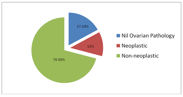
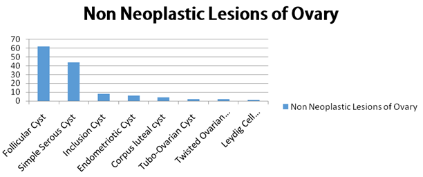
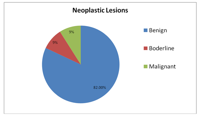
Amongst 151 cases studied, 129(70.9%) were non- neoplastic, 22(12%) were neoplastic and the remaining 31(17.1%) had nil ovarian pathology (Figure 1). Most of the non-neoplastic lesions of ovary were incidental findings. The most common non-neoplastic lesion found was follicular cysts (Figure 2). Among the 22 neoplastic ovarian lesions 18 cases were benign, 2 cases were at borderline and 2 cases were malignant (Figure 3). The neoplastic lesions that were diagnosed during ours study period were, Serous Cyst Adenoma, Cystic Teratoma, Granulosa Cell tumor (Table 1).
| Neoplastic | Benign | Borderline | Malignant |
| Serous Cyst Adenoma | 14 | 2 | 0 |
| Cystic eratoma(Dermoid Cyst | 4 | 0 | 0 |
| Granulosa Cell Tumor | 0 | 0 | 2 |
Table 1: Serous Cyst Adenoma, Cystic Teratoma, Granulosa Cell tumor.
Discussion
Ovarian cancer is said to be the sixth amongst the cancers in the world and the seventh leading cause of mortality among women worldwide [1]. Based on the Bupesh G, et al. Spectrum of Neoplastic & Non-Neoplastic Lesions of Ovary at SBMCH (A Tertiary Care Centre). Med J Clin Trials Case Stud 2019, 3(1): 000196.
cancer registries in India, the third most leading cancer among women is ovarian cancer. Among all gynecological malignancies, ovarian cancer has a very worst prognosis. The diagnosis is of ovarian cancers in usually at late & advanced stages & hence the 5year survival rate is less than 45% [2]. Incomplete or a poor compliance to chemotherapy to patients with ovarian cancer is one of the major factors, affecting survival from advanced stages. It was found that the second most common benign tumor after serous cystadenoma is mature teratoma and many of the malignant ovarian tumors usually present at earlier ages of life [3]. Ovarian cancers are said to be having a higher rate of occurrence in nulliparous women [4]. Obstetric history with minimum of one parity (pregnancy) reduces the risk of developing ovarian cancer by approximately 40–60%, and on additional parity (pregnancy) reduces the risk by another 10–15% [5, 6].
Conclusion
Based on the statistical analysis serous cyst adenoma has a more rate of incidence amongst ovarian neoplastic lesions at SBMCH, those with a benign entity tended to occur more common than malignant. Amongst the overall ovarian lesions at SBMCH during our study period, Non- neoplastic lesions occupy a higher rate of incidence.
References
-
Parkin DM, Bray F, Ferlay J, Pisani P (2005) Global cancer statistics, 2002. CA Cancer J Clin 55(2): 74- 108.
-
Jemal A, Siegel R, Ward E, Murray T, Xu J, et al. (2006) Cancer statistics, 2006. CA Cancer J Clin 56(2): 106- 130.
-
Basu P, De P, Mandal S, Ray K, Biswas J (2009) Study of ‘patterns of care’ of ovarian cancer patients in a specialized cancer institute in Kolkatta, eastern India. Indian J cancer 46(1): 28-33.
-
Mondal SK, Banyopadhyay R, Nag DR, Roychowdhury S, Mondal PK, et al. (2011) Histologic pattern, bilaterality and clinical evaluation of 957 ovarian neoplasms: A 10-year study in a tertiary hospital of eastern India. J Can Res Ther 7(4): 433-437.
-
Scully RE (1977) Ovarian tumours. A review. Am J Pathol 87(3): 686-720.
-
Stanley J Robboy, George L Mutter, Jaime Prat, Rex C Bentley, Peter Russell, et al. Robboy's Pathology of the Female Reproductive Tract. Bupesh G, et al. Spectrum of Neoplastic & Non-Neoplastic Lesions of Ovary at SBMCH (A Tertiary Care Centre). Med J Clin Trials Case Stud 2019, 3(1): 000196.
- Psychogenic Erectile Dysfunction in Late Adulthood: A Case Report on Clinical Intervention and Intimacy Restoration
- Clinical Trials on COVID-19 in 2025: A New Chapter in Global Health Research
- Innovations and Challenges in Contemporary Medical Clinical Trials: An Editorial Perspective
- Innovations and Challenges in Contemporary Medical Clinical Trials: A Critical Perspective
- Reimagining Clinical Trials: The Power of Continuous Feedback from Medical Reports
- Factors Influencing Brain Drain: Perspectives from a Medical School in Turkey