Bilateral Axillary Accessory Breast with Fibroadenoma: A Rare Case with Review
Supernumerary Breast or polymastia is a familiar discrepancy of the Breast and occurs along the embryonic milk line,extending between the axilla and groin. About 20% of accessory breast tissue occurs in the axilla, and approximately 67% of ectopic breast tissue occurs below the inframammary crease. The remaining (13%) can occur anywhere along the milk line. Itcan occur unilaterally or bilaterally. Here, we report an unusual case of fibroadenoma in the bilateral axillary Breast.
Introduction
Ectopic breast tissue (EBT), polymastia or supernumerary and accessory Breast are identical words used for extra breast tissue at more than two locations with or without a nipple. Supernumerary Breast or polymastia is a well-known variance of the Breast and mostly occurs along the embryonic milk line’s length extending between the axilla and groin [1]. The frequency of accessory Breast is 5.19% in women and 1.68% in men. The most commonly affected place is the axilla (particularly its inferior portion). All the pathological disease which affects normal breast tissue can affect EBT with a low incidence [2]. Fibroadenoma is a frequent benign disease of normal breast tissue. Fibroadenoma is a rare tumour of accessory breast tissue, and only a few cases have been reported in the literature. Among the alterations that affect the topical breast tissue, the fibroadenoma is most commonly found in the premenopausal period, a frequent cause of mass in young women, with higher incidence from 20 to 30 years old. It manifests itself as a nodular lesion, frequently unique, movable, with slow growth. At the mammography, a homogeneous, oval and confined node is observed. Polymastia cases have also been reported on other sites like the face, vulva and perineum, and the axilla [2]. These anomalies’ are of clinical significance includes their association with other congenital anomalies of the urinary and cardiovascular systems. The present article reports a case of fibroadenoma developing in the supernumerary breasts of both axillae in a 26-year-old woman [3]. Both pectoral breasts were normal clinically with normal mammograms. The histopathological examination of the excised samples showed a well-defined, capsulated intracanalicular type of fibroadenoma similar to normal breast tissue.
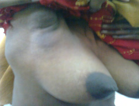
A 26-year-old female presented with bilateral subcutaneous axillary masses for the last 7 years, with discomfort in both axillary masses more so during the menstrual cycles. On physical examination, both axillary swellings were firm and slightly tender, measuring 5x3cms and 4x2.5cms in the right and left axillae, respectively (Figure 1). A small, tender, well-defined nodule of about 1x1.5 cm on the right side and about 1.5x1.5 on left sides was palpable on both sides. Mammograms of both pectoral breasts were normal. There was no associated urologic or cardiovascular abnormalities. Ultrasonography of the urogenital and cardiovascular system was normal. Both axillary breasts were excised under general anaesthesia, and histopathological report revealed a well-defined, capsulated intracanalicular fibroadenoma in both accessory breast tissues (Figure 2).

Figure1: Photo of B/L mass axillae.
Discussion
During breast development, the pectoral region’s two segments develop into normal breasts, and the remaining ectodermal thickening disappears. The accessory breast occurs in 0.4 to 6% of women due to normal developmental failure. First breast tissue develops in the pectoral region and rest of milk line involutes. If the remaining milk line does not involute, there is the formation of an accessory breast [3]. Approximately 6% are familial and are believed to signify an autosomal dominant trait with variable penetrance; the rest 94% are sporadic. The supernumerary nipple can be present at birth, but accessory Breast develops only after hormonal stimulation during puberty, pregnancy, or lactation. Accessory breast tissue arises from different sites, including the face, posterior neck, chest, buttock, vulva, hip, shoulder, posterior and lateral thigh, perineum, and the midback [4]. Two hypotheses can explain the occurrence of ectopic breast tissues in locations other than the milk line. Either it can represent a migratory arrest of breast primordium during chest wall development, or this occurrence can be attributed to its development from the modified apocrine sweat glands. Accessory breast tissue in the axilla is situated in the subcutaneous tissue and deep in the skin [5, 6]. It mixes with normal skin appendages gland and causes a diagnostic dilemma for pathologists. Both benign and malignant diseases can affect it as in a normal breast [7]. Cancer arising in the ectopic breast tissue has poorer prognosis due to difficult assessment, early lymph node involvement, and more difficult surgical excision.
Recently, EBT is classified as follows: [8]
- Polymastia: glandular breast tissue in an organized ductal system, communicating with overlying skin
- Polythelia: accessory nipples and/or areola. The presence of an areola only or patch of hair only may be further categorized as polythelia areolaris and polythelia pilosa, respectively
- Aberrant breast tissue: disorganized secretory tissue, unrelated to the overlying skin.
Kajava Y [9] old classification of supernumerary breast tissue as below: Class I - complete Breast with the nipple, areola, and glandular tissue. Class II - nipple and glandular tissue but no areola. Class III - areola and glandular tissue but no nipple. Class IV- glandular tissue only. Class V - nipple and areola but no glandular tissue (pseudomamma). Class VI- polythelia. Class VII- an areola only (polythelia areolaris). Class VIII - a patch of hair only (polythelia pilosa).
Polythelia or supernumerary nipple is the most frequent form of accessory breast tissue malformation that has been reported to be associated with nephrological, cardiovascular anomalies and rarely with esophageal atresia. The polythelia can be associated with urinary tract abnormalities such as supernumerary kidneys, renal formation failure, renal adenocarcinoma, hydronephrosis; polycystic kidney disease, duplicate renal arteries and ureteric stenosis and this connection can be partly clarified by the parallel development of mammary structure and genitourinary system [10]. However, no such reports are there concerning polymastia. However, as polythelia and polymastia may exist together, renal pathology should be ruled out in all polymastia cases through a physical examination, urine analysis, and renal ultrasound [5]. The differential diagnosis includes enlargement of lymph nodes, lipomas, tuberculosis, neuromas, the axillary tail of Spence, skin lesions such as epidermal inclusion cysts, sebaceous cyst, suppurative hidradenitis, vascular lesions and lesions arising from EBT [11].
An unusual case of juvenile fibroadenoma was reported by Borsook et al., in a 10-year-old female [12, 13, 14, 15, 16]. The patient was a known case of mitochondrial disease with a mutation in the POLG1 gene and presented with a left axillary mass of 10 months. Suspecting it to be a lymph node in this case too, the mass was excised in an attempt to rule out lymphoma. However, histopathological examination proved it to be a fibroadenoma [17].
| Year | Reference | Age of patient (yrs) | Location | Side | Size (mm) |
| 1982 | Khan, et al. [18] | 34 | Below the left Breast | R | 40 |
| 2000 | Aughsteen, et a. [19] | 28 | Axilla | R | ND |
| 2002 | Baisre, et al. [20] | 29 & 42 | Vulva | ND | ND |
| 2005 | Coras et al. [3] | 23 | Axilla | R | 20x20 |
| 2006 | Citralik, et al. [6] | 23 | Axilla | ND | ND |
| 2007 | Nayak, et al. [11] | 30 | Axillae | Both | ND |
| 2008 | Odike, et al. [21] | 34 | Axilla | R | ND |
| 2008 | Carter, et al. [22] | 45 | Vulva | ND | ND |
| 2009 | Cantu de Leon, et al. [23] | 19 | Vulva | R | 120 x 50 |
| 2009 | Lucas, et al. [24] | ND | Vulva | ND | ND |
| 2010 | Sawa, et al. [25] | 41 | Axilla | R | 38 |
| 2010 | Mukhopadhyay M, et al. [7] | 17 | Axilla | Left | 10x20x10 |
| 2011 | Zhang, et al. [26] | 18 | Vulva | ND | ND |
| 2011 | Senatore, et al. [27] | 21 | Axilla | L | 30 |
| 2012 | Val-Bernal, et al. [28] | 29 | Axilla | R | ND |
| 2012 | Dhaoui, et al. [29] | 28 | Vulva | ND | 30x30x20 |
| 2013 | Borsook J, et al. [17] | 10 | Left | Axilla | 75x50x35 |
| 2014 | Goyal S, et al. [1] | 23 | Left | Axilla | 10x10 |
| 2014 | Rong X, et al. [12] | 41 | Left | Axilla | 21x17 |
| 2015 | Kohli S, et al. [16] | 25 | Rt | Axilla | 40x30 |
| 2017 | Pooja Kamlesh Gajaria, et al. [5] | 37 | Rt | axilla | 20 x 15 |
| 2018 | Korumilli RK, et al. [30] | 48 | Axilla | R | 20x20 |
| 2019 | Balmiki P, et al. [31] | 20 | Ant. | Lt | 40x30 |
| Chest wall | |||||
| 2020 | Bruno Eduardo Pereira Laporte, et al. [2] | 23 | Axilla | R | 19x20 |
| 2020 | Krishna M, et al. [8] | 30 and 28 | Groin | Left | 40x30 |
| Axillae | Both | 35x35 | |||
| Current | Mukta, et al | 26 | axillae | Both | 10x15, 15x15 |
Table 1: Reports of cases of fibroadenoma in accessory breast tissue [2].
ND-not documented, R-Right, L- Left Table 1: Reports of cases of fibroadenoma in accessory breast tissue [2].
A combination of the mammogram, ultrasonogram, colour flow Doppler, fine-needle aspiration or core biopsy is helpful in final diagnosis before surgery. Surgery is done for cosmetic and psychological reasons. Liposuction can be done with the minimum scar.
Conclusion
It is most important to assess patients with supernumerary Breast and nipple for any associated anomalies with adequate surgical excision of the lesion. A
regular follow up of the treated patients is mandatory.
References
-
Goyal S, Bawa R, Sangwan S, Singh P (2014) Fibroadenoma of axillary ectopic breast tissue: A rare clinical entity. Clin Cancer Investig J 3(3): 242-244.
-
Pereira Laporte BE, Salgado HC, Monteze NM, Matheus de Castro Rangel J, Galvão Carvalho MA, Drumond Esperança S (2020) Fibroadenoma in axillary accessory breast: a case report. Mastology 30: e20200055.
-
Coras B, Landthaler M, Hofstaedter F, Meisel C, Hohenleutner U (2005) Fibroadenoma of the axilla. Dermatol Surg 31(9): 1152-1154.
-
Burdick AE, Thomas KA, Welsh E, Powell J, Elgart GW (2003) Axillary polymastia. J Am Acad Dermatol 49(6): 1154-1156.
-
Pooja Kamlesh Gajaria, Ujwala M Maheshwari (2017) Fibroadenoma in Axillary Ectopic Breast Tissue Mimicking Lymphadenopathy. J Clin Diagn Res 11(3): ED01-ED02.
-
Ciralik H, Bulbuloglu E, Arican O, Citil R (2006) Fibroadenoma of the ectopic Breast of the axilla: A case report. Pol J Pathol 57(4): 209-211.
-
Makhopadhyay M, Saha AK, Sarkar A (2010) Fibroadenoma of the ectopic Breast of the axilla. Ind J Surg 72(2): 143-145.
-
Krishna M, Khan A, Khan A (2020) Anamolies in milk line: Case report of two cases. Indian J Pathol Microbiol 63(2): 319-21.
-
Kajava Y (1915) The proportions of supernumerary nipples in the Finish population. Duodecim 1: 143-170.
-
Boia ES, Mittal A (2005) Oesophageal Atresia and Tracheeosophageal Malformations. Jurnalul Pediatrului VIII: 41-50.
-
Nayak S, Acharya B, Devi B (2007) Polymastia of axillae. Indian J Dermatol 52: 118-120.
-
Rong X, Zhu Q, Jia W, Ma T, Fang Y, et al. (2014) Fibroadenoma of the ectopic axillary breast tissue: sonographic appearances. Open Journal of Clinical Diagnostics 4: 205-211.
-
Shin SJ, Sheikh FS, Allenby PA, Rosen PP (2001) Invasive secretory (juvenile) carcinoma arising in the ectopic breast tissue of the axilla. Archives of Pathology and Laboratory Medicine 125(10): 1372–1374.
-
Breast Embryology: Overview, The Integument, The Embryologic Breast.
-
Craigmyle M (1984) The apocrine glands and the Breast. 1st (Edn.), Chichester: Wiley.
-
Kohli S, Garg P, Kharkwal S, Verma A (2015) Rare clinical entity- Fibroadenoma of ectopic breast tissue. Indian Journal of Basic and Applied Medical Research (1): 575- 578.
-
Borsook J, Thorner PS, Grant R, Langer JC (2013) Juvenile fibroadenoma arising in ectopic breast tissue presenting as an axillary mass. J Pediatr Surg Case Rep 1(10): 1359- 1361.
-
Khan T, James CR, White JE (1982) Tumours of extramammary breast tissue. J Natl Med Assoc 74(1): 37-38.
-
Aughsteen AA, Almasad JK, Al-Muhtaseb MH (2000) Fibroadenoma of the supernumerary Breast of the axilla. Saudi Med J 21(6): 587-589.
-
Baisre A, Heller DS, Lee J, Zheng P (2000) Fibroadenoma of the vulva. A report of two cases. J Reprod Med 47(11): 949-951.
-
Odike MAC, Orakwe JC, Oguejiofor OC, Odenigbo UC, Onyiaorah IV (2008) Axillary fibroadenoma mimicking lymphadenopathy. Niger J Clin Pract 11(1): 72-73.
-
Carter JE, Mizell KN, Tucker JA (2008) Mammary-type fibroepithelial neoplasms of the vulva: a case report and review of the literature. J Cutan Pathol 35(2): 246-249.
-
Cantú de Leon D, Perez Montiel D, Vázquez H, Hernández C, Cetina L, et al. (2009) Vulvar fibroadenoma: a common neoplasm in an uncommon site. World J Surg Oncol 7: 70.
-
Lucas EW Jr, Branton P, Mecklenburg FE, Moawad GN (2009) Ectopic breast fibroadenoma of the vulva. Obstet Gynecol 114(2): 460-462.
-
Sawa M, Kawai N, Sato M, Takeuchi T, Tamaki T, et al. (2010) Fibroadenoma of the axillary accessory breast: diagnostic value of dynamic magnetic resonance imaging. Jpn J Radiol 28(8): 613-7.
-
Zhang J, Chen Y, Wang K, Xi M, Yang K, et al. (2011) Prepubertal vulval fibroma with a coincidental ectopic breast fibroadenoma: report of an unusual case with literature review. J Obstet Gynaecol Res 37(11): 1720- 1725.
-
Senatore G, Zanotti S, Cambrini P, Montroni I, Pellegrini A, et al. (2010) Ectopic breast fibroadenoma. Case report. G Chir 31(3): 96-99.
-
Val-Bernal JF, González-Vela MC, De Grado M, Garijo MF (2012) Sclerotic fibroma (storiform collagenoma)-like stroma in a fibroadenoma of axillary accessory breast tissue. J Cutan Pathol 39(8): 798-802.
-
Dhaoui A, Nfoussi H, Kchir N, Haouet S (2012) Vulvar lactating adenoma associated with a fibroadenoma: common neoplasms in an uncommon site. Pan Afr Med J 13: 47.
-
Korumilli RK, Srikanth J, Muvva SH, Saka L (2018) Fibroadenoma of ectopic breast tissue in axilla: a case report. Int Surg J 5: 1592-1594.
-
Balmiki P, Kamlesh Mourya (2019 Sept) Fibroadenoma of ectopic breast tissue in the chest wall: a rare case report and review of the literature. Int Surg J 6(9): 3389- 3392.
- Psychogenic Erectile Dysfunction in Late Adulthood: A Case Report on Clinical Intervention and Intimacy Restoration
- Clinical Trials on COVID-19 in 2025: A New Chapter in Global Health Research
- Innovations and Challenges in Contemporary Medical Clinical Trials: An Editorial Perspective
- Innovations and Challenges in Contemporary Medical Clinical Trials: A Critical Perspective
- Reimagining Clinical Trials: The Power of Continuous Feedback from Medical Reports
- Factors Influencing Brain Drain: Perspectives from a Medical School in Turkey