Giant Primary Splenic Hydatid Cyst Diagnostic Dilemma A Case Report with Review
Primary extra hepatic hydatid cysts are rare, and primary splenic hydatid cysts even more so, constitute 2% to 3.5% of all hydatid cysts. We did a retrospective study from Jan 2022 to April 2023, and only two cases of hydatid cysts were recorded. There was only one case of isolated primary splenic hydatid cyst. The diagnosis was made per-operatively as ultrasonography and contrast-enhanced computed tomography were nonconclusive. Surgery was the mainstay of diagnosis and treatment.
Introduction
Hydatid disease is an ancient disease and has a worldwide distribution. It is more common in sheep and cattle-raising areas of the world [1]. Humans contract the disease by ingesting highly infective eggs of adult echinococcus harbouring in the small intestine of the definitive hosts like dogs and canine animals. The liver acts as the first filter in trapping the embryos, which then develop into hydatid cysts in 55 – 70% of cases [2] followed by the lungs being 2nd filter in 18-35% of cases. The incidence of unusual sites is about 8-10%, and various locations are like the spleen, psoas muscle, pelvic cavity, peritoneum, mesentery, brain, kidneys, bones, muscles and soft tissues [1, 3, 4, 5, 6, 7, 8, 9]. Hydatid cysts involving the spleen are rare (2.5%) [2]. Splenic hydatid, a rare entity, can occur primarily or in association with hepatic, pulmonary or multi-organ hydatidosis [3]. This giant primary splenic hydatid cyst case has been reported per the SCARE criteria [10].
Case Report
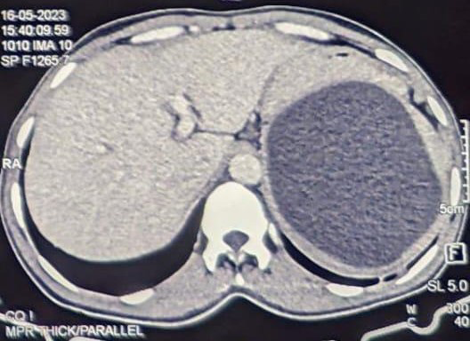
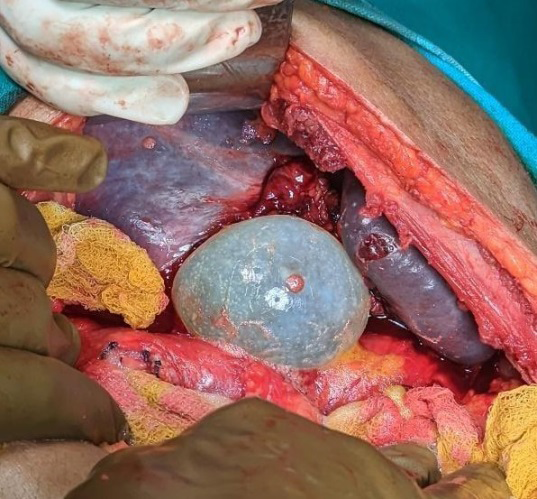
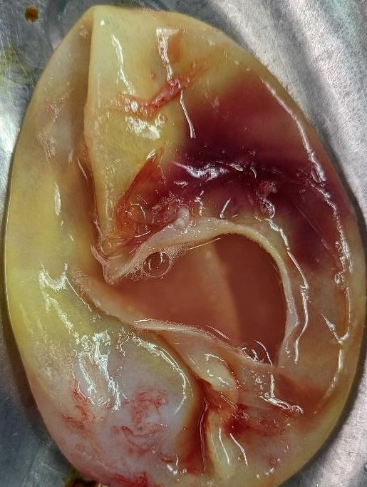
Presenting a case of a 33-year-old gentleman from Haryana who came to the outpatient department of General Surgery in our hospital with complaints of insidiously developed dull aching and continuous pain in the upper abdomen, left-sided more than right, which was non- radiating and without any aggravating or relieving factors, 15 days before date of admission. The patient did not give any associated history of fever, altered bowel habits or dyspnoea. The family had no cattle raising or farming history, and the patient had no known co-morbidities or any past surgical history. On clinical examination, a mildly tender lump which seemed to be arising from the spleen was palpable in the left hypochondrium, which was dull on percussion. There were abnormalities in the rest of the systemic examination. Investigative findings of ultrasound followed by CECT revealed a spleen of size 181mm with a well-defined focal hypodense area which measured 111x101x75mm (SI X AP X TR) in size, C.T. attenuation value ranging from 2-8HU (Figure 1). No parenchymal or peripheral enhancement was seen. Due to the large size of the symptomatic cyst, a curative surgical approach was planned, and the patient underwent total splenectomy after all necessary pre-operative preparations. Intraoperatively the cyst (Figure2), which was thick-walled and pale white suggestive of a hydatic cyst, was visualised which burst spontaneously inside the peritoneal cavity leading to the expulsion of the contents, which did not contain any daughter cysts and only clear fluid (Figure 3). The postoperative period under ICU management was uneventful, and the patient was discharged satisfactorily. The patient was given Pneumococcal polyvalent vaccination as a total splenectomy was done. Microscopic examination: sections from the spleen were consistent with chronic venous congestion spleen. Gamma gandy bodies are noted. Sections from cysts are consistent with hydatid cysts.
Discussion
Hydatid disease is prevalent in the Middle East and common in India, Africa, South America, New Zealand, Australia, Turkey and South Europe. Splenic cysts are classified as true cysts (primary) or pseudocysts (secondary) based on the presence of an epithelial lining. True cysts can be subdivided into parasitic (caused by echinococcus) and nonparasitic [4]. Nonparasitic true cysts are congenital or neoplastic. Congenital cysts can be epidermoid, dermoid, or endodermoid, present at a young age (children and young adults) and are commonly located in the upper pole of the spleen (as in this case). CA 19-9 and CEA levels are elevated in the epidermoid cyst’s contents and the patient’s serum. Pseudocysts are believed to develop after post-traumatic intraparenchymal or subcapsular splenic haematoma and occasionally after splenic infarcts or infections. Secondary cysts account for 75% of all nonparasitic splenic cysts [4]. Frequent abdominal imaging and the increasingly successful non-operative management of splenic injuries contribute to a rise in the incidence of nonparasitic splenic cysts, considered rare. Over two-thirds of the splenic cysts worldwide are parasitic hydatid cysts caused by Taenia Echinococcus. Sometimes hydatid cyst can present as a simple cyst without the classic serological and imaging features (as in this case) and later can lead to life-threatening complications like anaphylaxis [11].
The differential diagnosis for splenic hydatid cysts includes other splenic cystic lesions such as epidermoid cysts, pseudocysts, splenic abscesses, hematomas and cystic neoplasms of the spleen. [4/5] The differential diagnosis of splenic cyst consists of epidermoid and dermoid cysts, large solitary abscess or hematoma, cystic hemangiomas, intrasplenic pancreatic pseudocyst, and cystic neoplasm of the spleen (lymphangiomas), true cysts are very rare [12].
Epidemiology
Echinococcus granulosis is a member of the order Cestoda (flatworm) family Taenia. Man is an accidental intermediate host, as entering the larval forms into humans represents an end-stage in its life cycle. Consumption of contaminated vegetables or meat, which are not washed or scrubbed free of eggs, exposes man to the larval forms. An alternate mode of entry is direct contact with dogs whose fur has the eggs sticking to it. Once in the intestine of man, these embryonic forms enter the portal circulation and can spread to various organs, including the liver, lungs, pancreas, spleen, etc. About 10-15% of embryos escape from the liver and lung and are filtered into general circulation. A small fraction of these escaped embryos settle in the spleen (as in our case). The cyst may settle in the interior of the spleen or at its periphery under the capsule. Infection is usually acquired in childhood, but they mostly remain asymptomatic. The cyst grows at a rate of 0.3-1 cm per year, and it may take 5-20 years to grow into a sufficient size (3 – 35 cm) to cause symptoms of constant abdominal pain and a visible/palpable swelling in the abdomen. [5/13] The incidence of hydatid cysts of the spleen varies widely in sheep-raising countries. In India, the incidence of splenic hydatid cysts reported by different workers from different cities is shown in Table 1 [13].
| Central India | 6.3 |
| Delhi | 4.2 |
| Srinagar | 4.1 |
| Indore | 3.3 |
| Pondicherry | 2.7 |
| Ahmedabad | 2 |
Table 1: Incidence of hydatid cyst of the spleen in India Area Incidence (%).
People of all age groups and both sexes are affected with equal frequency. Diagnosis is usually established incidentally during the investigation of unrelated symptoms. When the cyst reaches an advanced size, the patient presents with a painless mass in the left hypochondrium. Some patients may present with complications such as infection of the cyst, rupture of the cyst into the peritoneal cavity, and fistula formation into hollow viscera like the colon or stomach [14]. Moderate eosinophilia of 6% or more is usually present. There are several serological tests to diagnose hydatidosis, like serum immune-electrophoresis, which is currently the most reliable, with a sensitivity of approximately 90%; indirect haemagglutination has a sensitivity of 85%4, ELISA and western blot analysis have also been used, in addition to eosinophilia, high IgE and IgM. The Cassoni skin test is sensitive but not specific. Serologic tests are helpful for the diagnosis, screening and post-op follow-up for recurrence.
Pre-operative diagnosis of this infection is mandatory as one of the common complications of hydatid disease is cyst rupture after trauma or spontaneous rupture due to increased intracystic pressure (as in this case). Splenic hydatid cysts can rupture into a hollow viscous, pleural cavity through the diaphragm or the peritoneal cavity. The cyst that ruptures into the peritoneal cavity may cause peritoneal irritation, urticaria, anaphylaxis, and even death. Therefore, hydatid cyst rupture needs immediate surgical intervention [15]. Since the condition closely resembles a soft tissue tumour on clinical examination, pre-operative radiological diagnosis is crucial to avoid a biopsy. Plain films are usually incapable of showing cysts within soft tissue. Pre-operative diagnosis of hydatid cysts can be made on ultrasound and confirmed by Computed Tomography (C.T.) scan (94- 96% and 100% sensitivity, respectively. Ultrasonography helps detect the cyst wall’s calcification, daughter cysts, cystic membranes, septa or hydatid sand [16]. In the present case, an ultrasound scan and CECT showed a large splenic cyst without typical daughter cysts.
Treatment strategies include PAIR (puncture, aspiration, injection, and re-aspiration), developed at the beginning of the 1980s and proved successful in various selected indications, also recommended by WHO [17]. Today, percutaneous treatment of liver hydatid cysts, a safe, easily applicable, and well-tolerated method, has become the most effective and reliable. In patients treated with the percutaneous treatment technique, a decrease in the cyst’s dimensions, solidification of the cyst contents, and irregularity in the walls of cysts are signs suggestive of a cure [13]. Until early 1970, open splenectomy was the standard treatment for benign splenic disorders, when fatal post-splenectomy sepsis was widely recognised.
Post Splenectomy infection (OPSI) syndrome comprises fulminating bacteraemia, disseminated intravascular coagulation, multiple organ failure, severe hypoglycaemia and rapid death. Its incidence is about 0.9 to 60%, and the mortality rate is about 50% [13]. So, tissue saving, partial splenectomy was introduced to reduce the chances of post- splenectomy sepsis, initially for trauma and later for most benign splenic diseases, including nonparasitic cysts and parasitic hydatid cysts. Spleen preservation should always be done, particularly in children. In the minimally invasive surgery era, laparoscopy is possible for benign splenic cysts and is superior to open splenectomy. These days partial splenectomy for nonparasitic/parasitic cysts is being done laparoscopically. Earlier, the main concern with laparoscopy surgery was the possibility of anaphylactic reaction and recurrence due to spillage. Despite all precautions, the incidence of spillage of scolex-rich fluid during surgery is about 5%–10%, which does not necessarily lead to dissemination. The incidence of recurrence is about 18%, possibly due to incomplete removal, spillage, or growth of small occult cysts that were missed initially. Recently, laparoscopy has become the gold standard in trained hands, and the enucleation of cysts with omentopexy can be done laparoscopically [18].
In the pre-operative period, medical treatment with albendazole is used to reduce the size and postoperatively to reduce the incidence of recurrence. It is the treatment of choice in patients who are unfit for surgery. Occult per- operative leaks are always possible, so medical therapy is mandatory in the postoperative period. Mebendazole and albendazole are a benzimidazole derivative. These drugs interfere with glucose absorption mechanisms through the parasite’s wall. According to the WHO guidelines for treating hydatid disease, chemotherapy is indicated for inoperable patients and those with multiple cysts scattered in many organs where surgery can be ineffective or hazardous.
Conclusion
Hydatid cyst of the spleen is rare, but a high suspicion should occur whenever a splenic cyst is encountered in clinical practice. Pre-operative diagnosis sometimes becomes challenging as radiological techniques cannot improve diagnostic accuracy. Hydatid disease should always be considered in the differential diagnosis of all cystic masses in the spleen, especially in endemic areas. If diagnosed early, this disease can be treated completely. In such cases, diagnostic laparoscopy followed by exploration and perioperative decisions is worth practising.
References
-
Cuschieri A, Steele RJC, Moosa AR (2000) Infected patients: Essential surgical practice. 4th (Edn.), Oxford. Butterworth-Heineann, pp: 157-160.
-
Goyal S, Pandit S, Raina R, Maurya S (2013) Our Initial Experience Of Laparoscopic Management of Liver Hydatid Disease in a Rural Medical College. Archives of Clinical and Experimental Surgery 2(1): 16-23.
-
Murtaza B, Gondal ZI, Mehmood A, Shah SS, Abbasi MH, et al. (2005) Massive splenic hydatid cyst. JCPSP 15(9): 568-570.
-
Hansen MB, Moller AC (2004) “Splenic cysts,” Surg Laparosc Endosc Percutan Tech 14(6): 316-22.
-
Karabicak, I, Yurtseven I, Yuruker SS, Ozen N, Kesim M (2009) Splenic hydatid cyst. Can J Surg 52: E209-E210.
-
Goyal S, Goyal S, Bhardwaj A (2016) Giant Primary Hydatid Cyst of the Spleen: A Rare Case Report with a Brief Literature Review. JMR 2(3): 59-61.
-
Sachar S, Goyal S, Goyal S, Sangwan S (2014) Uncommon Locations and Presentations of Hydatid Cyst. Annals of Medical and Health Sciences Research 4(3): 447-452.
-
Goyal S, Goyal S, Saini I (2015) Right Psoas Muscle Hydatid Cyst Causing Giant Hydronephrosis: A Case Report. International Medical Journal of Sifa University 2(3): 56-58.
-
Goyal S, Saini I, Goyal S (2015) Primary Retro Vesical Hydatid Cyst: Two Cases Report with Review. Open Acess Library Journal 2(7): E1579.
-
Agha RA, Franchi T, Sohrabi C, Mathew G (2020) SCARE group The SCARE 2020 statement: updating consensus surgical Case Report (SCARE) guidelines. Int J Surg 84: 226-230.
-
Rasheed K, Zargar SA, Telwani AA (2013) Hydatid cyst of the spleen: a diagnostic challenge. N Am J Med Sci 5(1): 10-20.
-
Merad Y, Derrar H, Zeggai A, Chadli M, Bemrah N, et al. (2021) Primary Splenic Cyst An Unexpected Diagnosis: A Case Report. Ann Med Surg (Lond) 65: 102293.
-
Bhandarwar AH, Katara AN, Bakhshi GD, Rathod MG, Quraishi AM (2008) A review of literature-Splenic hydatidosis. BHJ 44: 4.
-
Kanojiya R, Mittal A, Tantia R, Deen S, Dutt SC (2014) Primary Splenic Hydatid Cyst- A Rare Case. JMSCR 2(9): 2328-2331.
-
Belli S, Akbulut S, Erbay G, KoCer NE (2014) Spontaneous giant splenic hydatid cyst rupture causing fatal anaphylactic shock: A case report and brief literature review. Turk J Gastroenterol 25(1): 88-91.
-
Soufi M, Kharrasse G, Wafae K, Ismaili Z, Haroudi T, et al. (2015) Giant Isolated Hydatid Cyst of Spleen. J Spleen and Liver Research 1(1): 7- 11.
-
Maqbool, Anwar SF (2007) Hepatic hydatid cyst presenting as anaphylaxis. JCPSP 17: 224-25.
-
Khan FA, Ali S, Younis M, Bhat KA, Akhtar M (2014) Laparoscopic Enucleation and Omentopexy of a Primary Hydatid Spleen: a Case Report and Literature Review. J Minni Invasive Surg Scienc 3(4): e17108.
- Psychogenic Erectile Dysfunction in Late Adulthood: A Case Report on Clinical Intervention and Intimacy Restoration
- Clinical Trials on COVID-19 in 2025: A New Chapter in Global Health Research
- Innovations and Challenges in Contemporary Medical Clinical Trials: An Editorial Perspective
- Innovations and Challenges in Contemporary Medical Clinical Trials: A Critical Perspective
- Reimagining Clinical Trials: The Power of Continuous Feedback from Medical Reports
- Factors Influencing Brain Drain: Perspectives from a Medical School in Turkey