Small Bowel Perforation at the Duodeno-Jejunal Junction from a Migrated Biliary Stent: A Case Report
Background: The most common sites of reported bowel perforation secondary to migrated biliary stents were duodenal or colonic. However, small bowel perforations were reported in a few cases. Herein we present a case of small bowel perforation secondary to migrated biliary plastic stent at the duodeno-jejunal junction (DJ). We think this peculiar site is the first to be reported as a perforation site secondary to migrated biliary stent. Case Presentation: We present the case of female Egyptian patient with severe abdominal pain and air under diaphragm after Endoscopic Retrograde Cholangiopancreatography (ERCP) for Klatskin tumor. The patient underwent a midline laparotomy. A moderate collection of enteric fluid was encountered with migrated stent causing perforation of the small intestine at DJ. The stent was removed, and primary closure was done. The patient could not be extubated due to bad chest condition and admitted to ICU for 48 hours then extubated after improvement of the chest condition and ability of the patient to maintain good oxygen saturation in room air. Unfortunately, the patient developed massive pulmonary embolism on the fifth postoperative day and died. Conclusion: Suspicion of stent migration should be put in mind in any patient presented with abdominal pain with history of previous ERCP and biliary stenting. Endoscopic removal and mucosal repair are possible in select cases. This is still not available for DJ or small bowel. Further research and future innovations may overcome this challenge.
Introduction
Endoscopic Retrograde Cholangiopancreatography (ERCP) with biliary stent placement whether temporary or permanent is currently used for benign and malignant biliary obstruction [1, 2]. There are many biliary stents available according to size, design and material but mainly classified by material into two types: self-expandable metallic stents and plastic stents which are cheaper and easier to remove [3]. Although this advanced technology provides magic solutions for big biliary problems however, it’s not free from complications which may reach up to 10%. These complications include stent occlusion, cholangitis, pancreatitis, bleeding, duodenal perforation and stent migration [4]. Biliary stent migration can occur in 5-10% of cases whether proximal or distal. Distally migrated stent usually passes per rectum without any complications [5]. In less than 1% of patients, distal bowel perforation at any part of the small or large bowel can occur necessitating surgical or endoscopic intervention [6]. The most common sites of reported bowel perforation secondary to migrated biliary stents were duodenal or colonic. However, small bowel perforations were reported in a few cases [2]. Herein we present a case of small bowel perforation secondary to migrated biliary plastic stent at the duodeno-jejunal junction (DJ). We think this peculiar site is the first to be reported as a perforation site secondary to migrated biliary stent.
Case Presentation
Main Complaints
Diffuse severe abdominal pain.
History of Present Illness
We present the case of a 79-year-old Egyptian female, who presented with severe abdominal pain in the inpatient department of our hospital at 01/09/2023 with progressive course.
History of Past Illness
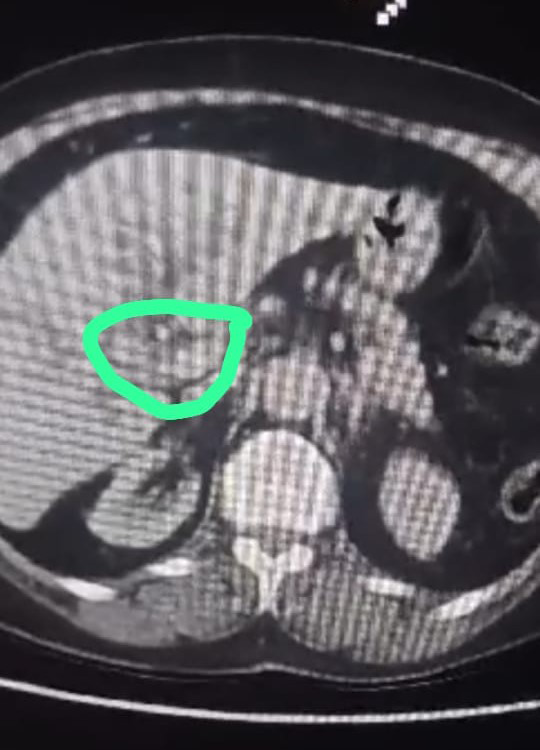
She initially presented on 19/08/2023 with obstructive jaundice. At the time she had an ultrasound that showed abnormal contracted shape GB with mud inside, dilated common bile duct (CBD) (1 cm) with no stones. Another ultrasound on 21/08/2023 revealed contracted GB upon tiny gravels, CBD dilated 13mm with moderate intra- hepatic biliary dilatation (IHBD), echogenicity grade II in both kidneys. CT on 25/08/2023 showed an ill-defined hypodense focal lesion seen involving the confluence of common hepatic hilum extended to the proximal portion of CBD, measures 3x1.7 cm and associated with moderate IHBD. After IV contrast injection, there was homogenous enhancement at the arterial phase. There were few peripancreatic LNs, the largest measures about 1 cm. The remaining portion of the CBD is not dilated. The gall bladder (GB) has markedly thickened and edematous wall with collapsed lumen with no dense stones. Features are those of intrahepatic biliary obstruction at the level of the common hepatic duct bifurcation with soft tissue mass lesion, the possibility of cholangiocarcinoma; Klatskin tumor, was highly suggested (Figure 1). She underwent a magnetic resonance cholangiopacreatography (MRCP) which did not add more information to the previous imaging studies. Following this she underwent ERCP on 29/08/2023 which revealed hilar stricture with marked IHBD, sphincterotomy was done with plastic stent (10 Fr, 10 cm, single external and single internal flap) insertion bypassing the hilar stricture. The patient developed melena the next day of ERCP, then upper endoscopy was done on 31/08/2023 showing no bleeding from the papilla and stent protruding from the papilla and in place. On the next day, the patient developed severe abdominal pain with surgical abdomen.
Past History
Her past medical history was significant for hypertension on medications and asthma but no history of previous operations or any manifestations related to gastro-intestinal problems.
Physical Examination
On initial evaluation the patient had temperature 38°C, pulse 110 beat per minute, blood pressure 95/60 mmHg. Her clinical examination revealed diffuse abdominal distension, tenderness and rigidity.
Laboratory Investigations
Laboratory investigations the day before ERCP were TB 13.4 mg/dL, DB 7.4 mg/dL, INR 1.1. Pre-laparotomy investigations were HB 7.1 gm/dL, Total Leucocyte Count 12.200 /µL, Platelet count 280.000 /µL, Na 138 mEq/L, K 2.9 mEq/L, Creatinine 1.9 mg/dL.
Imaging Investigations
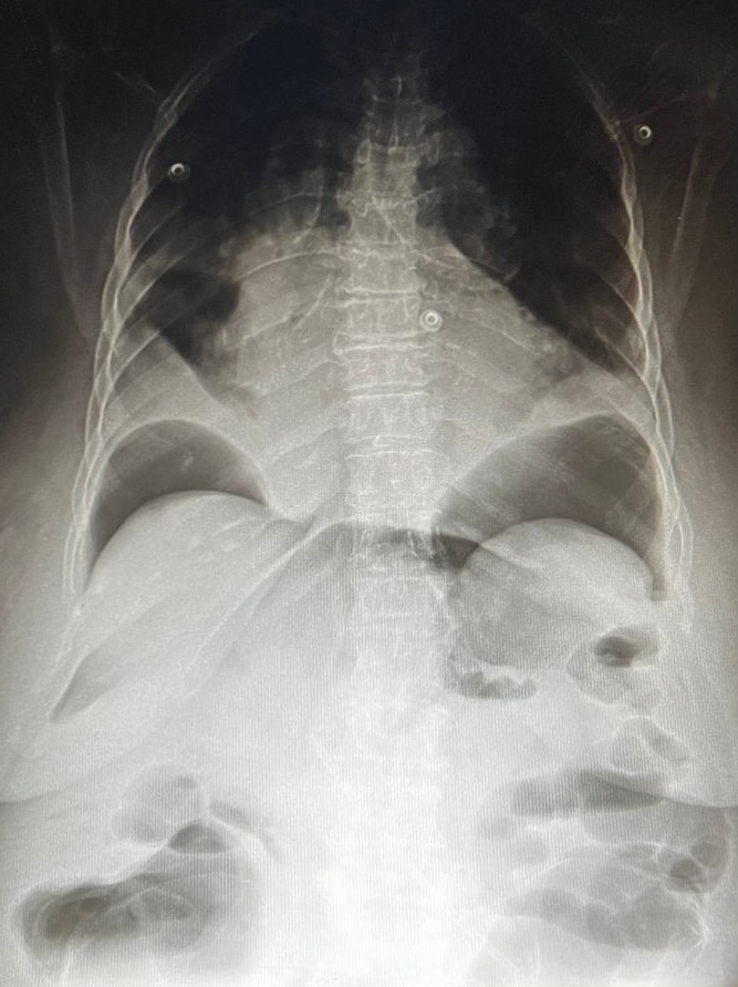
Plain X ray abdomen in erect position on 01/09/2023 showed marked amount of air under diaphragm (Figure 2). So, surgical intervention was decided.
Treatment
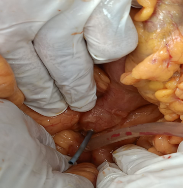
The patient underwent a midline laparotomy on 01/09/2023. Moderate collection of enteric fluid was encountered. Formal exploration of the small intestine showed migrated stent causing perforation of the small intestine at DJ (Figure 3). The stent was removed with trimming of the edges of the perforation and primary closure in two layers using Vicryl 3/0. The peritoneal cavity was washed with warm saline and pelvic tubal drain was left behind.
Outcome and Follow-Up
The patient could not be extubated due to bad chest condition and admitted to ICU for 48 hours then extubated after improvement of the chest condition and ability of the patient to maintain good oxygen saturation in room air. Laboratory investigations after ICU discharge were HB 8.5 gm/dL, Total Leucocyte Count 4900 /µL, Platelet count 290.000 /µL, Na 147 mEq/L, K 3.4 mEq/L, Creatinine 1.3 mg/ dL. Unfortunately, the patient developed massive pulmonary embolism on the fifth postoperative day and died.
Discussion
ERCP has predominantly become the preferred therapeutic option in management of benign or malignant obstruction of the pancreaticobiliary tract. It is most commonly utilized for the removal of CBD stones and relief of obstructive jaundice specially in high risk or inoperable cases replacing surgical choledochoduodenostomy [7]. Due to less morbidity of this technique when compared with surgical intervention, it became popular and a wide varieties of plastic and metal stents became available [8]. Plastic stents are commonly used due to easy insertion, easy removal and lower price, but have a higher complication rate including stent occlusion, stent fracture and migration whether proximal or distal [9, 10]. Migration rate ranges from 5% to 10%, with higher rates in plastic stents compared to others [10]. Distally migrated stents may be complicated by abscess formation, fistula, obstruction and bowel perforation in less than 1% of cases [11]. Stent migration may be attributed to long stent, distal benign biliary strictures which were dilated before stent insertion and if sphincterotomies were done during the procedure [12]. Therefore, use of multiple plastic stents in benign biliary strictures may reduce the risk of migration [13]. Colonic perforation as a complication of stent migration usually occurs in sigmoid colon. Intraluminal migrated stents can be retrieved endoscopically. However, in cases of perforations surgical intervention is indicated [1]. Diverticulosis, adhesions, hernia or strictures are common risk factors for perforation [6]. Surprisingly, none of these risk factors were present in our case. Most of the reported cases with bowel perforation from biliary stent migration documented duodenal or colonic perforation, with very few cases of small bowel perforation. Diffuse peritonitis and signs of sepsis are the presenting manifestations in those patients [2]. That is exactly what happened in our patient who presented with diffuse peritonitis and limited sepsis due to early surgical intervention. Several studies have reported that distal stent migration is more common in benign than in malignant biliary disease, which may be attributed to resolution of the stenosis after subsidence of inflammation [3]. However, in our case the migrated stent was placed for malignant biliary stricture (Klatskin tumor). Regardless of the patient’s clinical status, any migrated stent should be removed immediately [3], so, surgical intervention in the current case was not delayed. Endoscopic techniques in the form of over-the-scope clip were employed for some cases of bowel perforation from migrated stent but mainly in the duodenum or the distal colon [2, 14]. This was not feasible in our case where the bowel perforation was in the DJ, so, we went ahead to surgical intervention. A large volume retrospective study reported that the potential risk factors for stent migration are straight stents with large diameter, stent employed for more than one month and dilated common bile duct with diameter more than 1 cm [15]. All these potential risk factors were present in our cases except that the stent duration was about one week. To the best of our knowledge, this is the first reported case of small bowel perforation at the DJ secondary to biliary stent migration. Systematic review of literature for bowel perforation from migrated biliary stents recorded 81 cases with 93.6% of cases had plastic stent. The site of perforation was duodenal in 44.9%, colonic in 29.5%, and small bowel in 23.1% of cases. Surgical intervention was indicated in 60.3%, endoscopic removal in 34.6% of cases. Overall mortality reached 10%. Geographical distribution of cases was 48.7% from Europe, 26.9% from Asia and Middle East, 15.4% from the United States, 6.4% from Australia and 2.6% from South America [2].
Conclusion
Suspicion of stent migration should be put in mind in any patient presented with abdominal pain with history of previous ERCP and biliary stenting. Endoscopic removal and mucosal repair are possible in select cases. This is still not available for DJ or small bowel. Further research and future innovations may overcome this challenge.
Ethics Approval and Consent to Participate
Approval of Minia College of Medicine Institutional Ethics Committee was obtained. Written informed consent was obtained from the patient for all interventions.
Consent for Publication
Written informed consent was obtained from the patient for publication of her case.
Acknowledgements
We would like to thank our nurses in the endoscopy unit and operative theatre for their effort during operative work and postoperatively.
References
-
Kodia K, Huerta CT, Arora Y, Wickham C, Deshpande AR, et al. (2022) Minimally invasive management of an ascending colonic perforation secondary to distal biliary stent migration: a multidisciplinary, novel laparoendoscopic approach. J Surg Case Rep 2022(9): rjac404.
-
Zorbas KA, Ashmeade S, Lois W, Farkas DT (2021) Small bowel perforation from a migrated biliary stent: A case report and review of literature. World J Gastrointest Endosc 13(10): 543-554.
-
Namdar T, Raffel AM, Topp SA, Namdar L, Schmitt M, et al. (2007) Complications and treatment of migrated biliary endoprostheses: a review of the literature. World J Gastroenterol 13(40): 5397-5399.
-
Arhan M, Odemiş B, Parlak E, Ertuğrul I, Başar O, et al. (2009) Migration of biliary plastic stents: experience of a tertiary center. Surg Endosc 23(4): 769-775.
-
Wang X, Qu J, Li K (2020) Duodenal perforations secondary to a migrated biliary plastic stent successfully treated by endoscope: case-report and review of the literature. BMC Gastroenterol 20(1): 149.
-
Park TY, Hong SW, Oh HC, Do JH (2021) Colonic diverticular perforation by a migrated biliary stent: a case report with literature review. Medicine 100(52): e28392.
-
Adler DG, Lieb JG, Cohen J, Pike IM, Park WG, et al. (2015) Quality indicators for ERCP. Gastrointest Endosc 81(1): 54-66.
-
Güngör G, Okur N (2016) A Fatal Complication: Intestinal Perforation Secondary to Migration of a Biliary Stent. Pol J Radiol 81: 170-172.
-
Lam R, Muniraj T (2021) Fully covered metal biliary stents: a review of the literature. World J Gastroenterol 27(38): 6357.
-
Yilmaz Ö, Kiziltan R, Aydin O, Bayrak V, Kotan Ç, et al. (2015) A Rare Complication of Biliary Stent Migration: Small Bowel Perforation in a Patient with Incisional Hernia. Case Rep Surg 2015: 860286.
-
Jafferbhoy SF, Scriven P, Bannister J, Shiwani MH, Hurlstone P, et al. (2011) Endoscopic management of migrated biliary stent causing sigmoid perforation. Case Rep 2011: bcr0420114078.
-
Emara MH, Ahmed MH, Mohammed AS, Radwan MI, Mahros AM, et al. (2021) Biliary stent migration: why, how, and what? Eur J Gastroenterol Hepatol 33(7): 967- 973.
-
Daabiss M (2011) American Society of Anaesthesiologists physical status classification. Indian J Anaesth 55(2): 111-115.
-
Bureau MA, Gkolfakis P, Blero D, Pezzullo M, Devière J, et al. (2020) Lateral duodenal wall perforation due to plastic biliary stent migration: a case series of endoscopic closure. Endosc Int Open 8(5): E573-E577.
-
Kawaguchi Y, Ogawa M, Kawashima Y, Mizukami H, Maruno A, et al. (2014) Risk factors for proximal migration of biliary tube stents. World J Gastroenterol 20(5): 1318-1324.
- Psychogenic Erectile Dysfunction in Late Adulthood: A Case Report on Clinical Intervention and Intimacy Restoration
- Clinical Trials on COVID-19 in 2025: A New Chapter in Global Health Research
- Innovations and Challenges in Contemporary Medical Clinical Trials: An Editorial Perspective
- Innovations and Challenges in Contemporary Medical Clinical Trials: A Critical Perspective
- Reimagining Clinical Trials: The Power of Continuous Feedback from Medical Reports
- Factors Influencing Brain Drain: Perspectives from a Medical School in Turkey