Mind the Gap Familial Maxillary Midline Diastema and High Labial Frenum Attachment
In day today life clinicians can come across various dental malocclusions in human beings. Maxillary midline diastema is one among those and when this condition occurs it causes aesthetic impairment requiring orthodontic treatment in order to close the existing ugly looking gap between two central incisors. However, before performing this treatment, it is essential to rule out the underlying etiologic factors as this clinical entity is caused by various causative factors. In the present article, occurrence of maxillary midline diastema and high labial frenum attachment both in mother and daughter indicating genetic relation associated with it is shown.
Introduction
Maxillary midline diastema is an unesthetic appearing clinical entity observed in the maxillary arch and is characterised by presence of space between two central incisors. Although it most commonly observed in children during primary or mixed dentition period, but its existence in the permanent dentition or in adolescents and adults raises major concern among patient and parents and should be addressed immediately [1, 2]. Occurrence of midline diastema between two upper teeth in children during mixed dentition period which is also called as “ugly duckling stage” is usually resolved following the eruption of upper permanent canines. When it persists after the eruption of all permanent teeth, an orthodontic consultation is very important along with diagnosis of possible etiological factor [2].
The dental midline diastema is a clinical entity, which has a multitude of underlying etiological factors that are interdependent or independent [1, 2]. Various etiological factors have been suggested in the literature for the occurrence of maxillary midline diastema like physiological, developmental, self-limiting diastema, missing or undersized lateral incisor, familial, oral habits (thumb sucking, mouth breathing, tongue thrusting), ankylosed central incisor, flared or rotated central incisors, anodontia, macroglossia, generalized spacing, facial type, ethnic and familial characteristics, abnormal labial frenum, presence of midline pathology, tongue piercing habit, pathological tooth migration and presence of mesiodens [1, 5, 6, 7, 8, 9, 10]. The prevalence of maxillary midline diastema is reported as 1.6% to 13.6% or 25.6% among various general population studied and is more reported in females [3, 4]. The difference in the prevalence rates obtained from numerous epidemiological studies is may be due to increased number of different factors contributing to the development of midline diastema or due to the various definitions used to explain its presence and to gender and race sample difference [3, 4]. The purpose of this article is to present a case of maxillary midline diastema which occurred in three members of the same family belonging to Indian ethnicity depicting familial inheritance of the condition.
Case Report
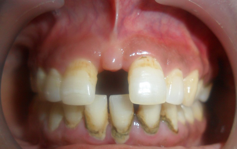
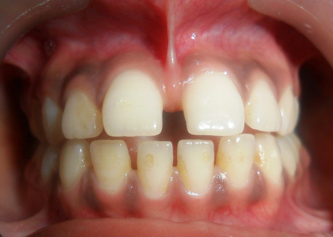
A 55-year-old female patient reported to a private dental clinic complaining of presence of wide gap in the upper front region between two teeth. Patient was moderately built and well-nourished and did not show signs and symptoms of systemic diseases or syndromic disorders. Intra oral examination was carried out which showed presence of maxillary midline diastema with the gap measuring about 12 mm (Figure 1). Class I molar relation was present with normal overjet and overbite. Patient oral hygiene was poor with presence of heavy amount of calculus over the teeth surfaces. A prominent maxillary labial frenum attachment was noticed between two central incisors. Following examination of this patient, her daughter also wanted to get her oral condition to be examined. She was of 17 years of age with well-nourished and well-built and with no systemic conditions. Intra oral examination showed complete permanent dentition, with class I molar relation, normal overjet and overbite. Her daughter also exhibited presence of maxillary midline diastema with a gap of 6 mm and also showed presence of prominent maxillary labial high frenal attachment (Figure 2). No other dental findings were observed apart from this. Upon eliciting the family history, it was revealed that this dental finding is present in her grandmother too. Therefore, based on clinical features, family history and literature search the present case was diagnosed as a familial maxillary midline diastema. Oral prophylaxis was carried out in both patient and patients were scheduled for further treatment.
Discussion
The word “diastema” is derived from a Greek word meaning “interval” and hence it is described as a “gap” or “space” seen between two or more consecutive teeth [5, 6, 7, 8]. This condition occurs more frequently in the median plane of the maxillary arch between the two central incisors and hence referred as median, central or midline diastema. According to famous author of the Orthodontic field, Angle, the dental midline diastema is described as ‘a common form of incomplete occlusion characterized by a space between the maxillary and less commonly mandibular central incisors’ [11]. Angle [11] also stressed on the functional and aesthetic implications of the midline diastema and suggested that the maxillary interdental diastema always creates an unpleasant appearance and interferes with speech depending on its width. Again in 1972 Andrews [12] in his classical article on “the six keys to normal occlusion” has stated in pointing the fifth principle showing that ‘the interdental diastemas should not exist and that all contact areas should be tight,’ so that the patient has “straight and attractive teeth as well as a correct overall dental occlusion.” Diastema is multifactorial clinical entity encompassing more than one underlying etiological factors and are interconnected [5, 6, 7, 8, 9, 10, 11, 12, 13, 14, 15]. Presence of maxillary midline represents an aesthetic and psychological impairment and distress. It is stated that interrelationship between the familial pattern of midline diastema and the microdontia, macroglossia, labial frenum has been shown in a 2016 study [9]. Dental literature showed that two subtypes of frenum such as papillary and papillary penetrating frenum are associated with maxillary midline diastema. Therefore, superior labial frenectomy should be delayed till the complete eruption of the permanent lateral incisors as this will spontaneously close the physiological upper diastema. However, there is controversial debate pertaining to corelation between maxillary midline diastema and highly attached frenum and frenectomy during early mixed dentition period.
Nagaveni et al in 2014 [2] conducted a prospective study including 3000 children and evaluated different types of maxillary labial frenum morphology based on Sewerin’s frenum typology. From this investigation authors reported that simple frenum was the most prevalent frenum types observed in all the three groups such as primary, mixed and permanent dentition followed by persistent tectolabial frenum and frenum with nodule. The prevalence of simple frenum was found to be increased with age, while the persistent tectolabial frenum decreased proportionally. In addition to this, authors also found that there was no statistically significant gender difference found with respect to the frenum morphology in all three groups [2]. Another prospective Indian study evaluated the presence of mesiodens in 2500 Indian children aged between 3 to 12 years of age by looking at the patient’s records and radiographic examinations. Authors detected mesiodens in 25 children and reported that the maxillary midline diastema was associated with mesiodens [6]. Later the Nagaveni NB reported numerous case series showing occurrence of different types of mesiodens resulting in development of maxillary midline diastema [4, 8, 10, 13].
Ranjbaran MA, et al. [7] recently evaluated the associations among the occurrence or types of maxillary canine impaction, labial frenum attachment types, lateral incisor anomalies, and midline diastema in patients with and without impaction. Based on the data obtained, authors concluded that buccal type of canine impaction is positively associated with papillary labial frenums and midline diastema is positively and independently associated with canine impaction, papillary frenums and abnormal lateral incisors [13]. In the present study a strong correlation was observed with existence of maxillary midline diastema and presence of high prominent labial frenal attachment and also showed familial occurrence of both clinical entities including occurrence of prominent labial frenum attachment and midline diastema. This finding clearly and strongly indicates the requirement of further research concentrating on genetic studies to predict the possibility of development of these two separate clinical entities.
Jaija AM, et al. [9] from their investigation found various etiological factors and segregated them into major and minor types. Authors finally summarized the findings of their investigation in the form checklist consisting of impact of different etiological factor of the maxillary midline diastema upon the diagnosis, treatment or retention protocol. They designed the checklist to highlight the intervention at the different stages of treatment for each etiological factor. Therefore, this checklist can be used as a guide during management of different stages of treatment towards maxillary midline diastema [14].
Conclusion
Occurrence of maxillary midline diastema is commonly seen clinical finding which needs early diagnosis as the treatment depends on many factors and includes a multi- disciplinary approach. Occurrence of familial maxillary midline diastema gives an indication for future research to formulate more appropriate therapeutic guidelines in the overall management of maxillary midline diastema.
References
-
Tadros S, Ben-Dov T, Cathan EO, Anglin C, April MM (2022) Association between superior labial frenum and maxillary midline diastema – a systematic review. Int J Pediatr Otorhinolaryngol 156: 111063.
-
Nagaveni NB, Umashankara KV (2014) Morphology of maxillary labial frenum in primary, mixed and permanent dentition of Indian children. J Craniomaxillo Dis 1: 5-10.
-
Awooda EM (2023) Twelve-year follow-up of laser frenectomy during early mixed dentition. Case Rep Dent, pp: 5525534.
-
Nagaveni NB (2023) Inversion of impacted mesiodens: Report of case series with literature review. Glob J Res Dent Sci 3(5): 7-12.
-
Jonathan PT, Thakur H, Galhotra A, Galhotra V, Gupta N (2018) Maxillary labial frenum morphology and midline diastema among 3 to 12-year-old school going children in Sri Ganganagar city: A cross-sectional study. J Indian Soc Pedod Prev Dent 36(3): 234-239.
-
Nagaveni NB, Sreedevi B, Praveen BS, Praveen RB, Vidyllatha BG, et al. (2010) Survey of mesiodens and its characteristics in 2500 children of Davangere city, India. Eur J Paediatr Dent 11(4): 185-188.
-
Ranjbaran MA, Aslani F, Jafari-Naeimi A, Rakshan V (2023) Associations among the occurrence or types of maxillary canine impaction, labial frenum attachment types, lateral incisor anomalies, and midline diastema in patients with and without impaction: A case-control study. Int Orthod 21(2): 100743.
-
Nagaveni NB, Umashankara KV, Sreedevi, Reddy BP, Radhika NB, el al. (2010) Multi-lobed mesiodens with a palatal talon cusp – A rare case report. Braz Dent J 21(4): 375-378.
-
Jaija AM, El-Beialy AR, Mostafa YA (2016) Revisiting the factors underlying maxillary midline diastema. Scientifica (Cairo), pp: 5607594.
-
Nagaveni NB (2023) Three-lobed (multi-lobed) incisoriform mesiodens with type I talon cusp – Report of a unique dental anomaly. Glob J Res Dent Sci 3(6): 7-10.
-
Angle EH (1907) Treatment of malocclusion of the teeth. 7th (Edn.), Philadelphia: S.S. White Dental Manufacturing Co, pp: 167.
-
Andrews LF (1972) The six keys to normal occlusion. Am J Orthod 62(3): 296-300.
-
Nagaveni NB, Umashankar KV (2023) Vertical, Intra- osseous impaction of permanent maxillary central incisor in association with multiple anomalies - Report of a rare case. EC Dental Science 22(9): 1-4.
-
Romero MF, Babb CS, Brenes C, Haddock FJ (2018) A multidisciplinary approach to the management of a maxillary midline diastema: A clinical report. J Prosthet Dent 119(4): 502-505.
-
Nuvvula S, Ega S, Mallineni SK, Almulhim B, Alassaf A, et al. (2021) Etiological factors for the midline diastema in children: A systematic review. Int J Gen Med 8(14): 2397-2405.
- Psychogenic Erectile Dysfunction in Late Adulthood: A Case Report on Clinical Intervention and Intimacy Restoration
- Clinical Trials on COVID-19 in 2025: A New Chapter in Global Health Research
- Innovations and Challenges in Contemporary Medical Clinical Trials: An Editorial Perspective
- Innovations and Challenges in Contemporary Medical Clinical Trials: A Critical Perspective
- Reimagining Clinical Trials: The Power of Continuous Feedback from Medical Reports
- Factors Influencing Brain Drain: Perspectives from a Medical School in Turkey