Sonographic Evaluation of Normal Placenta Thickness among Apparently Healthy Women in Second and Third Trimester Pregnancy
Some maternal fetal disorders can be detected or revealed for further investigation from data obtained from the placenta thickness. Suboptimal placenta thickness or enlarged placenta may be a pointer to disorder such as anaemia, hypertension or diabetes, growth retardation among others. Small placenta is associated with malnutrition and increased risk of chronic disease later in life. The aim of this study is to determine the normal thickness of placenta in Nigeria. Any placenta thickness outside this value should be further investigated for possible maternal or fetal anomalies. Four hundred subjects were selected by convenience sampling after passing the inclusion criteria, and educational status required. The abdomen of pregnant women with gestational age of 16 to 40 weeks referred from antennal clinic and who gave consent to participate in the study were scanned. The scan ruled out abnormalities and measured the gestational age using fermur length (FL) and Biparetal Diameter (BPD) to compare with menstrual gestational age (MGA). The placenta thickness was measured at the thickest point of the umbilical cord insertion into the placenta. Result showed that placenta thickness for GA of 16 to 40 weeks was from 15 to 39mm (SD: 5.62). The study suggests that pregnancy with placenta thickness outside this predictive thickness should be subjected to further investigation to rule out maternal or fetal abnormalities associated with enlarged placenta or subnormal placenta thickness.
Introduction
Accurate assessment of the placenta is not only important for evaluation of fetal nutrition but also for fetal health. The placenta has two components; the fetal portion developed from the chorionfrond sum (chorionic plates) and a maternal portion, the deciduasbasal is formed by the endometrial surface. The placenta is a fetal organ with important metabolic, endocrine and immunologic functions, besides being responsible for nutrition. It also provides respiration and secretion functions of the fetus. It provides physiological link between the pregnant woman and her fetus. The placenta develops from the chronic villi at the implantation site at 5th week of gestation and by 9th and 10th week the diffuse echoxture of the placenta start to appear on sonography. The growth and development of placenta controls fetal growth and development. Placenta transfers occur essentially by diffusion. According to, placenta serves as historical record of maternal and fetal health throughout pregnancy. Hence the knowledge of the placenta provides insight into pathology of pregnancy. Placental thickness is closely related to fetal wellbeing and may be a key factor in pre-natal outcomes. Large placenta is associated with hemolytic disease of new born, maternal diabetes mellitus, severe anaemia and intra-uterine fetal infections. Smallplacenta is associated with preeclampsia, chromosomal abanomarlities, severe maternal diabetes mellitus, chronic fetal infections and intra-uterine growth restriction. Second trimester placenta volumes measured by three dimensional ultrasound have been used to identify fetuses at risk of growth restriction. Ultrasound measurement of placental thickness is of prognostic value in identifying the occurrence of fetal growth restriction. Knowledge of the placental thickness is essential to determine the prognostic value of the fetal and meternal health status. Magnetic Resonance Imaging (MRI) has the advantage of demonstrating fetal and maternal anatomy without the need for distended bladder and its images are unaffected by maternal bowel gas and oligohydramnous, all of which degrade ultrasound images.
However, its high cost and resultant scarcity especially in economically disadvantaged areas, have continued to limits its obstetric roles. These factors have also limited the value of computerized tomography (CT). Ultrasound is cheap, readily available and provides virtually all the imaging outline of the fetus and the pregnant uterus. The modality is uniquely suited to the challenges presented by a moving baby. Ultrasound is safe for developing fetus throughout pregnancy. Placenta volume surface and thickness show progressive increase in values with increasing gestational age. However, placental thickness appears to be the simplest ultrasound parameters to evaluate gestational health status.
This study was undertaken to evaluate placental thickness at the thickest level of insertion of umbilical cord to establish a normal placental thickness for assessment of health status of the fetus and mother. Excess placenta thickness or reduced placenta thickness is associated with fetal or maternal disorders. This study is to determine normal healthy placenta thickness for the prediction of anomalies using placenta thickness.
Materials and Methods
The study was a prospective study carried out at Abia State Nigeria between December 2019 and November 2020. Four hundred and eighty pregnant women presented for ultrasonography. Four hundred candidates who passed the inclusion criteria were selected for the examination. The following women were selected - Women who presented for pregnancy ultrasound, women in the gestational age of 15 to 40 weeks, women with minimum academic level of equivalent of First School leaving certificate (FSLC) who can be sure of their last menstrual date, women who have regular menstrual cycle, women who were not on oral or injectable contraceptives at least 3 month prior to last normal menses, women whose clinical reports show no evidence of system illness, no diabetes, anaemia no hypertension, no sickle cell, no evidence of genetic history of abnormalities, non smokers, non-alcoholics referred for obstetric scanning from ante-natal clinic. The examination and research was explained to them. They were first scanned for fetal or maternal abnormality. A convenience sampling method was used to choose the candidates. Four hundred (400) pregnant women scanned with GE logic V5 expert (France) real time ultrasound machine with 3.0MHz - 5MHz curvilinear probe. A longitudinal and transverse abdominal Sean was carried out. The measurement of biparental diameter (BPD), Fermur length (FL) and abdominal circumference (AC) was taken for age comparison. The measurement of placental thickness were also taken and recorded. The LMP and gestational age (GA) were also recorded. The placental thickness was measured and the thickness level of the umbilical cord insertion with the placenta. The line of measurement is perpendicular order to the fetal and maternal surface. The distance measured in from fetal placental surface to the placental-endometrial surface[1, 2, 3, 4, 5, 6, 7].
Results
Prospective study of 400 pregnant women between the ages of 16 years to 40 was carried out. The age range of 21 years to 35 year had the highest frequency of 340 (85 %). The highest level of education was first degree and lowest level was first school Leaving Certificate (FSLC) (Table 1).
Distribution of Placenta. Distribution of Placenta thickness with menstrual gestational age (MGA).
| Fetal Placenta Thickness | MGA | |
|---|---|---|
| N | 400 | 400 |
| Mean | 28.23 | 30.8 |
| Median | 30 | 32 |
| Std Deviation | 5.46 | 5.62 |
| Minimum | 14 | 15 |
| Maximum | 40 | 41 |
Table 1: Shows marginal increases of the placenta thickness with weekly increases of the menstrual Gestational Age (MGA).
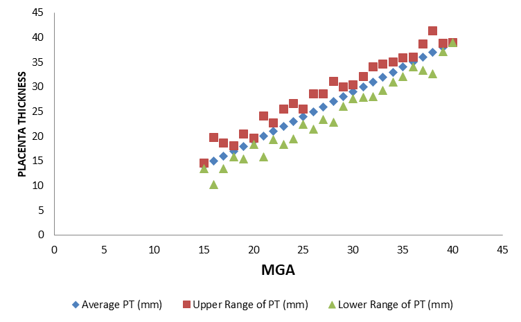
Distribution of placenta thickness (PT) standard Deviation: SD with Menstrual Gestational Age in weeks (Table 2).
| MGA (Weeks) | PT (mm) |
|---|---|
| 15 | 14 ± 0.58 |
| 16 | 15 ± 4.77 |
| 17 | 16 ± 2.58 |
| 18 | 17 ± 1.14 |
| 19 | 18 ± 2.52 |
| 20 | 19 ± 0.69 |
| 21 | 20 ± 4.16 |
| 22 | 21 ± 1.69 |
| 23 | 22 ± 3.57 |
| 24 | 23 ± 3.57 |
| 25 | 24 ± 1.57 |
| 26 | 25 ± 3.54 |
| 27 | 26 ± 2.62 |
| 28 | 27 ± 4.18 |
| 29 | 28 ± 1.96 |
| 30 | 29 ± 1.40 |
| 31 | 30 ± 2.08 |
| 32 | 31 ±3.00 |
| 33 | 32 ± 2.66 |
| 34 | 33 ± 2.00 |
| 35 | 34 ± 1.94 |
| 36 | 35 ± 0.99 |
| 37 | 36 ± 2.68 |
| 38 | 37 ± 4.29 |
| 39 | 38 ± 0.89 |
| 40 | 39 |
Table 2: Contain the mean values of placenta thickness which showed positive linear order with increasing GA.
Discussion
Increased or reduced placenta thickness is associated with some maternal or fetal disorders. This study set out to evaluate the normal placenta thickness as a marker for detection of possible gestational abnormalities. In this study there were 400 apparently healthy antenatal women between the ages of 16 years to 41 years. Minimum academic qualification was the first leaving school certificate, so that they could correctly be sure of their last menstrual period. From this study, there is a positive correlation between the normal ultrasonically measured placenta thickness in millimeters and fetal gestational age in weeks similar to the finding of The observed placenta thickness varies from 14mm to 38 mm. The linear relationship of placenta thickness to mean gestational age is represented as PT 1.357 = 0.874 MGA + 1.88 from the regression analysis of both parameters at 95 % confidence level R = 0.7613. This is also in line with the findings ofthis result shows that placenta grows in line with fetal growth. The placenta thickness in milli-meter is almost equivalent to weeks of gestation. Therefore obvious sharp differences in measurement between the placenta in millimeters and the GA in weeks can alert health provider that certain abnormalities might exist. Due to the closeness of the placenta thickness and GA a deviant placenta value should raise questions on fetal growth. Pregnancy dating by means of BPD (Biparietal Diameter) and Femur length (FL) can be cross-checked by the uses of placenta thickness measurement and observation on normal and abnormal fetal growth will be enhanced. This will be useful particularly in environment with history of late booking of pregnancy and in cases of unsure of date [8, 9, 10, 11]. In this study the mean placenta thickness in millimeters (mm) was not 2 units higher or lower than the number of week’s gestational age. The normal placenta thickness was determined to be between 15 to 39mm for GA of 16 to 40 weeks having a difference of 2 to 3 mm for the GA in weeks. There was positive linear relationship between placenta thickness (mm) and menstrual gestation Age (weeks).
References
-
Azpurua H, Funai EF, Coraluzz LM, Doherty LF, Sasson IE, et al. (2010) Determination of Placenta weight using two- Dimensional Sonography and Volumetric Mathematical Modeling. Am J Perinata 27(1): 151-155.
-
Browne PC, Hammer LH, Clarn WS (1997) Sonographic Fetal growth Curves from an Indigent Population in Atlanta, Georgia Singleton Pregnancies. Am J Perinatol 9(5-6): 467-476.
-
Lia JED, Bendon RW Normal and Abnormal Placenta Development.
-
Scot JR, Disaia PJ, Hammond CB, Spellacy WN (1994) Derforth’s Obstetrics and Gynecology; 7th (Edn.), Lippin Coth Philadephiapp: 49-65.
-
Evans DW, Connor PD, Hahn RG, Rodney WM, Arhear KL (1995) A Comparison of Manual and Ultrasound Measurements of Fund Height. J FarnPrac 40(3): 233- 236.
-
Kratochwil A (1980) Ultrasonic Diagnosis in Obstetrics and Gynecology. Med ProgTechnol7(4):157-167.
-
Mongelli M, Biswas A (2002) Menstrula age-dependent Systematic Error in Sonographic fetal Weight Esturiation: A Mathematical Model. J Clinical Ultrasound30(3): 139- 144.
-
Raio L, Ghezzi F, Cromi A, Nelle M, Dürig P, et al.(2004) The Thick Heterogenous (Jelly-like) Placenta: A strong Predictor of adverse pregnancy outcome. Prenat Diagn24(3):182-188.
-
Reis NSV, Brizot ML, Shultz R, Nomura RMY, Zugaib M (2005) Placenta lakes on Sonographic Examination, Correlation with Obstetric Outcomes and Pathologic Findings. J Chin Ultrasound 33(2): 67-71.
-
Shirish N, Wang E, Parry S (2012) Two-Dimensional Sonographic Placenta Measurement in the Prediction of Small for Gestational Age infants. Ultrasound Obstet Gynecol 40(6):674-679.
-
Zukam DT, Njock LA, Kenla A, Shu D, Doh A, et al. (2002) Fetal Ultrasound Biometry in a Camerounian Population: Study of Femur Length. Med Trop (Mar)62(5): 521-524.
- Teaching Cognitive Behavioral Therapy to Graduate Psychiatric Mental Health Nurse Practitioner Students: Utilizing A Mixed Methods Course Evaluation
- The Role of Pharmacogenetic Testing in Clinical Practice: A Path toward more Effective, Personalized and Cost-effective Care
- Implementing Screening with the GAD-7 in an Outpatient Mental Health Setting: A Quality Improvement Project
- Social Media and Health Promotion
- Impact of Covid-19 Pandemic on the Academic Performance and Attitude of Nursing Students towards E-Learning
- A Study to Assess the Knowledge Regarding Ill Effects of Tobacco Consumption on Health among Adolescents in Selected Higher Secondary School in Nadiad City