Antibacterial Activity of Cow’s Milk Silver Nanoparticles Synthesized across Various pH against Clinically Isolated MDR Staphylococcus aureus
The study aims to evaluate the antibacterial activity of silver nanoparticles (AgNPs) from pasteurized cow’s milk synthesized across various pH (3, 5, 7, 9 and 11) along with study of synergy with the selected antibiotics against the clinically isolated MDR Staphylococcus aureus (S. aureus). The results indicated successful synthesis of AgNPs across pH which were characterized using: a) UV–visible spectroscopy which displayed shifts in absorption peaks from 380 to 460 nm and b) Transmission Electron Microscopy (TEM): It confirmed the range of particles sizes between 10 to 100 nm. Antibiotic sensitivity test confirmed S. aureus as a potential MDR as it was resistant to over 80% of the antibiotics. Antibacterial studies of AgNPs (50 and 75 µl) across pH were performed using disc diffusion method vis-à-vis positive control (Antibiotics- Erythromycin, Ampicillin, Nalidixic acid, Cefazolin and Tetracycline) and negative control (cow’s milk+D.H2O). Results revealed AgNPs synthesized at pH 3, 7 and 9 exhibited 10-13 mm inhibition zones whereas AgNPs at pH 5 and 11 failed to exhibit any activity. The synergistic effect (antibiotics+AgNPs) exhibited greater sensitivity than its individual effects of either AgNPs or antibiotic in cases of 3,7 and 9 pH synthesized AgNPs. Synergistic effect of AgNPs was well observed with Erythromycin and Nalidixic acid followed by other antibiotics. Thus the results obtained indicated the possibility of cow’s milk synthesized AgNPs as an alternate to the current antibiotics to treat MDR bacteria.
Introduction
A resistance profile of MRSA (Methicillin Resistant Staphylococcus aureus) to various antibiotics is an increasing global threat. MRSA has been one of the major causal organism for both health care-associated (HA) and community associated (CA) infections worldwide. Of these, serious and lethal infections like sepsis [1], necrotizing pneumonia [2], necrotizing fasciitis [3] etc. are caused by CA-MRSA compared to the HA-MRSA [4]. This increase in the rate of infections is because of the evolutionary changes and epidemiologic expansion undergone by the organism. According to the reports of CDC (Centre of disease control and prevention) in 2017, 1, 19, 247 S. aureus bloodstream infections were reported with 19,832 associated deaths [5]. With the emergence and re-emergence of this pervasive drug- resistant pathogen, it has weakened the efficacy of currently available antibiotics and has increased the treatment costs.
Mechanisms developed by the organisms such as mutations, enzymatic degradation of antibiotics, alterations in antibiotic binding sites, alterations/loss of drug entry ports, increase in expression of efflux pumps in the bacterial cells are some of the major reasons for the development of drug resistance [6]. Clinicians and medical practitioners are therefore facing a greater therapeutic challenge in finding an effective anti- MRSA agent for combating these MDR’s. In an epoch where antibiotics seem undermined and beginning to capitulate to the mechanisms developed by microbes, the field of Nanotechnology holds a new line of defense. This technology helps to re explore the properties of known antibacterial compounds by size manipulation which in turn leads to alteration in their effect [7]. A great potential in nanoparticle research especially with respect to AgNPs have been realized due to their effective antimicrobial property since the age old times. Reports suggest that AgNPs have been obtained from several sources such as plants [8, 9], microorganisms [9], enzymes [10, 11], fruit juices [12] and animal products [13, 14, 15, 16, 17] and used as antibacterial agents. Amongst these sources, cow’s milk was one such potential source which was less explored, though the AgNPs synthesized from this exhibited good antifungal and antibacterial properties when synthesized at the neutral pH [13, 14, 17]. Several research works indicate that factors such as temperature, pH, ion concentration, reaction time etc. influence the synthesis of AgNPs. Marambio-Jones et al. have shown that these parameters greatly influence the antibacterial activity of AgNPs [18]. Amongst these pH is one such important factor which effects the synthesis and characterization of nanoparticles in multiple ways which includes: a) Variation in the charge on the metabolites. b) Influencing the redox reaction and binding between metal and the phytochemical capping agents. c) Affecting the shape and size of the nanoparticles by acidity and basicity of the reaction medium. d) Influencing the stability of nanoparticles [19].
Therefore, the present study aims to explore the antibacterial potential of pasteurized cow’s milk AgNPs synthesized across 3, 5, 7, 9 and 11 pH against the MDR S. aureus and such a study has been reported for the first time unlike other studies where effect of pH on antibacterial activity of AgNPs synthesized from plants and microbes as sources against non MDR bacteria have been reported [20, 21, 22, 23, 24].
Materials and Methods
Isolation of the MDR from Clinical Sample
The pus samples from infectious patients were collected from Kaade Multi Specialty hospital, Bangalore. They were streaked at right angles on selective media (Blood agar) and kept for incubation at 37˚C for 24 - 48 h. Colonies with highest colony forming units (106 CFU/ml) were picked. They were identified by observing the colony morphology, gram’s staining, and standard biochemical tests (Catalase, Oxidase, Indole, Citrate, Methyl red, Voges Proskauer) using the Bergey’s manual [25, 26]. Further, they were sub cultured on nutrient agar plates for further studies. Chemicals required for the study were procured from Sigma Aldrich.
Molecular identification was done using the 16S rRNA method. Genomic DNA was isolated using the standard method [27]. Amplification of the 16S rRNA gene was performed using the Forward primer: 5’-AGAGTTTGATCCTGGCTCAG-3’ and Reverse primer: 5’- ACGGCTACCTTGTTACGACTT - 3. PCR was performed in a total volume of 50 μl with 35 reaction cycles using DNA ladder (0.1, 0.2, 0.3, 0.6, 1, 1.5, 2.0, 2.5, 3.0, 3.5 Kb). The DNA was sequenced using Sangers sequencing. Obtained sequence was subjected to Basic Local Alignment for nucleotide sequence (BlastN) in the NCBI GenBank database (www.ncbi.nlm.nih.gov). Sequences with maximum query coverage, identity and score were used to determine the identity of the bacteria. Sequence of the isolated bacteria and reference sequences obtained from GenBank were subjected to ClustalW analysis for sequence alignment. Phylogenetic tree was constructed by UPGMA (unweighted pair group method with arithmetic mean) method using MEGA6 software. Further the sequence was submitted to GenBank using the Sequin tool to generate the accession number.
Synthesis and Characterization of AgNPs
Synthesis of AgNPs from pasteurized cow’s milk at pH 7 using silver nitrate (AgNO3) has been reported in our earlier work along with the detailed characterization with respect to UV/VIS spectroscopy, Scanning electron microscopy (SEM), Energy dispersive spectroscopy (EDS), Fourier transform infrared spectroscopy (FTIR) and Transmission electron microscopy (TEM) [17]. In the present study, AgNPs were synthesized using the same method used in our previous work, but at various pH such as 3,5,9 and 11 by addition of 1N NaOH and HCl respectively. This was done to find the most suitable pH for antibacterial activity. UV/ VIS spectroscopy was carried out to find out the spectral shifts and Transmission electron microscopy (TEM) was performed to know the variation in the size of AgNPs across the pH.
Antibiotic Sensitivity
Antibiotic sensitive assay was performed using Kirby- Bauer’s disk diffusion method with 25 standard antibiotics procured from Hi Media (Table 1) [28]. Media plates containing 20 ml of MHA (Mueller-Hinton agar) were prepared and inoculated with 200 μl of isolated bacterial culture. Standard antibiotic discs were placed on media and incubated for 24 h at 37°C [24]. Interpretation of the inhibition zones for susceptibility and resistance pattern were done according to CLSI reference standard [29].
a-z representations in Figure 2 Dosage (mcg) Antibiotic class Antibiotic
Macrolides Erythromycin-E a 15 23 14-22 13 - R
Standard measures of zones (mm)
Sensitive Intermediate Resistant Obtained zones Outcome
- Gentamycin- GEN b
- 10
- 15
- 13-14
- 12
- 8
- R
- Amikacin-AK c
- 30
- 17
- 15-16
- 14
- 14
- R
- Aminoglycosides
- Tobramycin- TOB d
- 10
- 15
- 13-14
- 12
- 6
- R
- Penicillin
- Penicillin-P e
- 10
- 29
- -
- 28
- -
- R
- Penicillin 2nd gen
- Oxacillin-OX f
- 1
- 18
- -
- 17
- -
- R
- Amino penicillin
- Ampicillin-AMP g
- 10
- 29
- -
- 28
- -
- R
- Vancomycin
- Vancomycin-VA h
- 30
- 17
- 15-16
- 14
- 12
- R
- Amoxyclav- AMC i
- 30
- 18
- 14-17
- 13
- -
- R
- Sulfonamides
- Co-Trimoxazole-
- COT j
- 25
- 16
- 11-15
- 10
- 6
- R
- Cephalosporin
- 1st gen
- Cefazolin-CZ k
- 30
- 18
- 15-17
- 14
- -
- R
- Cephalosporin
- 2nd gen
- Cefoxitin-CX l
- 30
- 18
- 15-17
- 14
- -
- R
- Cephalosporin
- 3rd gen
- Ceftriaxone- CTR m
- 30
- 21
- 14-20
- 13
- -
- R
- Cephalosporin
- 4th gen
- Cefexime-CFM n
- 5
- -
- -
- -
- -
- R
- Lincosamide
- Clindamycin-CD o
- 2
- 21
- 15-20
- 14
- 20
- R
- Fluoroquinolone s 1st gen
- Nalidixic Acid- NA p
- 30
- -
- -
- -
- -
- R
- Fluoroquinolone s 2nd gen
- Ciprofloxacin- CIP q
- 5
- 21
- 16-20
- 15
- -
- R
- Tetracycline
- Doxycycline-DO r
- 30
- 14
- 11-13
- 10
- 18
- S
- Tetracycline-TE s
- 30
- 19
- 15-18
- 14
- 22
- R
- Rifampicin
- Rifampicin-RIF t
- 5
- 20
- 17-19
- 16
- 20
- S
- Linezolid
- Linezolid-LZ u
- 30
- 21
- -
- 20
- 22
- S
- Teicoplanin-TEI v
- 30
- 14
- 11-13
- 10
- 10
- R
- Chloramphenico l
- Chloramphenicol
- -C w
- 30
- 18
- 13-17
- 12
- 16
- IS
- Nitrofurans
- Nitrofurantoin-
- NIT x
- 300
- 17
- 15-16
- 14
- 18
- S
- Monobactams
- Aztreonam-AT y
- 30
- -
- -
- -
- -
- R
Table 1: Antibiotics used in the study and their standard inhibition zones [29].
Table 1: Antibiotics used in the study and their standard inhibition zones [29]. ‘-‘ indicates absence of inhibition zones ,R : Resistant, S: Sensitive, IS-Intermediate sensitive Methodology for antibacterial activity was similar to antibiotic sensitivity procedure with the treatments mentioned below (Refer a to d). AgNPs synthesized at pH 3,5,7,9 and 11 were used in Set A, B,C,D and E respectively mentioned under i, ii, iii, iv and v sub headings in Figure 6. a. AgNPs -50 and 75 μl concentration. b. Erythromycin, Ampicillin, Nalidixic acid, Cefazolin and Tetracycline- Represented as Ab1, Ab2, Ab3, Ab4, Ab5 in Set A to Set E under i, ii, iii, iv and v labels respectively [Figure 6]. c. Respective antibiotics + AgNPs (50 and 75 μl). d. Milk + distilled water (negative control).
Treatment c indicates the study of the synergistic activity of AgNPs with antibiotics. It is studied by calculating the fold increase obtained using the formulae *C=B2-A2/A2 where A and B are the inhibition zones (mm) obtained for antibiotic alone and AgNPs + antibiotics, respectively. In case of no zone of inhibition, diameter of the disk (6 mm) was taken for the calculation purpose [25].
Results and Discussions
Isolation of the MDR from Clinical Sample
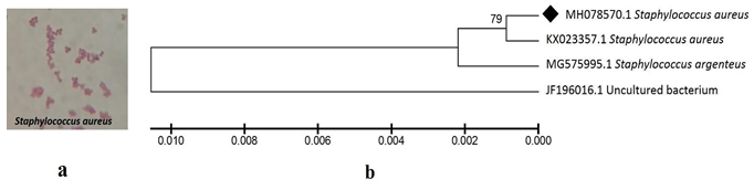
Colonies which had greater than 106 CFU/ml on blood agar was considered as the major infection causing organism. One such major colony was picked. Gram staining results revealed they were gram positive. Colony morphological studies indicated they were round and smooth, found in clusters, shiny, raised and white to golden yellow tinged colonies. Biochemical tests showed positive results towards catalase, methyl red and Voges Proskauer. Negative results were obtained for oxidase, indole and citrate tests. Based on these organism was identified to be Staphylococcus sp. Further 16S rRNA gene amplification result indicated a single band of 1.5 Kb (100 ng intensity). The 16S rRNA sequence of the organism showed 79% identity with S.aureus (KX023357.1) (Figure 1). Based on this it was identified as S.aureus. Further the sequence was submitted to GenBank and MH078570 was the accession number generated.
Antibiotic Sensitivity
Amongst 25 antibiotics tested for sensitivity only 5 antibiotics exhibited sensitivity, 12 antibiotics failed to show any zone of inhibition and thus were resistant, 8 other antibiotics exhibited a mild zone of inhibition but were still considered to be resistant because the zone diameter were under resistant category according to the CLSI guidelines (Table 1 ). Overall, S.aureus showed resistance to 20 antibiotics which indicated that this was a Multi drug resistant organism (Table 1, Figure 2).
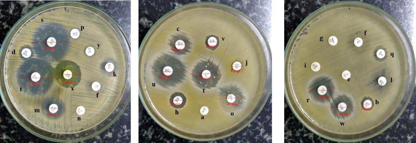
Yuan, et al. have studied the antibiotic sensitivity of S.aureus across 14 antibiotics. Out of these, 9 antibiotics such as by vancomycin tetracycline, erythromycin, rifampicin, oxacillin, norfloxacin, doxycycline, ciprofloxacin, gentamycin which are common to our study have shown similar resistance profiles in the present study [31].
Synthesis and Characterization of AgNPs
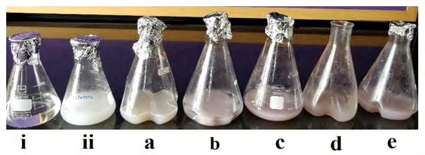
Results of synthesis and characterization of AgNPs from pasteurized cow’s milk at pH 7 has been reported in our previous study [17]. In the present study, color change from white to brown in experimental flasks a, b, c, d and e (Figure 3) indicated AgNPs synthesis across 3,5,7,9 and 11 pH respectively. It was observed that intensity of brown color increased towards basic pH. A similar observation was made by Kredy, et al. in case of AgNPs synthesized from Lawsonia inermis extract across pH 4,7 and 9 [20]. Muthu et al. have reported the synthesis of AgNPs from Cassia auriculata flowers at pH at 3, 4, 5, 6, 7, 8 and 9 and have found that AgNP synthesis occurred rapidly at neutral pH when compared to acidic pH which is in par with the present results [21].
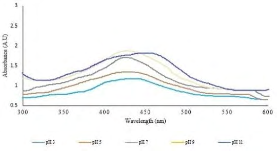
UV-Vis spectroscopy revealed that absorbance increased with increase in pH from 3 to 9 where sharp and narrow peaks were observed at pH 7 and 9. This could be due to the enhanced bioavailability of functional groups in milk at this pH which promoted the synthesis of AgNPs. At low pH, small with broadening SPR band was formed which indicated the formation of larger sized nanoparticles. Decrease in absorbance at pH 11 could due to formation of unstable and agglomerated particles (Figure 4). Absorbance increase could be attributed to the decrease in particle size observed towards basic pH. Smaller particles predominantly absorb light and have sharp peaks near to smaller wavelength, while larger sized particles exhibit increased scattering and have broader peaks towards longer wavelengths. Similar observations were made by Khalil et al. and Vanaja et al. in AgNPs synthesized from olive leaf and Coleus aromaticus leaf extract across various pH respectively [22, 23].
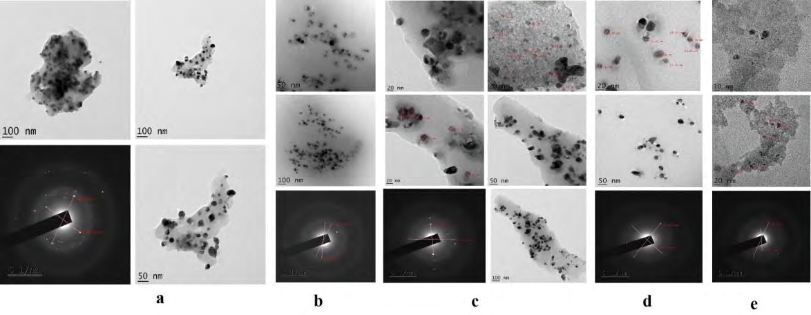
Transmission electron microscopy revealed that change in pH affected the size of the synthesized nanoparticles, as it has the ability to modify the charge present on the organic moieties and biomolecules. Figure 5 clearly indicates that as pH changes from 3 to 11, size of the particles decreased from 100 nm to 10-20 nm which is in par with the observations made in case of the spectral shifts. Similar observations were obtained by Muthu et al. where they have shown that size of AgNps (synthesized from Cassia auriculata flower extract) reduced towards high pH [21]. Another study made by Khalil et al. have also made similar observations of decrease in particle size from pH 3 to pH 8 with regular spherical shape observed in all cases. Their study reports that the alkaline pH environment enhanced the reduction and stabilizing capability of AgNPs [22].
Antibacterial Activity
![Figure 6: a. AgNPs -50 and 75 μl concentration. b. Erythromycin, Ampicillin, Nalidixic acid, Cefazolin and Tetracycline- Represented as Ab1, Ab2, Ab3, Ab4, Ab5 in Set A to Set E under i, ii, iii, iv and v labels respectively [Figure 6]. c. Respective antibiotics + AgNPs (50 and 75 μl). d. Milk + distilled water (negative control).](/fulltextimages/8033/fig_6.png)
Figure 6: Set A, B, C, D and E show representative image for Antibacterial activity of AgNPs across various pH 3,5,7,9 and 11 respectively against MDR S.aureus. Antibiotics (Positive control) : Ab1 (ERY), Ab2(AMP), Ab3(NA), Ab4(CZ), Ab5(TET) in i,ii,iii,iv,v across Set A to E respectively; 1- Negative Control (cow’s milk+D.H2O), 2, a- 50 µl AgNPs, 3,b- 50 µl AgNPs + respective antibiotics, 4,c-75 µl AgNPs, 5,d-75 µl AgNPs + respective antibiotics.
| Treatments | pH 3 | pH 5 | pH 7 | pH 9 | pH 11 |
|---|---|---|---|---|---|
| Control (Milk+D.H20) | 0 | 0 | 0 | 0 | 0 |
| 50 µl AgNPs | 9.36 ± 1.18 | 0 | 9.63 ± 1.48 | 9.51 ± 2.37 | 0 |
| Ab (ERY) | 0 | 0 | 0 | 0 | 0 |
| Ab (AMP) | 7.45 ± 0.31 | 7.58 ± 0.35 | 7.87 ± 0.18 | 7.72 ± 0.36 | 7.58 ± 0.31 |
| Ab (NA) | 0 | 0 | 0 | 0 | 0 |
| Ab (CZ) | 10.86 ± 0.71 | 10.00±0.78 | 11.5 ± 0.82 | 11.50 ± 0.81 | 10.20±0.46 |
| Ab (TE) | 0 | 0 | 0 | 0 | 0 |
| 50 µl AgNPs + ERY | 9.48 ± 0.50 | 0 | 11.48 ± 1.52 | 9.48 ± 0.50 | 0 |
| Fold increase | 1.49 | - | 2.66 | 1.50 | - |
| 50 µl AgNPs + AMP | 9.76 ± 0.32 | 7.12 ± 0.19 | 11.48 ± 1.52 | 10.96 ± 1.00 | 7.42 ± 0.20 |
| Fold increase | 0.71 | - | 1.12 | 1.01 | - |
| 50 µl AgNPs + NA | 9.51 ± 0.57 | 0 | 9.98 ± 0.99 | 9.96 ± 0.12 | 0 |
| Fold increase | 1.51 | - | 1.76 | 1.75 | - |
| 50 µl AgNPs + CZ | 13.20 ± 0.93 | 9.98±1.10 | 13.17 ± 0.37 | 13.91 ± 1.36 | 10.35±1.10 |
| Fold increase | 0.47 | - | 0.41 | 0.46 | - |
| 50 µl AgNPs + TE | 10.28 ± 1.25 | 0 | 12.00 ± 1.21 | 10.54 ± 0.31 | 0 |
| Fold increase | 1.93 | - | 3.00 | 2.08 | - |
| Treatments | pH 3 | pH 5 | pH 7 | pH 9 | pH 11 |
| 75 µl AgNPs | 10.50 ± 0.88 | 0 | 10.89 ± 0.57 | 10.72 ± 0.93 | 0 |
| Ab (ERY) | 0 | 0 | 0 | 0 | 0 |
| Ab (AMP) | 7.45 ± 0.31 | 7.58 ± 0.35 | 7.87 ± 0.18 | 7.72 ± 0.36 | 7.58 ± 0.31 |
| Ab (NA) | 0 | 0 | 0 | 0 | 0 |
| Ab (CZ) | 10.86 ± 0.71 | 10.00±0.78 | 11.5 ± 0.82 | 11.50 ± 0.81 | 10.20±0.46 |
| Ab (TE) | 0 | 0 | 0 | 0 | 0 |
| 75 µl AgNPs + ERY | 10.60 ± 0.40 | 0 | 12.50 ± 0.51 | 10.78 ± 0.53 | 0 |
| Fold increase | 2.12 | - | 3.34 | 2.22 | - |
| 75 µl AgNPs + AMP | 11.01 ± 1.07 | 7.25 ± 0.1 | 11.75 ± 1.79 | 11.76 ± 0.77 | 7.39 ± 0.35 |
| Fold increase | 1.18 | - | 1.22 | 1.32 | - |
| 75 µl AgNPs + NA | 10.53 ± 1.43 | 0 | 11.73 ± 0.32 | 10.51 ± 0.56 | 0 |
| Fold increase | 2.08 | - | 2.82 | 2.06 | - |
| 75 µl AgNPs + CZ | 13.08 ± 1.83 | 10.00±0.64 | 13.77 ± 0.40 | 13.91 ± 0.34 | 10.42±1.00 |
| Fold increase | 0.45 | - | 0.43 | 0.46 | - |
| 75 µl AgNPs + TE | 10.90 ± 1.02 | 0 | 12.38 ± 1.20 | 10.62 ± 0.94 | 0 |
| Fold increase | 2.30 | - | 3.25 | 2.13 | - |
Table 2: Inhibition zones recorded (mm ± S.D) across various pH. Note: All assays were performed in triplicates and standard devi
Table 2 provides details of inhibition zones of AgNPs recorded against MDR S.aureus along with their fold increase values. AgNPs synthesized at pH 3,7 and 9 showed inhibition zones ranging between 9.0-9.60 mm on treatment with 50 µl of AgNPs and 10.5-10.8 mm on treatment with 75 µl of AgNPs respectively against the MDR S. aureus. AgNPs at pH 5 and 11 failed to show any zones. Inhibition zones obtained with 75 µl AgNPs was greater than 50 µl AgNPs by 1-1.3 folds.
Overall it indicated that AgNPs exhibited good antibacterial activity at neutral pH (7) followed by basic pH (9) and acidic pH (3). Results of antibacterial activity of AgNPs at pH 9 is in par with the study reported by Kredy et al. [20]. They have synthesized AgNps from Lawsonia inermis extract and have observed maximum synthesis at pH 9 along with potential antibacterial activity against S. aureus along with several other gram positive and gram negative bacteria. Studies made by Khalil et al 2014 have indicated alkaline pH of 8 has resulted in faster formation of AgNPs from olive leaf extract and have reported good antibacterial activity against S. aureus along with few other gram positive and gram negative bacteria [22].
Results of UV-Vis spectroscopy suggested that AgNPs formed at pH 11 were unstable due to which a fall in absorbance was observed. This could be one of the reason why antibacterial activity was not observed at this pH. The reason for no antibacterial activity at pH 5 could be attributed to the fact that this pH is not favoring the bio availability of the organic moieties present in milk which play a key role in capping and stabilizing the synthesized nanoparticles. These moieties could be greatly present at highly acidic, neutral and highly basic pH. It is also evident from the Table 2 that wherever antibiotics failed to inhibit, AgNPs have shown potential antibacterial activity. Negative control (Milk+D. H20) showed no inhibition zones across various pH.
Amongst the 5 antibiotics tested Erythromycin, Nalidixic acid and Tetracycline were the three antibiotics which displayed no inhibition zones across any pH. Ampicillin exhibited 7.4-7.8 mm inhibition zone across all pH tested. Cefazolin exhibited 10.8-11.5 mm zones across all pH tested. The observed zones with respect to ampicillin and cefazolin were considered resistant according to standard zones provided in CLSI measurements (Table 1). Overall all 5 antibiotics were observed to be resistant across all pH against this MDR.
Synergistic activity of AgNPs (50 and 75 µl) synthesized at all pH with the 5 antibiotics were studies based on the fold increase obtained (Table 2). At pH 5 and 11, since there were no inhibition zones recorded by AgNPs no synergy was recorded with any antibiotic. In case of Ampicillin and Cefazolin zones obtained with AgNPs at pH 5 and 11 were due to the antibiotic alone. AgNPs (50 µl) synthesized at pH 3 showed highest synergy with TE by exhibiting 1.93-fold increase followed by ERY, NA, AMP and CZ with fold increase of 1.49, 1.51, 0.71 and 0.47 respectively. A similar pattern in synergy was obtained on treatment with 75 µl AgNPs with antibiotics where fold increases recorded were 2.30, 2.08, 2.12, 1.18, 0.45 for TET, ERY, NA, AMP and CZ respectively.
AgNPs (50 µl) synthesized at pH 7 with antibiotics displayed maximum synergy with respect to TE with fold increase of 3 followed by ERY, NA, AMP and CZ with 2.66, 1.76,1.12 and 0.41-fold increase respectively. AgNPs 75 µl with antibiotics yielded highest synergy with Erythromycin with 3.34 increase in fold followed by TE, NA, AMP and CZ with 3.25, 2.82, 1.22 and 0.43-fold increase respectively.
pH 9 synthesized AgNPs at 50 µl with antibiotics showed highest synergy with TE with 2.08-fold increase followed by NA, ERY, AMP and CZ with fold increase of 1.75, 1.50, 1.01 and 0.46 respectively. AgNPs at 75 µl with antibiotics yielded highest synergy with ERY by showing 2.22 increase in fold followed by TE, NA, AMP and CZ with 2.13, 2.06, 1.32 and 0.46-fold increases respectively. Similar observations were made by Kredy et al. where they have reported highest antibacterial activity of Lawsonia inermis AgNPs against S.aureus at Ph 9 [20]. Overall it can be observed that AgNPs + Antibiotics displayed greater inhibition zones than antibiotics alone which show the synergistic effect.
The values obtained for the inhibition zones and fold increases recorded at various pH clearly indicates that AgNPs synthesized at pH 7 exhibits highest antibacterial activity followed by AgNPs synthesized at pH 9 and pH 3. Results also indicate that AgNPs at 75 µl exhibited greater inhibition zones and greater synergy with antibiotics compared to AgNPs at 50 µl. Also the highest synergy was observed with TE, ERY and NA at all pH compared to AMP and CZ.
Conclusions/Summary
AgNPs were successfully synthesized and characterized at pH 3,5,7,9 and 11 from cow’s pasteurized milk and the antibacterial activity of these AgNPs were successfully explored against the MDR S.aureus in this study. It was found that AgNPs synthesized at pH 3,7 and 9 exhibited antibacterial activities and showed the synergistic effects with the chosen antibiotics against the MDR. The antibacterial activity observed at pH 9 AgNPs was consistent with earlier reports of Husam et al. and Lakappa et al. [20, 24]. These results obtained were indicative of an ecofriendly, cost effective and an alternate approach to treat the MDR bacteria. Also, the study reveals the importance of pH in exhibiting the antibacterial activity; the smallest nanoparticles were synthesized at alkaline pH compared to the acidic pH. To our knowledge, this is the first case to establish the influence of pH treatments on antibacterial activities of AgNPs from pasteurized cow’s milk against clinically isolated MDR bacteria. However further research involving purification of AgNPs and in vivo studies needs to be called off for it to be considered as a potential therapeutic antibacterial agent against the MDR bacteria.
References
-
Mongkolrattanothai K, Boyle S, Kahana MD, Daum RS (2003) Severe Staphylococcus aureus infections caused by clonally related community-acquired methicillin- susceptible and methicillin-resistant isolates. Clin Infect Dis 37(8): 1050-1058.
-
Gillet Y, Issartel B, Vanhems P (2002) Association between Staphylococcus aureus strains carrying gene for Panton-Valentine leukocidin and highly lethal necrotising pneumonia in young immunocompetent patients. Lancet 359(9308): 753-759.
-
Miller LG, Perdreau Remington F, Rieg G (2005) Necrotizing fasciitis caused by community-associated methicillin-resistant Staphylococcus aureus in Los Angeles. N Engl J Med 352(14): 1445-1453.
-
Cosgrove SE, Sakoulas G, Perencevich EN, Schwaber MJ, Karchmer AW, et al. (2003) Comparison of mortality associated with methicillin-resistant and methicillin- susceptible Staphylococcus aureus bacteremia: a meta- analysis. Clin Infect Dis 36(1): 53-59.
-
Kourtis AP, Hatfield K, Baggs J, Yi Mu, Isaac See, et al. (2019) Vital Signs: Epidemiology and Recent Trends in Methicillin-Resistant and in Methicillin-Susceptible Staphylococcus aureus Bloodstream Infections — United States. MMWR Morb Mortal Wkly Rep 68(9): 214-219.
-
Jose MM, Cesar AA (2016) Mechanisms of Antibiotic Resistance. Microbiol Spectr 4(2): 10.
-
Thill A, Zeyons O, Spalla O, Chauvat F, Rose J, et al. (2006) Cytotoxicity of CeO2 nanoparticles for Escherichia coli. Physicochemical insight of the cytotoxicity mechanism. Environ Sci Technol 40(19): 6151-6156.
-
Anupam R, Onur B, Sudip S, Amit KM, Yilmaz MD (2019) Green synthesis of silver nanoparticles:biomolecule- nanoparticle organizations targeting antimicrobial activity. RSC Advances 9: 2673- 2702.
-
Barros CHN, Fulaz S, Stanisic D, Tasic L (2018) Biogenic Nanosilver against Multidrug- Resistant Bacteria (MDRB). Antibiotics 7(3): 69.
-
Willner I, Baron R, Willner B (2006) Growing metal nanoparticles by enzymes. J Adv Mater 18(9): 1109- 1120.
-
Ashraf S, Chatha MA, Ejaz W, Janjua HA, Hussain I (2014) Lysozyme-coated silver nanoparticles for differentiating bacterial strains on the basis of antibacterial activity. Res Lett 9(1): 565.
-
Hungund BS, Dhulappanavar GR, Ayachit NH (2015) Comparative Evaluation of Antibacterial Activity of Silver Nanoparticles Biosynthesized Using Fruit Juices. J Nanomedice Nanotechnology 6(2): 271.
-
Lee KJ, Park SH, Govarthanan M, Hwang PH, Seo YS, et al. (2013) Synthesis of silver nanoparticles using cow milk and their antifungal activity against phytopathogens. Mater Lett 105: 128-131.
-
Hegazi A, Hamdy Elshazly E, Abdou AM, Abd Allah F, Eman H, et al. (2014) Potential antibacterial properties of silver nanoparticles conjugated with cow and camel milks. Glob Vet 12(6): 745-749.
-
Rajapandiyan K, Shanthi S, Murugan AM, Muthu GA, Ranjit Singh AJA (2011) Azadirachta indica - cow urine extract, a novel controlling agent towards clinically significant Multi Drug Resistant Pathogens. Journal of Applied Pharmaceutical Science 1(10): 107-113.
-
Karthika R, Sevarkordiyone SP (2015) Synthesis and characterization of silver nanoparticles using aqueous extract goat faecal pellets. International journal of Current Science and Research 1(1): 1-7.
-
Athreya AG, Shareef MI, Gopinath SM (2018) Antibacterial Activity of Silver Nanoparticles Isolated from Cow’s Milk, Hen’s Egg White and Lysozyme: A Comparative Study. Arabian Journal for Science and Engineering 44: 6231- 6240.
-
Marambio Jones C, Hoek E (2010) A review of the antibacterial effects of silver nanomaterials and potential implications for human health and the environment. J Nanopart Res 12(5): 1531-1551.
-
Halima R, Archna (2016) A review on green synthesis of silver nanoparticle, characterization and optimization parameters. Int J Res Eng Technol 5(15): 49-52.
-
Kredy HM (2018) The effect of pH, Temperature on the green synthesis and biochemical activities of silver nanoparticles from Lawsonia inermis extract. J Pharm Sci & Res 10(8): 2022-2026.
-
Muthu K, Priya S (2017) Green synthesis, characterization and catalytic activity of silver nanoparticles using Cassia auriculata flower extract separated fraction. Spectrochimica Acta Part A: Molecular and Biomolecular Spectroscopy 179: 66-72.
-
Khalil M, Ismail EH, El Baghdady KZ, Mohamed D (2014) Green synthesis of silver nanoparticles using olive leaf extract and its antibacterial activity. Arabian Journal of Chemistry 7(6): 1131-1139.
-
Vanaja M, Rajeshkumar S, Paulkumar K, Gnanajobitha G, Malarkodi C, et al. (2013) Kinetic study on green synthesis of silver nanoparticles using Coleus aromaticus leaf extract. Advances in Applied Science Research 4(3): 50-55.
-
Anigol LB, Charantimath JS, Gurubasavaraj PM (2017) Effect of Concentration and pH on the Size of Silver Nanoparticles Synthesized by Green Chemistry. Organic & Medicinal Chem IJ 3(5): OMCIJ.MS.ID.555622
-
Habib F, Rind R, Durani N, Bhutto AL, Buriro RS, et al. (2015) Morphological and Cultural Characterization of Staphylococcus aureus Isolated from Different Animal Species. J Appl Environ Biol Sci 5(2): 15-26.
-
Holt JG, Krieg NR, Sneath PHA, Staley JT, Williams ST (1994) Bergey’s Manual of Determinative Bacteriology. Williamsons and Wilkins.
-
Athreya AG, Shareef MI, Gopinath SM (2020) Silver nanoparticles from cow’s milk to combat multi drug resistant gram negative bacteria from clinical isolates- Proceedings of Indian National Academy-Biological Science 90: 863-871.
-
Bauer AW, Kirby WM, Sherris JC, Turck M (1966) Antibiotic susceptibility testing by a standardized single disk method. Am J Clin Pathol 45(4): 493-496.
-
Wayne PA (2017) CLSI. Performance standards of antibiotic disc susceptibility testing, 27th (Edn.). CLSI Supplement M100.
-
Jyoti K, Baunthiyal M, Singh A (2016) Characterization of silver nanoparticles synthesized using Urtica dioica Linn. leaves and their synergistic effects with antibiotics. J Radiat Res Appl Sci 9(3): 217-227.
-
Yuan YG, Ling Peng Q, Gurunathan S (2017) Effects of Silver Nanoparticles on Multiple Drug-Resistant Strains of Staphylococcus aureus and Pseudomonas aeruginosa from Mastitis-Infected Goats: An Alternative Approach for Antimicrobial Therapy. International Journal of Molecular Sciences 18(3): 569.
- Solution-Processed Chiral Perovskites for Biomedical Applications
- Nanotechnology in Health Chemistry and Medicine: Current Challenges and Future Directions
- Human Exposure to Micro- and Nanoplastics: Pathways, Toxicity, and Intervention Strategies
- Exosome Nanomedicine for Cancer Therapy
- Micro and Nanoplastics–Plastisphere, Biotoxicity, Impact on Human Health, and Mitigation Strategies
- Process Validation of Cefixime Powder for Suspension Dosage Form, 50 mL