Veterinary Nanomedicine Delivery System for Mastitis
Inflammatory disease of cow and buffalo is known as Bovine mastitis caused by different type of infectious and non-infectious etiological agents. About 95% of the intramammary infections are mainly caused by Staphylococcus aureus, Streptococcus agalactiae, also by Streptococcus dysgalactiae, Streptococcus uberis and Escherichia coli. Mastitis is global problem, which adversely affects health of animals, quality of milk and economics of milk production. It is the most common cause for the use of antibacterial drugs in lactating dairy cattle and results in presence of above-the-limit residues in antibacterial agent’s marketed milk. Antibacterial therapy of bacterial induced disease has been incriminated for resistance in bacteria; a major drawbacks of utilizing conventional antimicrobial agents. There is need of advanced technology to address the issue. Application of nanotechnology in the field of antibiotics contained in nano-sized carriers for drug delivery has explored along with their effectiveness in the treatment of infectious disease, including antibiotic resistant ones with minimal side effects. This review summarizes need of advanced nanotechnology in treating most economically important disease bovine mastitis. Particularly, antimicrobial nanoparticles, nanocarriers and nanoantibiotics are emerging as new nanomedicine tool to tackle the current or upcoming challenges in infectious disease.
Introduction to Mastitis
Mastitis is the most and more complex disease condition mainly because of multiple causative agents, poorly understanding of the early immune response and complications associated with mammary epithelial cell damage by both the agents and the host factors. Mastitis is defined as inflammation of mammary gland parenchyma caused by microorganism; usually bacteria that process of invade the udder, multiply and produce toxins that are harmful to the mammary gland. Mastitis is identified by physical, chemical and usually bacteriological changes in milk. There is also pathological changes in glandular tissues of the udder and affects the quality and quantity of milk. It can have an infectious or non-infectious etiology [1, 2, 3, 4].
Types of Mastitis
Mastitis can be divided into two categories; clinical and sub-clinical. It is a disease that has a different level of intensity (Table 1) and may be caused by different types of organisms.
| Types of Mastitis | Characteristic Symptoms |
|---|---|
| Acute clinical | Inflammation of the teat, fever above 39°C, weak and dejected animal, lack of appetite. Drastic drop in milk yield. Often follows calving and, less seriously, after cow goes dry. |
| Hyperacute clinical | Swollen, red, painful quarter. Milk passes with difficulty. Fever over 41°C. Cow has no appetite, shivers and loses weight quickly. Lactation often stops. |
| Subacute clinical | No apparent change in udder, presence of flaky particles in milk, especially in initial ejection. Subject appears healthy. |
| Subclinical | No symptoms. 15 to 40 cases for every clinical case. Milk appears normal. Only change is detection of pathogenic agent in analysis and increased somatic cell count. Mostly caused by Staphylococcus aureus. |
| Chronic | Repeated but mild clinical attacks, generally without fever. Lumpy milk, quarters sometimes swollen. Quarter may become hard (fibrous indurations). Antibiotic treatments often do not work. |
| Gangrenous | Affected quarter is blue and cold to the touch. Progressive discoloration from the tip to the top. Necrotic parts drop off. Cow often dies. |
| Contagious | Mastitis caused by bacteria such as Staphylococcus aureus and Streptococcus agalactiae, and other of which other infected cows are the main source. |
| Environmental | Mastitis caused by bacteria such as coliforms (e.g. E. coli) and streptococci (S. uberis, S. disgalactiae, S. bovis), of which the main source is a contaminated environment, i.e. manure. |
Table 1: Different levels of mastitis disease.
Clinical (Visible) Mastitis
• Mild signs flakes or clots in the milk may have slight swelling of infected quarter. • Severe signs secretion abnormal, hot and swollen quarter or udder; cow may have a fever, rapid pulse, loss of appetite, dehydration and depression, death may occur.
Subclinical (Invisible) Mastitis
- Somatic cell count (SCC), of the milk is elevated.
- Bacteriological culturing of milk shows bacteria in the milk.
Somatic Cell Count (SCC) is the Number of Leukocytes or White Blood Cells per Milliliter of Milk
- Normal milk has less than 200,000 cells per milliliter.
- An elevated Somatic Cell (SCC) is an indication of inflammation in the udder.
- Bulk tank SCC gives an indication of the level of a sub- clinical mastitis and the loss of milk production in a herd due to mastitis.
Why Mastitis is Important?
Mastitis is commercially important disease of dairy cattle because it affects 38% of the total direct costs of the common production diseases (reference). Mastitis is a global problem as it adversely affects animal health, quality of milk and economics of milk production. Every country including developed ones suffers huge financial losses due to mastitis. It is the most important complicated and deadly disease of dairy animals. It is responsible for heavy economic losses due to decreased milk yield (up to 70%), also milk discard after treatment (9%), cost of veterinary services (7%) and premature culling (14%) [5, 6]. One of the study report proved that the annual economic losses due to mastitis were increased 114 folds in India in last 4 decades from 1962 (INR 529 million/annum) to 2001 (INR 60532 million/annum) [7, 8]. Subclinical mastitis was found more important in India. Estimates vary from 10-50% of the milk producing cows and 5-20% of the milk producing buffaloes compared to around 1-10% in case of clinical mastitis [9]. Though India is the highest milk producer in the world, the efficiency of the process remains low. Per capita production of milk still remains half of the world average. The estimated loss due to mastitis is nearly INR 16,702 millions per annum in India. Strategic intervention is necessary to fully harness the full potential of milk production in India. The annual economic loss because of mastitis even in developed countries like United States of America is nearly $1.5 to 2.0 billion.
Microorganisms Causing Mastitis
There are large numbers of microorganisms present on and in cow udder as well as on the teat. Several of them are the part of the normal flora and do not cause mastitis; examples include Staphylococcus hyicus, Staphylococcus epidermis and Corynebacterium bovis. They may even protect the udder from infections caused by pathogenic bacteria. Several other microorganisms may cause infection in the mammary gland. There are mainly contagious and environmental microorganisms. Contagious microorganisms are the main source of the infected cows and mainly survive and proliferate on the skin and teat wounds. It consists of Streptococcus agalactiae, Staphylococcus aureus, Streptococcus dysgalactiae and environmental microorganisms like Escherichia coli and other coliforms, Streptococcus uberis (Table 2 & Figure 2). Presence of these microorganisms in soil, bedding, and water indicates high degree of contamination. Nearly 95% of intramammary infections are caused by Streptococcus agalactiae, also Staphylococcus aureus, Streptococcus dysgalactiae, Streptococcus uberis, and Escherichia coli, while remaining 5% are caused by other organisms [10, 11, 12, 13].
| Species | Main Source | Living Conditions | Symptoms | Preventive Treatment |
|---|---|---|---|---|
| Streptococcus agalactiae | Infected cows | Infected quarter and udder only | Mild fever for about 24 hours | Wash udders after milking, reduces problem by 50% Cull infected cows |
| Staphylococcus aureus | Infected cows | On abnormal udder and teat, milkers, vagina, tonsils | Often quite acute for a few days after calving. May be fatal. Quarter swells and turns purple. Quickly affects entire system. In chronic state, udder hardens, aqueous secretion, eventual atrophy of the quarter. Intermediate form produces granular secretion. Milk hotter than normal. | Wash udders after milking, reduces problem by 50% Cull infected cows |
| Streptococcus dysgalactiae | Infected cows | Infected quarter, injuries | Pronounced swelling of one or more quarters. Milk highly abnormal. High fever in serious cases. | Wash udders after milking, reduces problem by 50% Cull infected cows |
| Streptococcus uberis | Contaminated environment | On cow’s skin, mouth, ground | Pronounced swelling of one or more quarters. Milk highly abnormal. High fever in serious cases. Affects mostly dry cows and heifers. | Wash teats only, dry well with disposable paper towels for each cow Supply generous bedding |
| Escherichia coli | Contaminated environment | Ground, bedding (sawdust and shavings), manure, water | Often very serious. May lead to loss of quarter or even death. Thin yellow secretions, with granular texture resembling bran. Often high fever. | Wash teats only, dry well with disposable paper towels for each cow |
| Corynebacterium pyrogene | Certain insects | Humid valleys, wooded areas | Pronounced systematic reaction due to toxins caused by bacteria. Often more than one quarter affected. They become hard, produce thick smelly secretion like cheese and difficult to eliminate. Followed by abscess that bursts, releasing creamy pus, and tissue loss. | Supply generous bedding |
Table 2: Main microorganisms involved in mammary infections, their characteristics and prevention treatment.
Development of Mastitis
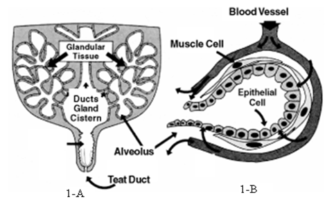
The basic knowledge of mammary gland, anatomy and physiology is necessary to understand the development of mastitis. The mammary gland is made up of a teat cistern, gland cistern, and glandular tissue (Figure 1A). Glandular tissue consists of millions of microscopic sacs known as alveoli (Figure 1B). Each alveolus is lined with milk producing epithelial cells, which is surrounded by muscle cells that contract and squeeze milk from alveolus during milking. Blood vessels transfer nutrients to each alveolus whereas epithelial cells convert nutrients into milk. Milk accumulates in the alveolar spaces, milk ducts and, while accumulated fluid is removed through the teat ducts during milking.
![Figure 2: Structure of _S._ _uberis_, _S._ _agalactiae_, _S._ _dysgalactiae_, _Klebsiella_, _S._ _aureus_, _E._ _coli_. In the development of mastitis, microorganisms enter the quarter and multiply while some microorganisms enter the udder from the other part of body, but this theory is under debate. The steps involved in mastitis infection shown below [14].](/fulltextimages/10557/fig_2.png)
Figure 2: Structure of S. uberis, S. agalactiae, S. dysgalactiae, Klebsiella, S. aureus, E. coli. In the development of mastitis, microorganisms enter the quarter and multiply while some microorganisms enter the udder from the other part of body, but this theory is under debate. The steps involved in mastitis infection shown below [14].
In the first step, there is contact with the microbe and then, the number of microorganisms multiplies near the orifice or sphincter of one or several teats. At this step, hygiene and milking habits play an important role in preventing the microbes from entering the quarter.
In the second step, microbes enter into the teats; this may be inadvertently facilitated by the milking machine, particularly at the end of milking. Structures and conditions like injuries, injured keratin inside teat or teats whose openings are too large may be easily invaded. At this point adjusting milking machines and preventing injuries are critical.
In the later, immune response of the cow develops in which the first line of defense is to send white blood cells i.e. leucocytes to eliminate the microbes that have penetrated the teat. If the response is insufficient then microbes multiply and the cow shows other immune responses such as a fever. The effectiveness of the cow’s immune system depends on the many factors.
Conventional Antibiotics and Antimicrobial Agents
Infectious diseases are the leading cause of death worldwide in twenty first century. The decreases in morbidity and mortality from the infectious diseases over the last century were possible due to an introduction of antimicrobial agents. So resistance to antibiotics has been reaching a critical level, antimicrobial agents in general and antibiotics in particular is still the main reason for low mortality and morbidity both in human beings and animals [15, 16, 17]. Over the last few decades, antibacterial drugs have been used to treat the mastitis. There is a fair degree of concern regarding inappropriate use of antibiotics as mastitis treatment is the most common cause of over-the- legal-limit antibacterial residues in milk available in the market [18]. The use of antibacterial therapy for bacterial induced disease in cattle had been incriminated as a catalyst for resistance in bacteria isolated from treated animals, other animals within the herd, and food derived from cattle for the human use [19]. Bacterial resistance to antibacterial drugs was reported soon after antimicrobial drugs were accepted for use in both human and veterinary medicine. The bacteria, which cause mastitis, are mostly resistant to drugs like streptomycin, penicillin, ampicillin, cloxacillin, amoxicillin and neomycin. Data revealed that 30-fold difference in the mean inhibitory concentration (MIC) values among species of streptococci for the used drug can exist (Table 3) [20, 21]. Approximately three decades ago Jones, et. al observed that S. aureus had very high MIC values for the drugs like penicillin and ampicillin resulting from beta-lactamase inactivation of the drugs. Beta-lactamase is an enzyme that breaks down ‘Beta-Lactam’, a specific heterocyclic ring structure in the penicillin family of antibiotics. Beta-lactamase production is induced in some bacteria, when it was exposed to the Beta- lactam drug. The MIC values (Tables 4 & 5) and disk diffusion results Table 6 shows that S. aureus are most commonly resistant to ampicillin and other penicillin drugs. Attempts have been made to address the issue of bacterial resistance to antimicrobial drugs by discovering new antibiotics and chemically modifying existing ones. But, there is no assurance that the development of new antimicrobial drugs can catch up to the microbial pathogen’s fast and frequent development of resistance in a time. Drug resistant infections caused by both Gram-positive and Gram-negative bacterial pathogens are growing and the continued evolution of antimicrobial resistance threatens health of animals and human beings by seriously compromising our ability to treat serious infections.
| 1990 | 1997 | 2002 | 1998 | 1984 | 1987 | 1993 | |||
|---|---|---|---|---|---|---|---|---|---|
| Ampicillin | --- | --- | 0.5, | -1 | 2 | --- | --- | --- | |
| Ceftiofur | --- | 0.5, | -1 | 2.0, | -0 | 1 | --- | --- | --- |
| Cephalothin* | --- | 0.25, | -0.25 | 1.0, | -1 | 8 | --- | --- | 0.58 |
| Cloxacillin | --- | 4.0, | -0.5 | --- | --- | 0.25 | 0.87 | 0.45 | |
| Erythromycin** | > 256, (4), 2 | 1.0, | -0.25 | 8.0, | -0 | 4 | 0.27 | 0.16 | 0.34 |
| Gentamicin | --- | --- | --- | --- | --- | 3.13 | 3.33 | ||
| Novobiocin | --- | 4.0, | -2 | --- | --- | --- | --- | --- | |
| Oxacillin | --- | --- | 1.0, | -1 | 16 | --- | --- | --- | |
| Penicillin | < 1, (< 1), <1 | .125, | < (0.06) | 0.25, | -1 | 2 | 0.07 | 0.25 | 0.11 |
| Pirlimycin | --- | --- | 8.0, | -0 | 4 | --- | --- | --- | |
| Tetracycline | 32, (8), 0 | --- | 16.0, | -16 | --- | ||||
| Chloramphenicol | --- | --- | --- | --- | 3.27 | 2.5 | 3.3 |
Table 3: Summary of studies using MIC data for Streptococci MIC90 φ.
| 1997 | 2000 | 1967 | 1983 | 2000 | 1986 | 1997 | 1997 | 1984 | 1987 | 1993 | |
|---|---|---|---|---|---|---|---|---|---|---|---|
| Ampicillin | --- | --- | --- | 0.5 | 1 | --- | 4 | 0.5 | --- | --- | --- |
| Ceftiofur | 2 | --- | --- | --- | 1 | --- | 1 | 1 | --- | --- | --- |
| Cephalothin* | 0.25 | 0.75 | --- | --- | 1 | --- | 0.5 | 0.25 | --- | --- | 1.21 |
| Cloxacillin | 0.5 | --- | 0.07 | --- | 0.5 | --- | --- | 0.25 | 0.4 | 0.22 | |
| Erythromycin** | 0.5 | 0.75 | --- | 0.5 | 0.5 | 0.5 | 64 | 0.5 | 3.28 | 3.46 | 3.21 |
| Neomycin | --- | --- | --- | 8 | 2 | 1 | --- | --- | 0.41 | 0.55 | 0.39 |
| Novobiocin | 0.5 | --- | 1.25 | --- | 0.5 | --- | --- | --- | --- | --- | |
| Oxacillin | --- | 0.5 | --- | 0.25 | 1 | --- | 1 | 0.25 | --- | --- | --- |
| Penicillin | 0.13 | 1.5 | > 100 | 0.25 | 0.5 | -- | 16 | 0.25 | (47.8%)# | (33.3%)# | (10.1%)# |
| Pirlimycin | 1 | --- | --- | --- | 1 | --- | 64 | 0.5 | --- | --- | --- |
| Streptomycin | --- | --- | 5 | 8 | 4 | --- | --- | --- | --- | --- | |
| Tetracycline | --- | --- | 0.6 | 2 | 0.5 | --- | --- | --- | --- | --- |
Table 4: Summary of MIC data for S. aureus from dairy cows MIC90 (µg/ml).
| 1998 Denmark | 1998 New Zealand | 1995 | |
|---|---|---|---|
| Ampicillin | 0.5 | --- | --- |
| Ceftiofur | 1 | 2 | 1 |
| Cephalothin | 0.5 | 0.5 | 0.5 |
| Cloxacillin | --- | 0.5 | 0.5 |
| Erythromycin | 0.5 | 0.5 | 0.5 |
| Novobiocin | --- | 1 | 0.5 |
| Oxacillin | 0.5 | --- | --- |
| Penicillin | --- | --- | 0.14 |
| Pirlimycin | 0.5 | 1 | 0.5 |
| Enrofloxacin | --- | 0.25 | 0.5 |
| Tetracvcline | --- | --- | 0.6 |
Table 5: Summary of MIC data for S. aureus from heifers MIC90 (µg/ml).
| 1988 | 1986 | 1971 | 1982- 83 | 1996 | 2000 | 2000 | 1988 | 1995 | 1994- 2001 | 1994- 2000 | |
|---|---|---|---|---|---|---|---|---|---|---|---|
| Ampicillin | 7 | 54 | 83 | --- | --- | --- | 34.9 | 49.6 | |||
| Ceftiofur | --- | --- | --- | --- | --- | --- | --- | 0.2 | |||
| Cephalothin* | 0 | 1 | 3.5** | 0** | 0** | 0 | 0 | 0.3 | 2 | 0.1 | 0.2 |
| Cloxacillin | --- | 20 | --- | --- | --- | --- | 6.8 | --- | |||
| Erythromycin | 0.3 | 6 | 3*** | 9*** | 12*** | 17 | 11.6 | 4.1 | 2.6 | 14.9 | |
| Gentamicin | 0 | 1 | --- | 3.4 | --- | --- | --- | 1.1 | |||
| Novobiocin | 0 | 7 | 0 | 3 | 1 | --- | --- | --- | --- | 21.8 | --- |
| Oxacillin | 0 | --- | 42 | 0 | 1.5 | 0 | --- | 0.6 | |||
| Penicillin | 7 | 57 | 38** | 75** | 51** | 75 | 40 | 31.8 | 50.7 | 32.6 | 40.6 |
| Pirlimycin | --- | --- | --- | 7.7 | --- | 13.6 | 2.1 | ||||
| Streptomycin | 6 | 36 | 26 | --- | --- | --- | --- | --- | --- | --- | --- |
| Tetracycline | 0 | 8 | 21 | 8 | 9 | 17 | --- | 7 | 11.7 | 22.6 | 8.5 |
| * Or cephapirin |
Table 6: Summary of studies using disk diffusion for S. aureus % of Isolates Resistant.
Need of Nanotechnology in Medicine
Nanotechnology is the area of science and technology which deals with developing and producing extremely small structures, platforms, tools and devices by controlling the arrangement of individual molecules in the overall particle structure. Such structures often called nanoparticles has been used in a wide variety of applications including material science, electronics, solar batteries, cosmetics, medical diagnostics, imaging and delivery of drugs to the target tissue. Nano-medicine i.e. the use of nanotechnology in treatment is focused to the monitor and potentially repair, reconstruct and control of human biological systems at the molecular/cellular level, using engineered nano devices and nanostructures.
Nanoantibiotics
Nanomaterials, which either exhibit antimicrobial activity by themselves or elevate the effectiveness and safety of antibiotics administration, are known as “nanoantibiotics” and their capability of controlling infections in vitro and in vivo has been explored and demonstrated in last few years [22, 23, 24].
Antimicrobial nanomaterials products to which microbial pathogens may not be able to develop resistance as well novel nanosized platforms, are good candidates for antibiotics delivery. For example, it has been suggested in recent studies that some metal nanoconstructs such as silver nanoparticles possess antimicrobial activities, which could be utilized in controlling infectious diseases [24, 25]. Antimicrobial nanoparticles (NPs) offer many distinctive advantages over the conventional antibiotics such as reduction of acute toxicity, reduction of drug resistance, cost effectiveness [26, 27]. Different types of nanosized drug carriers are available to effectively administer antibiotics by enhancing pharmacokinetics and accumulation, while reducing the adverse effects of antibiotics. Nanoparticles are retained much longer in the body when injected outside the systemic circulation than small molecule antibiotics, so the sustained therapeutic effects can be achieved easily. The safety profiles of nanoparticles and nanosized antibiotics drug carriers, particularly upon long-term exposure, safety factor and therapeutic effects must be considered [28]. Antibiotics delivery using nanomaterials offers certain advantages such as [29, 30, 31]: 1) Controllable and relatively uniformly distribution in the target tissue, 2) Improved drug loading, 3) Sustained and controlled release, 4) Improved patient-compliance due to sustained release, 5) Minimized side effects due to better targeting, and 6) Enhanced cellular internalization.
Impact of Nanomedicine to Control of the Infectious Diseases
Nanotechnology is increasingly being investigated and used in immunization and delivering antimicrobial drugs in particular in overcoming antibiotics-resistant pathogens and in imaging. Nanomedicine has been investigated as a promising alternative to the conventional delivery systems of antibiotic [32] Allaker. This section introduces application of nanotechnology and various challenges in controlling infectious diseases, diagnosis of bacterial resistance, delivery of antimicrobial agents, and vaccination in the area of veterinary medicine.
Nanotechnology-Assisted Detection of Antimicrobial Infection and Resistance: Bacteria show the high sensitivity and reproducibility but still conventional diagnostic methods for a microbial infection require sample preparation and long readout time [33]. Advanced nanomaterials with unique electrical, magnetic, luminescent, and catalytic properties should be potentially enable fast, sensitive and cost-effective diagnosis as well as fast and rapid determination of the susceptibility and resistance of anti-bacterial drugs in short time [34, 35].
Antibody conjugated nanoparticles have been shown to trigger the signals for the bioanalysis and enumeration of highly pathogenic bacterias like E. coli O157:H7, which results in highly selective, convenient, and rapid detection of single bacterium in 20 minute in laboratory [36].
It was demonstrated that nanoparticles with specific Raman spectroscopic fingerprints could differentiate antibiotic resistant bacteria, such as MRSA (Methicillin- Resistant S. aureus, MRSA) from non-resistant strains in microarray based systems by detecting single-nucleotide polymorphisms [37]. The magnetic nanoparticle can be used as very sensitive and efficient tools in the detection of microbial infections. Super-magnetic iron oxide nanoprobes have been used in the identification of Mycobacterium avium spp. paratuberculosis (MAP) and the quick quantification of MAP with high sensitivity in milk and blood [38]. In addition, recent studies in nanotechnology have be demonstrated the feasibility of achieving fast and reliable pharmaceutical and biopharmaceutical assays for microbial infections without any sample preparations in opaque media like blood and milk [39, 40].
Role of Nanotechnology in Antimicrobial Action and Treatment of Infectious Diseases: Nanoparticles made up of metal and metal oxide produce reactive oxygen species (ROS) under UV light. This property could potentially be used in antimicrobial formulations, preservative concoctions and dressings. Furthermore, nanosized silver, zinc and their metal oxide particles have been reported to be effective in inactivating different microorganisms [41, 42].
Antibiotics prepared with polymeric nanoparticles have demonstrated enhanced antimicrobial activities as well as anti-MRSA activities when compared with non polymerized forms of N-methylthio β-lactams and penicillin [43]. Gold (Au) nanoparticles of Vancomycin have also exhibited enhanced antimicrobial activities against VRE (Vancomycin Resistant E. Coli) strains and E. coli strains [44]. Recently selective killing of target bacteria by irradiating Au nanoparticle- attached bacterial surface with a laser has been successfully demonstrated. In this technology, effective and irreparable damage occurs to the bacterium bound with Au nanoparticles of different sizes conjugated with antiprotein A antibodies [45]. Different type of antimicrobial agents can be effectively co-administered using various nanoparticles. A wide range of lipophilic and hydrophilic antibiotics can be conjugated inside, outside or on the surface of polymeric nanoparticles or nano-capsules. Major pharmacokinetic characteristics of antibiotics, including improved solubility, controlled release, sustained release and specific site-targeted delivery, can be effectively achieved by employing appropriate nanocarriers or nanotechnology [23].
Use of Nanotechnology in Vaccination and Prevention of Infectious Diseases: Application of nanoparticles as novel therapeutics, adjuvants and colloidal vaccine carriers for immunization has been investigated. Particulate systems exhibit unique similarity in sizes with bacteria and viruses. The micro and nanoparticles are relatively the same sizes as of bacteria and viruses that the immune system recognizes easily [46]. Various particulate vaccine carrier properties like size, chemical composition, charge, and surface characteristics and also be tuned for enhanced uptake by mononuclear phagocytic system (MPS) for immune presentation of antigens, and stimulation of antigen presenting cells (APs) [47, 48]. For example, amphipathically coated nanoparticles smoothly deliver antigens to dendritic cells (DCs) that simultaneously organize cellular and humoral immunity [49]. In-vivo studies explored mixtures of nanoemulsions with either whole viruses like influenza a virus or proteins, (e.g., recombinant Bacillus anthracis [B. anthracis] protective antigens) as a potential vaccines [50, 51]. Such vaccine does not require cold storage and can be administered via mucosal routes, which would be particularly suitable for vaccination in many developing countries.
Nanomaterials used in Preparation of Antimicrobial Dosage Forms
Antibacterial nanoparticles or nanocarriers include metals, metal oxides and naturally occurring antibacterial substances, different types of polymers, lipids, surfactant- based nanoemulsions and carbon-based nanomaterials. Unique physical and chemical properties particularly large surface area to volume ratios of nanoparticles are believed to contribute to enhanced antimicrobial effect. A new study had been demonstrated that naturally occurring bacteria should not develop antimicrobial resistance to metal nanoparticle. In addition to treating microorganism with nanoparticles, the particles can themselves be prepared by bacterial cells or enzymes. In recent studies, intracellular or extracellular synthesis of metallic nanoparticles e.g. cadmium sulfide [CdS], gold [Au], and silver [Ag] using microbial cells or enzymes has been investigated as novel biosynthetic nanoparticles [52, 53, 54, 55].
The antimicrobial nanoparticles act through various mechanisms [56, 57] as shown in Figure 3:
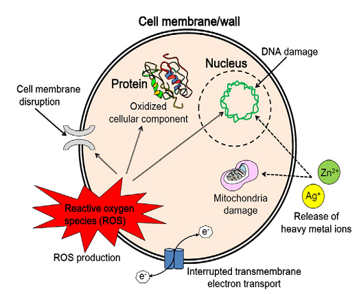
1) Photocatalytic production of reactive oxygen species (ROS) which damage the cellular and viral components. 2) Compromising the bacterial cell wall and cell membrane. 3) Interruption of energy transduction. 4) Inhibition of enzyme activity and DNA synthesis.
- Silver (Ag) NPs: Among the various types of metallic and metal oxide nanoparticles, Silver (Ag) nanoparticles have confirmed as the most effective against bacteria, viruses, and many other eukaryotic microorganisms [58, 59, 60]. Silver (Ag) nanoparticles access the respiratory chain and cell division that finally lead to cell death, while simultaneously releasing silver ions that increase bactericidal activity [61]. The antimicrobial activity of silver (Ag) nanoparticles is inversely proportional to size of the nanoparticles [62, 63]. Concomitant use of silver (Ag) nanoparticles with antibiotics, e.g. penicillin G, amoxicillin, erythromycin, and vancomycin, resulted in improved and synergistic antimicrobial effects against Gram-positive and Gram-negative bacteria. e.g., E. coli and S. aureus [64, 65].
- Zinc Oxide (ZnO) NPs: Some nanoparticles made of metal oxides such as Zinc oxide are stable under rough processing conditions and have targeted toxicity to bacteria with less effect on human or animal cells. Zinc oxide nanoparticles (ZnO NPs) are relatively nontoxic and safe to humans and biocompatible; they are being investigated as drug carriers and have antibacterial activity against important food borne pathogens, e.g. enterotoxigenic E. coli and E. coli O157:H7 [66, 67]. It has several advantages over silver nanoparticles (Ag NPs) including low production cost, UV-blocking properties, and a white appearance resulting in light reflection properties useful in sunscreen applications [68]. The nanoparticle of ZnO multilayer deposited on cotton fabrics exhibited excellent antibacterial activity against S. aureus [69]. It is also believed that nanoparticles of ZnO destruct lipids and proteins of the bacterial cell membrane, resulting in a leakage of intracellular contents and eventual death of bacterial cells. Furthermore, generation of hydrogen peroxide and Zn+2 ions are proposed as major contributing mechanisms antibacterial activity of zinc oxide nanoparticles [70]. Polyvinyl alcohol (PVA)-coated zinc oxide nanoparticles have shown increased membrane permeability, cellular internalization, and intracellular structural changes.
- Titanium Dioxide (TiO2) NPs: Titanium dioxide nanoparticles are the most studied for photocatalytic antimicrobial activity among all the metal/metal oxide/ sulphide nanoparticles [71]. TiO2 nanoparticles have strong antibacterial activity upon receiving irradiation with near-UV and UV-A. The concentration required to kill bacteria varies in the range of 100-1000 ppm of nanomaterial. The potency of antimicrobial effect also depends on the size of the TiO2 nanoparticles as well as intensity and wavelength of the light source. A recent study reported the antibacterial efficiency of TiO2 nanoparticles are in the decreasing order of E. coli > P. aeruginosa > S. aureus > E. faecium > C. albicans. The antibacterial activity is apparently determined by the complexity and the density of the cell membrane or cell wall. In addition it was also reported that the photocatalytic antimicrobial efficiency of TiO2 nanoparticles were in the decreasing order as described virus > bacterial wall > bacterial spore, mainly depending on the thickness of microbial membrane/ sheath structure [72]. The photocatalytic antibacterial activity of TiO2 is mainly related to the production of ROS like free hydroxyl radicals and peroxide [73]. Photocatalysis of TiO2 generates hydroxyl radicals that are very potent oxidants with a broad reactivity. The microbial surface is the primary target of the initial oxidative attack. Damaged membrane structure affects many important biological functions like selective permeability of the membrane, respiration, and oxidative phosphorylation. Irradiation independent bacterial death also shows other unknown nonphotocatalytic antimicrobial pathway of cell death by TiO2 nanoparticles [74]. Ag/(C, S)-TiO2 nanoparticles indicated to have strong and light-independent antimicrobial activities as well against both E. coli and B. subtilis spores, probably because of the synergistic effect of bactericidal activity of Ag and TiO2 [75].
• Gold (Au) NPs: A variety of nanoparticles including near infrared light absorbing gold (Au) nanoparticles, nanoshells, nanorods, nanocages have been applied to treat bacterial infection by irradiation with suitable wavelengths [76]. Antimicrobial activity of gold nanoparticles (Au NPs) is mainly mediated by strong electrostatic attractions to the negatively charged bilayer of cell membrane [77]. Gold nanoparticles (Au NPs) conjugated with antimicrobial agents and antibodies have been investigated to determine selective antimicrobial effects, for example, selective killing of S. aureus by gold nanoparticles (Au NPs) conjugated with anti-protein A antibodies, which target the bacterial surface [78]. Strong laser-induced hyperthermic effects accompanied by bubble-formation around clustered gold nanoparticles (Au NPs) have been observed and the process effectively damaged bacteria. Many studies have noted strong antimicrobial agents effects against Gram-positive and Gram-negative bacteria and also including antibiotic-resistant strains, by gold/ drug nanocomposites, e.g., Gold nanoparticles coated with antibiotics such as streptomycin, neomycin, and gentamicin [79]. In a new study, chitosan-capped gold nanoparticles (Au NPs) coupled with ampicillin exhibited a 2-fold increase in antimicrobial activity when compared with that of free ampicillin. Therefore, gold nanoparticles (Au NPs) are promising and effective delivery platform for antibiotics therapy in treating serious bacterial infections at a lower antibiotics dosage and with fewer side effects. Several other antimicrobial nanomaterials with their mechanism of action are given in Table 7.
| Nanomaterial | Antimicrobial mechanism |
|---|---|
| Ag NPs | Release of Ag+ ions; disruption of cell membrane and electron transport; DNA damage |
| ZnO NPs | Intracellular accumulation of NPs; cell membrane damage; H O production; release of Zn2+ ions 2 2 |
| TiO NPs 2 | Production of ROS; cell membrane and wall damage Antibacterial agent; |
| Au NPs | Interaction with cell membranes; strong electrostatic attraction |
| Chitosan | Increased permeability and rupture of membrane; chelation of trace metals; enzyme inactivation |
| Fullerenes | Destruction of cell membrane integrity; enhancing activity of infiltrating neutrophil |
| CNTs | Cell membrane damage by ROS; oxidation of cell membrane proteins and lipids |
| NO-releasing NPs | NO release and production of ROS |
| Nanoemulsions | Membrane disruption; disruption of the spore coat |
Table 7: Nanomaterial and their antimicrobial mechanism.
Nanoparticle as Efficient Antimicrobial Drug Delivery: • Liposomes: Liposomes are nano to micro sized vesicles consist of a phospholipid bilayer with an aqueous core. After the Doxil approved as first liposomal drug by the Food and Drug Administration in 1995, liposomes have been studied as promising clinically acceptable delivery carrier of enzymes, proteins and drugs [80, 81]. Liposomes are one of the most widely used antimicrobial drug delivery vehicles, because of vehicles lipid bilayer structure mimics the cell membrane and can easily fuse with infectious microbes [82]. Furthermore, both hydrophilic and lipophilic antimicrobial drugs can be encapsulated and retained in aqueous core and in the phospholipids bilayer, respectively, without chemical modifications. For the application of liposomes as antimicrobial drug delivery platforms, a number of parameters such as the physico-chemical properties of lipids, drugs to be loaded, particle size of liposomes and polydispersity, surface charge (zeta-potential), stability in storage (shelf-life), and reproducibility and feasibility in large scale production should be considered [83]. Liposomes, following conjugation with stealth materials like e.g., polyethylene glycol, PEG on the surface of liposomes, resulted in enhanced in vivo stability. The platform can also be used for targeted delivery of antimicrobial drugs when tethered with various targeting ligands such as antibody, antibody segments, aptamers, peptides and small molecule [84, 85]. In one of the studies it was reported that, benzyl penicillin-encapsulating cationic liposomes completely inhibited growth of S. aureus strain at lower drug concentrations for shorter exposure times when compared with free drugs [86]. Liposomal amikacin explored altered distribution in tissue and significantly extended half-life (blood 24.5 h, tissue 63–465 h) [87]. Liposomal gentamicin and ceftazidime exhibited prolonged blood circulation and enhanced localization at the site of infection [88]. Vancomycin and teicoplanin encapsulated liposomes resulted in significantly enhanced elimination of intracellular MRSA infection [89]. Liposomal carriers for antimicrobial drug delivery are shown in Table 8.
| Encapsulated Antibiotics | Target microorganism | Mechanism for improved therapeutic effects |
|---|---|---|
| Streptomycin | Mycobacterium avium | Increased antimicrobial activity by drug encapsulation; targeted delivery to the site of bacterial multiplication |
| Ciprofloxacin | Salmonella dubli | Decreased mortality of animals; distribution of liposomes to all areas of infection |
| Vancomycin or Teicoplanin | MRSA | Enhanced drug uptake by macrophages; enhanced intracellular antimicrobial effect |
| Ampicillin | Micrococcus luteus and Salmonella typhimurium | Increased stability; activity against |
| Amikacin | Gram-negative bacteria | Prolonged drug residence in tissue and plasma; reduced renal clearance and excretion |
| Gentamicin | Klebsiella pneumoniae | Increased survival rate of animal models; increased therapeutic efficacy |
| Polymyxin B | Pseudomonas aeruginosa | Decreased bacterial colony count in lung; decreased lung injury caused by bacteria; increased bioavailability |
| Benzyl penicillin | Staphylococcus aureus | Lower drug concentrations and shorter time of exposure |
Table 8: Liposome nanocarriers for antimicrobial drug delivery.
• Solid Lipid (SL) NPs: Solid lipid nanoparticles offer the best of traditional solid nanoparticles and liposomes while avoiding their disadvantages [90, 91]. SLNPs explored improved bioavailability and targeted delivery of antimicrobial drugs via parenteral, topical, ocular, pulmonary, and oral administration routes [92, 93, 94]. When SLNPs applied onto the skin, it tend to adhere to the surface and form a dense hydrophobic film that is occlusive and have a long residence time on the stratum cornea [95, 96]. Furthermore, enhanced transdermal diffusion of water-insoluble azole antifungal drugs such as clotrimazole, miconazole, econazole, oxiconazole, and ticonazole was noted when they were applied in the encapsulating form in solid lipid nanoparticles [97, 98]. The principle can be utilized in various formulations for oral administration and can also be used as antimicrobial drug delivery [99]. SLNPs can also be used for prolonged ciprofloxacin release, particularly in ocular and skin infections when used topically [100]. SLNPs investigated for antimicrobial drug delivery shown in Table 9.
| Encapsulated Antibiotics | Target microorganism | Mechanism for improved therapeutic effects |
|---|---|---|
| Tobramycin | Pseudomonas aeruginosa | Increased drug bioavailability |
| Ketoconazole | Fungi | Prolonged drug release; high physical stability |
| Rifampicin, isoniazid, Pyrazinamide | Mycobacterium tuberculosis | Increased drug bioavailability and residence time; decreased administration frequency |
| Econazole nitrate | Fungi | Controlled drug release profile; high encapsulation efficiency; enhanced drug penetration through stratum corneum |
| Ciprofloxacin Hydrochloride | Gram-negative bacteria, Gram- positive bacteria, and mycoplasma | Prolonged drug release |
Table 9: Solid Lipid Nanoparticle for antimicrobial drug delivery.
One of the reasons why traditional antibiotics have low effectiveness in bovine mastitis despite in-vitro susceptibility of the bacteria is the low intra-cellular uptake of the drug especially to the udder phagocytes [101]. Delivery of antibiotics and other antibacterial agents through nanoparticles and liposomes can actually help in cellular uptake of the drug as well as protect the drug from bovine physiological microenvironment. Antibiotics have been incorporated in polymeric nanoparticles as well as liposomes and drug-loaded delivery systems have been proved to enhance the intra-cellular delivery of antibiotics [102, 103]. Incorporating antibiotics from different families is possible in nanoparticulate formulations [104] while protecting the drugs from degradation and prolonging the release. All these characteristics make the use of nanoparticles in the treatment of bovine mastitis extremely appropriate. Metal and polymeric and solid lipid nanoparticles have been shown to be effective in detection [105, 106] and treatment of bacterial infection of bovine mastitis [107]. Advantages of Nanoantibiotics
1) Nanocarriers and nanovehicles can be formulated to be stimuli-responsive e.g., chemical, magnetic field, heat, and pH for targeted drug delivery and biological sensors [108, 109]. For example, freeze-dried amoxicillin in formulation with chitosan and polyvinyl pyrrolidone for acid-responsive release of antibiotics [110] is particularly useful for treating abscess which are frequently acidic and lowers the effect of conventional antimicrobial therapy. 2) Nanoparticles can be molecularly modified to possess versatile physico-chemical properties in order to minimize side effects. The systemic administration of traditional antimicrobial agents often produces side effects such as hepatotoxicity of cephalosporins, ototoxicity and nephrotoxicity of aminoglycosides. Nanocarriers can reduce these side effects by reducing the dose required and localizing the drug in the carrier depots [111, 112, 113]. 3) Nanoparticulate based antimicrobial drug delivery is promising in overcoming resistance to traditional antibiotics developed by different types of bacteria. 4) Administration of antimicrobial agents using nanoparticles can enhance therapeutic index, prolong drug circulation i.e., extended half-life, and achieve controlled drug release, improving the overall pharmacokinetics. Different studies explored greater efficacy of antimicrobial nanoparticles than the use of antibiotics alone. For example, vancomycin-capped Au NPs exhibited 64-fold enhanced efficacy against VRE strains and Gram-negative bacteria. E.g. E. coli over vancomycin alone. 5) Antimicrobial nanoparticles can be developed and administered in convenient and cost-effective ways by different routes with lowered administration frequency. 6) Nanoparticle-based antimicrobial drug delivery can achieve enhanced solubility and suspension of drugs and concomitant delivery of multiple agents for synergistic antimicrobial effects. Products available in the market for the treatment of bovine mastitis shown in Table 10 & Table 11.
| Intramammary products | Company | Company Active ingredients per tube |
|---|---|---|
| Ampiclox LC | Jurox Pty Ltd | Ampicillin 75mg, cloxacillin 200mg |
| Cepravin LC | Intervet/Schering-Plough Animal Health | Cefuroxime 250mg |
| Clavet LC | Norbrook Laboratories Aust P/L | Clavulanic acid 50mg, amoxycillin |
| Lactaclox LC | Norbrook Laboratories Aust P/L | Ampicillin 75mg, cloxacillin 200mg |
| Mastalone Blue | Pfizer Animal Health | Oxytetracycline 185mg, oleandomycin 100mg, neomycin 100mg |
| Maxalac LC | Jurox Pty Ltd | Cefuroxime 250mg |
| Noroclox LC | Norbrook Laboratories Aust P/L | Cloxacillin 200mg |
| Orbenin LC | Pfizer Animal Health | Cloxacillin 200mg |
| Intramuscular products | Company | Active ingredients |
| Mamyzin | Boehringer Ingelheim Pty Ltd | Penethamate hydriodide 5g |
| Penethaject | Bomac Animal Health Pty Ltd | Penethamate hydriodide 5g |
Table 10: Products available in the market used in the treatment of Mastitis:
| Antibiotic product | Company | Active ingredient per tube |
|---|---|---|
| Ampiclox Dry Cow | Jurox Pty Ltd | Ampicillin 250mg, cloxacillin 500mg |
| Bovaclox DC LA | Norbrook Laboratories Aust P/L | Ampicillin 300mg, cloxacillin 600mg |
| Cepravin Dry Cow | Intervet/Schering-Plough Animal Health | Cephalonium 250mg |
| Elaclox DCX | Norbrook Laboratories Aust P/L | Cloxacillin 600mg |
| Juraclox LA 600 | Dry Cow Jurox Pty Ltd | Cloxacillin 600mg |
| Noroclox 500 | Norbrook Laboratories Aust P/L | Cloxacillin 500mg |
| Orbenin Enduro | Pfizer Animal Health | Cloxacillin 600mg |
| Non-antibiotic product | ||
| Teatseal | Pfizer Animal Health | Bismuth subnitrate 2.6g |
Table 11: Antibiotic products available in the market:
Challenges and Future Opportunities
Socio-economics challenges, Management challenges, Milking Equipment challenges, Environment challenges, Pathogens challenges, Diagnostics challenges, Therapy challenges, Immunology challenges.
Opportunities like antibiotics development, vaccines development, bacteriocins, herbal therapy, immunotherapy, and nanoparticle technology development [114, 115, 116, 117, 118, 119, 120].
Conclusion
Increasing rate of mastitis in domestic animal’s like cows and buffaloes is a matter of broad discussion. More strategic research is required in this field to control the mastitis. Bovine mastitis is still big challenge to the field of veterinarians and mastitis researchers. The emergence of resistance to antibiotics acquired from and by microbial variants is a serious threat in the process of treatment against infectious diseases like mastitis. The development of antibiotic resistance is a universal challenge that is being addressed by pharmaceutical companies and academic researchers alike. The unique physicochemical properties of various nanomaterials have the promise of making existing antimicrobial agents more effective and offer new types of antibacterial agents. Overcoming antimicrobial resistance by utilizing properties of nanoparticles as antibiotic carriers seems to hold great promise. Various nanoparticles have been explored as efficient antibiotics delivery vehicles that also protect antimicrobial drugs from a resistant mechanism. Most importantly, nanoparticles enable combining multiple independent and potentially synergistic approaches on the same platform, in order to increase the antimicrobial activity and overcome resistance to antibiotics [120, 121, 122, 123, 124].
To develop efficient, safe, cost-effective, and targeted therapy for bovine mastitis in an era of developing antibiotics resistance requires interdisciplinary knowledge and innovative tools of microbiology, immunology, biomaterials, polymers, and nanotechnology.
Conflict of interest statement
None of the authors of this paper has a financial or personal relationship with other people or organizations that could inappropriately influence or bias the content of the paper.
References
-
Sharma N, Gautam A, Upadhyay SR (2006) Role of antioxidants in udder health: A review. Ind J Field Vet 2: 73-76.
-
Sharma N, Srivastava AKT, Bacic G, Jeong DK, Sharma RK (2012) Epidemiology. In: Bovine Mastitis. Satish Serial Publishing House India, pp: 231-312.
-
Bradley AJ (2002) Bovine mastitis: An evolving disease. Vet J 164(2): 116-128.
-
Radostits OM, Gay CC, Blood DC, Hinchkliff KW (2000) A textbook of the Veterinary medicine. WB Saunders USA, pp: 1877.
-
Sharma H, Maiti SK, Sharma KK (2007) Prevalence, etiology and antibiogram of microorganisms associated with sub-clinical mastitis in buffaloes in drug, Chhattisgarh state. Int J Dairy Sci 2: 145-151.
-
Kossaibati MA, Esslemont RJ (1997) The costs of production diseases in dairy herds in England. Vet J 154(1): 41-51.
-
Dhanda MR, Sethi MS (1962) Investigation of Mastitis in India. Icar Res Series No 35 India.
-
Dua K (2001) Incidence, etiology and estimated economic losses due to mastitis in Punjab and in India- An update. Indian Dairyman 53: 41-48.
-
Yathiraj S, Bhat MN, Deepti BR, Upendra HU, Murlidhara A (2007) Pattern of bacterial isolates from mastitis cases in cows. ISVM India, 26.
-
Kader MA, Samad MA, Saha S, Taleb MA (2002) Prevalence and etiology of sub clinical mastitis with antibiotic sensitivity to isolated organisms among milch cows in Bangladesh. Ind J Dairy Sci 55: 218-223.
-
Sudhan NA, Singh R, Singh M, Soodan JS (2005) Studies on prevalence, etiology and diagnosis of sub clinical mastitis among cross bred cows. Indian J Anim Res 39: 127-130.
-
Chahar A, Gahlot AK, Tanwar RK, Fakhruddin (2008) Evaluation of different screening tests for diagnosis of sub clinical mastitis in cattle. Indian J Vet Med 28(2): 91- 93.
-
Kumar M, Goel P, Sharma A, Kumar A (2009) Prevalence of sub clinical mastitis in cows at a Goshala. India, pp: 4.
-
Sharma N, Vohra V (2001) An update on bovine mastitis in India. India, pp: 20-24.
-
Cohen ML (2000) Changing patterns of infectious disease. Nature 406: 762-767.
-
Gold HS, Moellering RC (1996) Antimicrobial-drug resistance. N Engl J Med 335(19): 1445-1453.
-
Walsh C (2000) Molecular mechanisms that confer antibacterial drug resistance. Nature 406: 775-778.
-
Erskine RJ (1996) Why do antibiotic residues in milk happen?. Michigan Dairy Review 1: 16.
-
Berghash SR, Davidson JN, Armstrong JC, Dunny GM (1983) Effects of antibiotic treatment of nonlactating dairy cows on antibiotic resistance patterns of bovine mastitis pathogens. Antimicrob Agents Chemother 24: 771-776.
-
Owens WE, Ray CH, Watts JL, Yancy RJ (1997) Comparison of success of antibiotic therapy during lactation and results of antimicrobial susceptibility tests for bovine mastitis. J Dairy Sci 80(2): 313-317.
-
Rossitto PV, Ruiz L, Kikuchi Y (2002) Antibiotic susceptibility patterns for environmental streptococci isolated from bovine mastitis in central California dairies. J Dairy Sci 85(1): 132-138.
-
Kim JS, Kuk E, Yu KN, Kim JH, Park SJ, et al. (2007) Antimicrobial effects of silver nanoparticles. Nanomedicine 3(1): 95-101.
-
Abeylath SC, Turos E (2008) Drug delivery approaches to overcome bacterial resistance to β-lactam antibiotics. Expert Opin Drug Deliv 5(9): 931-949.
-
Rai M, Yadav A, Gade A (2009) Silver nanoparticles as a new generation of antimicrobials. Biotechnol Adv 27(1): 76-83.
-
Schaller M, Laude J, Bodewaldt H, Hamm G, Korting HC (2004) Toxicity and antimicrobial activity of a hydrocolloid dressing containing silver particles in an ex vivo model of cutaneous infection. Skin Pharmacol Physiol 17(1): 31-36.
-
Pal S, Tak YK, Song JM (2007) Dose the antibacterial activity of silver nanoparticles depend on the shape of the nanoparticle? A study of the gram-negative bacterium _Escherichia coli_. Appl Environ Microbiol 27(6): 1712- 1720.
-
Weir E, Lawlor A, Whelan A, Regan F (2008) The use of nanoparticles in anti-microbial materials and their characterization. Analyst 133(7): 835-845.
-
Allaker RP, Ren G (2009) Potential impact of nanotechnology on the control of infectious disease. Trans R Soc Trop Med Hyg 102(1): 1-2.
-
Mansour HM, Rhee YS, Wu X (2009) Nanomedicine in pulmonary delivery. Int J Nanomedicine 4: 299-319.
-
Sosnik A, Carcaboso AM, Glisoni RJ, Moretton MA, Chiappetta DA (2010) New old challenges in tuberculosis: potentially effective nanotechnologies in drug delivery. Adv Drug Deliv Rev 62(4): 547-559.
-
Santos Magalhães NS, Mosqueira VC (2010) Nanotechnology applied to the treatment of malaria. Adv Drug Deliv Rev 62(4): 560-575.
-
Talyor PW, Stapleton PD, Luzio JP (2002) New ways to treat bacterial infections. Drug Discov Today 7(21): 1086-1091.
-
Kaittanis C, Santra S, Perez JM (2010) Emerging nanotechnology-based strategies for the identification of microbial pathogenesis. Adv Drug Deliv Rev 62(4): 408-423.
-
Jain KK (2007) Applications of nanobiotechnology in clinical diagnostics. Clin Chem 53(11): 2002-2009.
-
Rosi NL, Mirkin CA (2005) Nanostructures in biodiagnostics. Chem Rev 105(4): 1547-1562.
-
Look M, Bandyopadhyay A, Blum JS, Fahmy TM (2010) Application of nanotechnologies for improved immune response against infectious diseases in the developing world. Adv Drug Deliv Rev 62(4): 378-393.
-
Li M, Hu B, Li J, Chen R, Zhang X, et al. (2009) Extractive electrospray ionization mass spectrometry toward in situ analysis without sample pretreatment. Anal Chem 81(18): 7724-7731.
-
Basu M, Seggerson S, Henshaw J, Jiang J, Cordna R, et al. (2004) Nano-biosensor development for bacterial detection during human kidney infection: use of glycoconjugate-specific antibody-bound gold nanowire arrays (GNWA). Glycoconj J 21(8): 487-496.
-
Grossman HL, Myers WR, Vreeland VJ, Bruehl R, Alper MD, et al. (2004) Detection of bacteria in suspension by using a superconducting quantum interference device. Proc Natl Acad Sci 101(1): 129-134.
-
Tully E, Hearty S, Leonard, OKennedy R (2006) The development of rapid fluorescence-based immunoassays, using quantum dot-labelled antibodies for the detection of Listeria monocytogenes cell surface proteins. Int J Biol Macromol 39(1-3): 127-134.
-
Huang Z, Zheng X, Yan D, Yin G, Liao X, et al. (2008) Toxicological effect of ZnO nanoparticles based on bacteria. Langmuir 24(8): 4140-4144.
-
Muhling M, Bradford A, Readman JW, Somerfield PJ, Handy RD (2009) An investigation into the effects of silver nanoparticles on antibiotic resistance of naturally occurring bacteria in estuarine sediment. Mar Environ Res 68(5): 278-283.
-
Turos E, Reddy G, Greenhalgh SK, Ramaraju P, Abeylath SC, et al. (2007) Penicillin-bound polyacrylate nanoparticles: restoring the activity of β- lactam antibiotics against MRSA. Bioorg Med Chem Lett 17(12): 3468-3472.
-
Gu H, Ho PL, Tong E, Wang L, Xu B (2003) Presenting vancomycin on nanoparticles to enhance antimicrobial activities. Nano Lett 3(9): 1261-1263.
-
Zharov VP, Mercer KE, Galitovskaya EN, Smeltzer MS (2006) Photothermal nanotherapeutics and nanodiagnostics for selective killing of bacterial targeted with gold nanoparticles. Biophys J 90(2): 619-627.
-
Peek LJ, Middaugh CR, Berkland C (2008) Nanotechnology in vaccine delivery. Adv Drug Deliv Rev 60(8): 915-928.
-
Singh M, Chakrapani A, OHagan D (2007) Nanoparticles and microparticles as vaccine-delivery systems. Expert Rev Vaccines 6(5): 797-808.
-
Rice Ficht AC, Arenas Gamboa AM, Kahl McDonagh MM, Ficht TA (2010) Polymeric particles in vaccine delivery. Curr Opin Microbiol 13(1): 106-112.
-
Johansen P, Raynaud C, Yang M, Colston MJ, Tascon RE, et al. (2003) Anti-mycobacterial immunity induced by a single injection of M. leprae Hsp65-encoding plasmid DNA in biodegradable microparticles. Immunol Lett 90(2-3): 81-85.
-
Myc A, Kukowska Latallo JF, Bielinska AU, Cao P, Myc PP, et al. (2003) Development of immune response that protects mice from viral pneumonitis after a single intranasal immunization with influenza A virus and nanoemulsion. Vaccine 21(25-26): 3801-3814.
-
Bielinska AU, Janczak KW, Landers JJ, Makidon P, Sower LE, et al. (2007) Mucosal immunization with a novel nanoemulsion-based recombinant anthrax protective vaccine protects against Bacillus anthracis spore challenge. Infect Immun 75(8): 4020-4029.
-
Holmes JD, Smith PR, Evans Gowing R, Richardson DJ, Russell DA, et al. (1995) Energy-dispersive X-ray analysis of the extracellular cadmiumsulfide crystallites of Klebsiella aerogenes. Arch Microbiol 163(2): 143-147.
-
Basavaraja S, Balaji SD, Lafashetty A, Rajasab AH, Venkataraman A (2008) Extracellular biosynthesis of silver nanoparticles using the fungus Fusarium semitectum. Mater Res Bull 43(5): 1164-1170.
-
Balaji DS, Basavaraja S, Deshpande R, Mahesh D, Prabhakar BK, et al. (2009) Extracellular biosynthesis of functionalized silver nanoparticles by strains of Cladosporium cladosporioides fungus. Colloids Surf B Biointerfaces 68(1): 88-92.
-
Saravanan M, Nanda A (2010) Extracellular synthesis of silver bionanoparticles from Aspergillus clavatus and its antimicrobial activity against MRSA and MRSE. Colloids Surf B Biointerfaces 77(2): 214-218.
-
Maness PC, Smolinski S, Blake DM, Huang Z, Wolfrum EJ, et al. (1999) Bactericidal activity of photocatalytic TiO2 reaction: toward an understanding of its killing mechanism. Appl Environ Microbiol 65(9): 4094-4098.
-
Rabea EI, Badawy ME, Stevens CV, Smagghe G, Steurbaut W (2003) Chitosan as antimicrobial agent: applications and mode of action. Biomacromolecules 4(6): 1457- 1465.
-
Sharma VK, Yngard RA, Liu Y (2008) Silver nanoparticles: green synthesis and their antimicrobial activities. Adv Colloid Interface Sci 145(1-2): 83-96.
-
Huang WC, Tsai PJ, Chen YC (2009) Multifunctional Fe3O4@Au nanoeggs as photothermal agents for selective killing of nosocomial and antibiotic-resistant bacteria. Small 5(1): 51-56.
-
Chamundeeswari M, Sobhana SS, Jacob JP, Kumar MG, Devi MP, et al. (2010) Preparation, characterization and evaluation of a biopolymeric gold nanocomposite with antimicrobial activity. Biotechnol Appl Biochem 55(1): 29-35.
-
Klasen HJ (2000) Historical review of the use of silver in the treatment of burns. I Early uses Burns 26(2): 117- 130.
-
Sondi I, Salopek Sondi B (2004) Silver nanoparticles as antimicrobial agent: a case study on E. coli as a model for gram-negative bacteria. J Colloid Interface Sci 275(1): 177-182.
-
Raimondi F, Scherer GG, Kötz R, Wokaun A (2005) Nanoparticles in energy technology: examples from electrochemistry and catalysis. Angew. Chem Int Ed Engl 44(15): 2190-2209.
-
Shahverdi AR, Fakhimi A, Shahverdi HR, Minaian S (2007) Synthesis and effect of silver nanoparticles on the antibacterial activity of different antibiotics against _Staphylococcus aureus_ and _Escherichia coli_. Nanomedicine 3(2): 168-171.
-
Fayaz AM, Balaji K, Girilal M, Yadav R, Tech M, et al. (2010) Biogenic synthesis of silver nanoparticles and their synergistic effect with antibiotics: a study against gram- positive and gram-negative bacteria. Nanomedicine 6(1): 103-109.
-
Roselli M, Finamore A, Garaguso I, Britti MS, Mengheri E (2003) Zinc oxide protects cultured enterocytes from the damage induced by _Escherichia coli_. J Nutr 133(12): 4077-4082.
-
Brayner R, Ferarri Iliou R, Brivois N, Djediat S, Benedetti MF, et al. (2006) Toxicological impact studies based on _Escherichia coli_ bacteria in ultrafine ZnO nanoparticles colloidal medium. Nano Lett 6(4): 866-870.
-
Dastjerdi R, Montazer M (2010) A review on the application of inorganic nano-structured materials in the modification of textiles: focus on anti-microbial properties. Colloids Surf B Biointerfaces 79(1): 5-18.
-
Ugur SS, Sarıışık M, Aktaş AH, Uçar MC, Erden E (2010) Modifying of cotton fabric surface with nano-ZnO multilayer films by layer-by-layer deposition method. Nanoscale Res Lett 5: 1204-1210.
-
Sawai J (2003) Quantitative evaluation of antibacterial activities of metallic oxide powders (ZnO, MgO and CaO) by conductimetric assay. J Microbiol Methods 54(2): 177-182.
-
Gelover S, Gómez LA, Reyes K, Leal MT (2006) A practical demonstration of water disinfection using TiO2 films and sunlight. Water Res 40(17): 3274-3280.
-
Kuhn KP, Cahberny IF, Massholder K, Stickler M, Benz VW, et al. (2004) Disinfection of surfaces by photocatalytic oxidation with titanium dioxide and UVA light. Chemosphere 53(1): 71-77.
-
Choi JY, Kim KH, Choy KC, Oh KT, Kim KN (2007) Photocatalytic antibacterial effect of TiO2 film formed on Ti and TiAg exposed to Lactobacillus acidophilus. J Biomed Mater Res B Appl Biomater 80(2): 353-359.
-
Adams LK, Lyon DY, Alvarez PJ (2006) Comparative eco-toxicity of nanoscale TiO2, SiO2, and ZnO water suspensions. Water Res 40(19): 3527-3532.
-
Hamal DB, Haggstrom JA, Marchin GL, Ikenberry MA, Hohn K, et al. (2010) A multifunctional biocide/ sporocide and photocatalyst based on Titanium dioxide (TiO2) codoped with silver, carbon, and sulfur. Langmuir 26(4): 2805-2810.
-
Sekhon BS, Kamboj SR (2010) Inorganic nanomedicine- part 2. Nanomedicine 6(5): 612-618.
-
Johnston HJ, Hutchison G, Christensen FM, Peters S, Hankin S, et al. (2010) A review of the in vivo and in vitro toxicity of silver and gold particulates: particle attributes and biological mechanisms responsible for the observed toxicity. Crit Rev Toxicol 40(4): 328-346.
-
Pissuwan D, Cortie CH, Valenzuela SM, Cortie MB (2010) Functionalised gold nanoparticle for controlling pathogenic bacteria. Trends Biotechnol 28(4): 207-213.
-
Grace AN, Pandian K (2007) Antibacterial efficacy of aminoglycosidic antibiotics protected gold nanoparticles-a brief study. Colloids Surf A 297(1-3): 63-70.
-
Lian T, Ho RJ (2001) Trends and developments in liposome drug delivery systems. J Pharm Sci 90(6): 667- 680.
-
Torchilin VP (2005) Recent advances with liposomes as pharmaceutical carriers. Nat Rev Drug Discov 4: 145- 160.
-
Zhang L, Pornpattananangku D, Hu CM, Huang CM (2010) Development of nanoparticles for antimicrobial drug delivery. Curr Med Chem 17(6): 585-594.
-
Lasic DD (1998) Novel applications of liposomes. Trends Biotechnol 16(7): 307-321.
-
Maruyama K, Kennel SJ, Huang L (1990) Lipid composition is important for highly efficient target binding and retention of immunoliposomes. Proc Natl Acad Sci 87(15): 5744-5748.
-
Alphandary HP, Andremont A, Couvreur P (2000) Targeted delivery of antibiotics using liposomes and nanoparticles: research and applications. Int J Antimicrob Agents 13(3): 155-168.
-
Kim HJ, Jones MN (2004) The delivery of benzyl penicillin to _Staphylococcus aureus_. J Liposome Res 14(3-4): 123- 139.
-
Gangadharam PR, Ashtekar DA, Gborf N, Goldstein JA, Debs RJ, et al. (1991) Chemotherapentic potential of free and liposome encapsulated streptomycin against experimental Mycobacterium avium complex infections in beige mice. J Antimicrob Chemother 28(3): 425-435.
-
Bakker Woudenberg IA, Kate MT, Stearne Cullene LE, Woodle MC (1995) Efficacy of gentamicin or ceftazidime entrapped in liposomes with prolonged blood circulation and enhanced localization in Klebsiella pneumoniae- infected lung tissue. J Infect Dis 171(4): 938-947.
-
Onyeji CO, Nightingale CH, Marangos MN (1994) Enhanced killing of methicillinresistant _Staphylococcus_ _aureus_ in human macrophages by liposome-entrapped vancomycin and teicoplanin. Infection 22(5): 338-342.
-
Müller RH, Mäder K, Gohla S (2000) Solid lipid nanoparticles (SLN) for controlled drug delivery-a review of the state of the art. Eur J Pharm Biopharm 50(1): 161-177.
-
Wissing SA, Müller RH (2003) Cosmetic applications for solid lipid nanoparticles (SLN). Int J Pharm 254(1): 65- 68.
-
Bargoni A, Cavalli R, Zara GP, Fundarò A, Caputo O, et al. (2001) Transmucosal transport of tobramyin incorporated in solid lipid nanoparticles (SLN) after duodenal administration to rats. Part II—tissue distribution. Pharmacol Res 43(5): 497-502.
-
Cavalli R, Gasco MR, Chetoni P, Burgalassi S, Saettone MF (2002) Solid lipid nanoparticles (SLN) as ocular delivery system for tobramycin. Int J Pharm 238(1-2): 241-245.
-
Videira MA, Botelho MF, Santos AC, Gouveia LF, Lima JJ, et al. (2002) Lymphatic uptake of pulmonary delivered radiolabelled solid lipid nanoparticles. J Drug Target 10(8): 607-613.
-
Pandey R, Khuller GK (2005) Solid lipid particle-based inhalable sustained drug delivery system against experimental tuberculosis. Tuberculosis 85(4): 227-234.
-
Souto EB, Wissing SA, Barbosa CM, Müller RH (2004) Development of a controlled release formulation based on SLN and NLC for topical clotrimazole delivery. Int J Pharm 278(1): 71-77.
-
Souto EB, Müller RH (2008) Cosmetic features and applications of lipid nanoparticles (SLN, NLC). Int J Cosmet Sci 30(3): 157-165.
-
Sanna V, Gavini E, Cossu M, Rassu G, Giunchedi P (2007) Solid lipid nanoparticles (SLN) as carriers for the topical delivery of econazole nitrate: in-vitro characterization, ex-vivo and in-vivo studies. J Pharm Pharmacol 59(8): 1057-1064.
-
Gupta AK, Cooper EA (2008) Update in antifungal therapy of dermatophytosis. Mycopathologia 166(5-6): 353-367.
-
Pouton CW (2000) Lipid formulations for oral administration of drugs: nonemulsifying, self- emulsifying and ‘self-microemulsifying’ drug delivery systems. Eur J Pharm Sci 11(2): 93-98.
-
Jain D, Banerjee R (2008) Comparison of ciprofloxacin hydrochloride-loaded protein, lipid, and chitosan nanoparticles for drug delivery. J Biomed Mater Res B Appl Biomater 86(1): 105-112.
-
Gruet P, Maincent P, Berthelot X, Kaltsatos V (2001) Bovine mastitis and intramammary drug delivery: review and perspectives. Advanced Drug Delivery Reviews 50(3): 245-259.
-
Alving CR (1988) Macrophages as targets for delivery of Liposome-encapsulated antimicrobial agents. Adv Drug Deliv Rev 2(1): 107-128.
-
Muller R (1991) Colloidal Carriers for Controlled Drug Delivery and Targeting: Modification, Characterization and _in-vivo_ Distribution. Wiss Verl-Ges Stuttgart Germany.
-
Henry Michelland S, Alonso MJ, Andremont A (1987) Attachment of antibiotics to antibiotics to nanoparticles: preparation, drug release and antimicrobial activity in vitro. Int J Pharm 35(1-2): 121-127.
-
Spain E, Kojima R, Kaner RB, Gordan GW, Grady JO, et al. (2011) High sensitivity DNA detection using gold nanoparticle functionalized polyaniline nanofibres. Biosensors and Bioelectronics 26(5): 2613-2618.
-
Munjawar LH, Moyers A, Norde W, Van Amerongen A (2013) Rapid Mastitis detection assay on porous nitrocellulose membrane slides. Analytical and Bioanalytical Chemistry 405: 7469-7476.
-
Wang XF, Zang SL, Zhu, LY, Xie SY, Dong Z, et al. (2012) Enhancement of antibacterial activity of tilmicosin against _Staphylococcus aureus_ by solid lipid nanoparticles in vitro and in vivo. The Veterinary Journal 191(1): 115-120.
-
Souto EB, Muller RH (2005) SLN and NLC for topical delivery of ketoconazole. J Microencapsul 22(5): 501- 510.
-
Sandhiya S, Dkhar SA, Surendiran A (2009) Surendiran, Emerging trends of nanomedicine-an overview. Fundam Clin Pharmacol 23(3): 263-269.
-
Dufresne MH, Garrec DL, Sant V, Leroux JC, Ranger M (2004) Preparation and characterization of water- soluble pH-sensitive nanocarriers for drug delivery. Int J Pharm 277(1-2): 81-90.
-
Risbud MV, Hardikar AA, Bhat SV, Bhonde RR (2000) pH-sensitive freeze-dried chitosan-polyvinyl pyrrolidone hydrogels as controlled release system for antibiotic delivery. J Control Release 68(1): 23-30.
-
Moghimi SM, Hunter AC, Murray JC (2005) Nanomedicine: current status and future prospects. FASEB J 19(3): 311-330.
-
Hetrick EM, Shin JH, Stasko NA, Johnson CB, Wespe DA, et al. (2008) Bactericidal efficacy of nitric oxide- releasing silica nanoparticles. ACS Nano 2(2): 235-246.
-
Goodman CM, McCusker CD, Yilmaz T, Rotello VM (2004) Toxicity of gold nanoparticles functionalized with cationic and anionic side chains. Bioconjug Chem 15(4): 897-900.
-
Griggs DJ, Hall MC, Jin YF, Piddock LJV (1994) Quinolone resistance in veterinary isolates of Salmonella. J Antimicrob Chemother 33(6): 1173-1189.
-
Jones A, Higgs TM, Neave FK, Smith A (1967) The sensitivity of bovine staphylococci, streptococci and corynebacterium to cloxacillin and various other antibiotics. J Dairy Res 34(3): 249-255.
-
Li Q, Mahendra S, Lyon DY, Brunet L, Liga MV, et al. (2008) Antimicrobial nanomaterials for water disinfection and microbial control: potential applications and implications. Water Res 42(18): 4591-4602.
-
Liu Y, He L, Mustapha A, Li H, Hu ZQ, et al. (2009) Antibacterial activities of zinc oxide nanoparticles against _Escherichia coli_ O157:H7. J Appl Microbiol 107(4): 1193-1201.
-
Nanda A, Saravanan M (2009) Biosynthesis of silver nanoparticles from _Staphylococcus aureus_ and its antimicrobial activity against MRSA and MRSE. Nanomedicine 5(4): 452-456.
-
Piddock LJV (1996) Does the use of antimicrobial agents in veterinary medicine and animal husbandry select antibiotic-resistant bacteria that infect man and compromise antimicrobial therapy. J Antimicro Chemother 38(1): 1-3.
-
Singh M, Chaudry MA, Yadava JN, Sanyal SC (1992) The spectrum of antibiotic resistance in human and veterinary isolates of _Escherichia coli_ from 1984-1986 in northern India. J Antimicrob Chemother 29(2): 159-168.
-
Turos E, Shim JY, Wang Y, Greenhalgh K, Kumar Reddy GS, et al. (2007) Antibiotic-conjugated polyacrylate nanoparticles: new opportunities for development of anti-MRSA agents. Bioorg Med Chem Lett 17(1): 53-56.
-
White DG (1999) Use and misuse of antimicrobials in veterinary medicine.
-
Zhou J, Xu N, Wang ZL (2006) Dissolving behavior and stability of ZnO wires in biofluids: a study on biodegradability and biocompatibility of ZnO nanostructures. Adv Mater 18(18): 2432-2435.
- Solution-Processed Chiral Perovskites for Biomedical Applications
- Nanotechnology in Health Chemistry and Medicine: Current Challenges and Future Directions
- Human Exposure to Micro- and Nanoplastics: Pathways, Toxicity, and Intervention Strategies
- Exosome Nanomedicine for Cancer Therapy
- Micro and Nanoplastics–Plastisphere, Biotoxicity, Impact on Human Health, and Mitigation Strategies
- Process Validation of Cefixime Powder for Suspension Dosage Form, 50 mL