Development of Nanocolloidosome Based Formulations and Characterization
Microencapsulation, 30 year old field is still growing with development of new materials and active ingredient. Encapsulation is an imperative technology used to deliver the component to targeted site in an intact manner with no effect from surrounding environment. Colloidosome a novel class of microcapsule generated from the concept Pickering emulsion which is used for production of microcapsule by fixing the particle assembly at interface. Colloidosomes are hollow spherical capsule developed from controlled self-assembly of colloidal particles on emulsion droplets which reduces total interfacial energy and stabilize the structure. In this droplet act as templates for self-assembly of colloidal particles which develops stable form as per their shapes, surface properties, mass and electrical charge. Different types of emulsions are used to develop the various colloidosomes such as aqueous colloidosomes, hairy colloidosomes, nanoparticle colloidosomes, layer by layer colloidosomes and non-spherical colloidosomes. Structural characterization exhibited that the colloidosomes are most stable structure and intrinsic porosity of colloidosomes can be used for controlled and targeted drug delivery. Study of packing of particle and 3D image revealed the presence of hexagonal and pentagonal patch like soccer ball and C60 fullerenes on the surface of colloidosomes which gives stability without collapsing the structure. Increase in concentration of small particle increases attraction between the colloidosome that causes flocculation. Stability of colloidosomes affected by time required for saturation of large particle. Applications of colloidosomes are mainly useful as encapsulating agent and in controlled drug delivery. Colloidosomes are also valuable in tumor therapy, antifungal, antimicrobial therapy and in DNA delivery. The flexibility in formulation of colloidosomes will be helpful in various applications in future.
Introduction
Nanoscience and nanotechnology have captured the attention of many researchers in the last few years and upcoming years hold a promise to be golden age for this technology. Pharmaceutical nanotechnology is the most sophisticated and specialized field which has the potential to revolutionize pharmaceutical industry in future. Drug carrier systems are mainly generated with aim of changing the distribution of active compound and to enhance its pharmacological efficacy. Colloidal science and technology has explored the expanding field of vesicular carrier system in biomedical area. Vesicles are used as vehicles in drug delivery system and are important in diagnosis, cosmetics, immunology, membrane biology, food supplements as well as in genetic engineering. Vesicles act for targeting and transport of active agents, and play important role in modeling biological membranes. Various researches have explored the safe and effective colloidal delivery of vesicles in parenteral, oral and transmucosal immunization [1].
The present article, reviews a flexible approach towards the development of such a hollow elastic capsule. The capsule surface consists only of closely packed layer of colloidal particles, which are linked together to develop a solid shell, the size of which is easily adjusted from nanometer to micrometer to handle the permeability. These capsules are known as “Colloidosomes” analogous to liposomes. The review also explores the encapsulation process, types and properties, methods of preparation, evaluation and stability studies of colloidosomes.
Nanocolloidal drug delivery system consists particles in the range between 1-1000 µm such as liposomes, niosomes [2], lipospheres [3], transferosomes [4], dendrimer, carbon nanotubes, proteasomes, antigen cochleates, virus like particles and virosomes [5]. Vesicular drug delivery system provides an easy technique to deliver the drug to the site of action which reduces toxicity and side effects of the drug. It also improves the bioavailability of poorly soluble drugs thus reducing the cost of therapy. This delivery system is also used as sustained release and to delay in drug elimination of rapidly metabolized drug. It also overcomes to issue of rapid degradation, instability, insolubility and is broadly applied in the areas of brain targeting; tumor targeting, gene delivery and protein delivery [6]. In colloidal systems, nanoparticles [7, 8] and microspheres have also gained importance [9]. Recent advances in nanotechnology have shown excellent feasibility for enhancing the efficacy of bioactive compounds and neutraceuticals. Advanced technology has exhibited great potential for phytochemicals, herbal extracts, as well as poorly adsorbed, poorly soluble cosmetics [10].
Efficient encapsulation of several active ingredients such as drugs, vitamins, proteins, dyes, biomaterials, cosmetics, as fillers in catalyst and waste removal has become possible due to colloidosomes which is used as advanced technique of vesicular system [11, 12].
Encapsulation System Overview
Encapsulation is an imperative technology used in developing products such as food, cosmetics and pharmaceuticals. It is a widely used tool to deliver the component to a target medium in an intact manner with no effects of surrounding environment. The most prominent example of encapsulation can be found in nature such as, egg shells, angiospermic plant seeds and sea shells. There are several other microscopic examples of encapsulation available in surrounding which include bacterial spores, pollen grains, diatoms, radiolarian, coccolithopores, and diatoms [13, 14]. One of the example of microencapsulation is cell where the semipermeable membrane protects and allows to pass molecule by selective permeability. Microencapsulation is useful to increase the effectiveness [15, 16] and also for controlled or triggered release. Application of microencapsulation also finds its use in food industry, textile industry, agriculture, cosmetics [17] and adhesives, examples like electronic ink, herbicides, fertilizers, dyes, and perfumes for textile and scratch and sniff prints [18], taste and odor masking.
The 30 years old field of microencapsulation is still growing with development of new materials and active ingredients [19]. Applications involve advanced technology like phase change material for thermal energy storage [20] and encapsulation of enzymes, vitamin, plant and animal cell [21]. Normally, an emulsion comprises two immiscible liquids, in which one liquid is dispersed as small droplets into other liquid but [22] this is not stable emulsion. The large area of interface among the two phases must be created and maintained. This type of instability drives to reduce the interfacial area by coalescence of oil droplets and results into complete separation of phases [23, 24]. Addition of solid particles that get adsorbed at the oil water interface to form densely packed layer which reduces coarsening [25]. The layer of particles surrounding the drop produces electrostatic repulsion and stabilizes the drop against the coalescence. This type of solid stabilized emulsions known as Pickering Emulsions [26]. Application of Pickering emulsion for the production of microcapsule has been explored largely by fixing the particle assemblies at interface using several techniques that consist of electrostatic binding with polyelectrolytes sintering gelation chemical cross linking and polymerization [27, 28, 29, 30, 31, 32]. In this type, the droplet acts as templates for self-assembly of colloidal particles and in self-assembly the colloidal particle develops in a stable form as per their shapes, surface properties, mass and electrical charge. These types of capsules are known as colloidosomes by their analogy with liposomes.
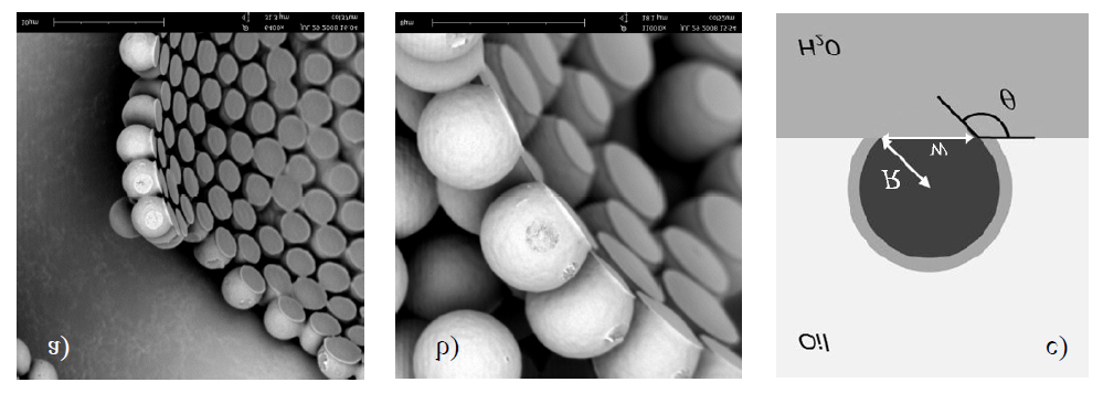
Colloidosomes, a novel class of microcapsule has been generated with high promise as a microencapsulation vehicle. The shell of collloidosomes made up of partially fused or coagulated single colloidal particles at interface of emulsion droplets [33, 34, 35]. The particle gets self-assembled to the surface of droplets to reduce the interfacial energy. Velev, et al. [36] developed such types of structures for the first time by templating latex particle adsorbed on the surface of octanol in water emulsion and by removal of oil after fusing the particle monolayers [37]. Yi, et al. [38]. Caruso, et al. [39] and Caruso, et al. [40] generated same structures by templating water in oil emulsions and templated solid nanoparticles on the surface of solid microparticles based on electrostatic attraction and assembling multilayer shells of positively or negatively charged polyelectrolytes and nanoparticles. The final colloidosomes are produced by removal of colloidal particles by assembly of polymer latex colloidal particles into shell by partial fusion of shell in water in oil emulsion drops. Hairy colloidosomes were formulated by Paunov VN, et al. [41] the shell of which was made up of microrod particles. They fabricated novel colloidosome capsules consisting of aqueous gel core and shells of polymeric microrods by templating water in oil emulsions, which were stabilized by rod like particles continued by aqueous phase gelling, dissolution of oil phase in ethanol and the obtained colloidosome microcapsule was redispersed in water. Figure 1 illustrates the formation of Colloidosome from oil in water emulsion [12].
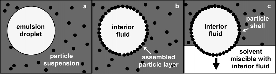
Figure 1: Shell formation: a) Emulsion droplets prepared in particle suspension. b) Particles adsorb at the interface of emulsion droplet to minimize the total interfacial energy. It forms a layer of particles that encapsulates the droplet. After the self-assembly, layers get stabilized to form solid shells. c) Shells are transferred to a solvent miscible with the fluid inside the shell. After removing the droplet interface it form porous capsule. Capsule remains intact due to shell stabilization.
NanoColloidosome is a versatile technique which provides efficient encapsulation to control the size, mechanical strength, compatibility and permeability. It has several advantages as compared to liposome and polymersome such as mechanical stability of colloidal shell, size flexibility, trigger release ability, choice of encapsulated material, high potential of controlling the permeability of entrapped species, selective and timely release, mechanical strength allows to withstand mechanical load, 100 % encapsulation efficiency, highly monodispersed colloidosomes can be produced in large quantity by microfluidic device [42], compatibility with sensitive materials like cells and biomolecules, allow to design release mechanisms. In vivo tracking is possible by doping the shell with metallic particles detectable in MRI or X-ray and makes it easy to develop from wide range of materials like organic, inorganic and polymeric materials. However, there are some limitations in the development of colloidosomes, the main problem being the poor yield of particles. Ineffective formation of shell locking in colloidosome may cause coalescence and they may get transferred to water. Lastly, there is loss of colloidosomes in large quantity on the transfer from organic to water media [43].
Colloidosomes are hollow, spherical capsules developed from controlled self-assembly of colloidal particles on emulsion droplets. These colloidal particles get adsorbed on emulsion droplet to minimize the total interfacial energy which also act as bridge between particles, lock them together and stabilize the structure. So, the colloidosomes are classified on the basis of emulsion used for the formulation. They are classified as follows:
- Water-in-oil emulsion based colloidosomes
- Oil-in-water emulsion based colloidosomes
- Water-oil-water emulsion based colloidosomes. The nature of colloids can also classify the colloidosomes. Classifications on the basis of nature of colloids are as follows:
- Aqueous or oily gel core colloidosomes
- 2. Hairy Colloidosomes
- Nanoparticle colloidosomes
- Layer by layer colloidosomes
- Non-spherical colloidosomes
Method of Preparation
Emulsion Based Colloidosome
Water-in-Oil Emulsion Based Colloidosomes: In presence of colloidal particles, aqueous phase is emulsified in oil to develop water-in-oil emulsion. These colloidal particles get adsorbed on the surface of droplets to minimize the total interfacial energy. Addition of polycations locks these particles together by Vander Walls forces or by sintering the particles [44]. The obtained water-in-oil based colloidal dispersion is transferred into water by two different ways i.e. centrifugation and filtration technique. The centrifugation technique is carried out by diluting obtained colloidal dispersion with organic solution (ethanol, dodecane) and offered to centrifuge to separate them from supernatant. The obtained colloidosomes of water core are redispersed in water after washing with ethanol and water.
In another technique, colloidal dispersion filtered through hydrophobic milipore membranes, then water containing small amount of ethanol is poured on the membrane to remove the oil interface and to suspend the colloidosomes in water. These water-in-oil emulsion based colloidosomes (water core colloidosome) are used as encapsulating agents for dyes and drugs mainly because of their tunable properties and mechanical resistance to their shell [33]. Oil-in-Water Emulsion Based Colloidosomes: In this type, oil is emulsified in aqueous phase containing particles and surfactant to develop o/w type emulsion. Colloidal particles are mainly used to stabilize droplet interface. The obtained colloidal dispersion is added to organic phase (ethanol), and is centrifuged to separate it from supernatant, washed with ethanol and finally redispersed in water [45]. Water-Oil-Water Emulsion Based Colloidosomes: In this type, oil phase consists of pendant drop of aqueous suspension of latex particle. Multiple infusion approach offers to adsorb the closely packed particle monolayer on the drop interface. Densely coated and adsorbed particles on the pendant water drop in oil phase are transferred through planar oil-water interface which are free of particles to form a giant pendant colloidosome. Developed colloidosomes are supported by water-oil-water film coated by latex particles bridges both surface [46].
Preparation On The Basis Of Nature Of Colloids
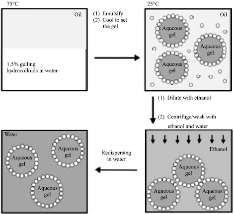
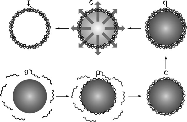
Aqueous or Oily Gel Core Colloidosomes: In this type of colloidosomes, a hot aqueous solution of agarose is emulsified in oil in presence of solid particles. Developed w/o emulsion is stabilized by solid particles and allowed to cool to form the agarose gel. The obtained aqueous gel microcapsule is diluted with ethanol and centrifuged to separate them from the supernatant. Microcapsules are washed with ethanol and water, then redispersed in water and finally giant colloidosomes formed. This technique is used to develop the colloidosome in different diameter size [33]. The basic technique is illustrated in Figure 2 [41].
Hairy Colloidosomes
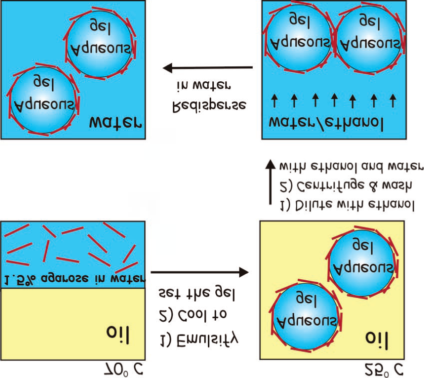
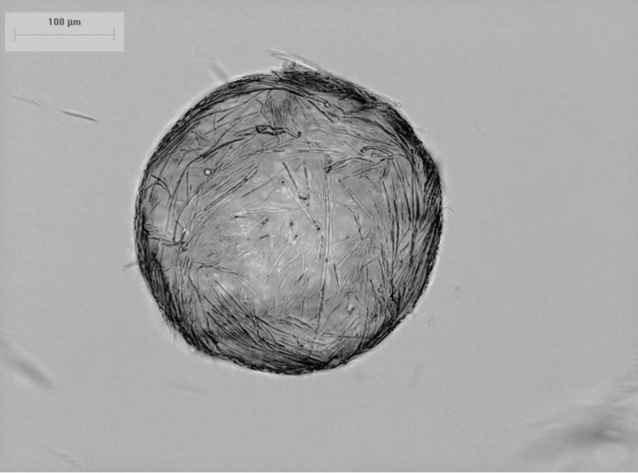
In this technique, colloidosome microcapsule prepared with shells of polymeric microrods. In presence of rod like polymeric particles, a hot aqueous solution of agarose emulsified in oil to develop stabilized w/o emulsion. This emulsion system is cooled off to set the agarose gel. The aqueous gel of microcapsule is diluted with ethanol, centrifuged to separate them from the supernatant, then washed with ethanol and water and is redispersed into water. The gel core of the colloidosome supports the particle shell and offers the microcapsules enough stiffness to be separated from oil phase by centrifugation technique. Basic method is illustrated in Figure 3 and constructed hairy colloidosomes microcapsules prepared from SU-8 microrod shown in Figure 4 [47].
Nanoparticle Colloidosomes
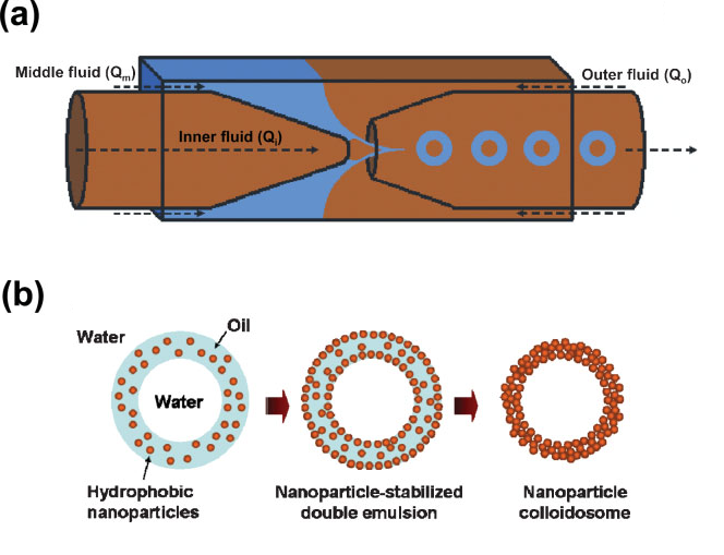
Water-oil-water emulsions can also be used to develop the nanoparticle colloidosome with selective permeability. Nanoparticle colloidosomes developed by using monodispersed double emulsion act as templates to supply robust and precisely tuned structure and compositions. Glass capillary microfluidic devices (Figure 5a) are used to develop monodispersed W/O/W double emulsion with shell geometry. Hydrophobic silica nanoparticles dispersed in oil shell stabilizes the droplets and it forms colloidosomes when oil solvent removed. The size of double emulsion and developed colloidosome can be tuned separately by controlling the flow rate of each fluid. Advantage of nanoparticle colloidosomes over the conventional method is that, there is no need to transfer the colloidosomes from oil to aqueous phase. Also, it is possible to develop the composite colloidosomes by adding different materials into the oil phase. The thickness of colloidal shell which is most critical parameter in colloidosomes can be controlled by changing the dimension of double emulsion. The nanoparticle colloidosomes shows selective permeability to different sizes of molecules, which is main characteristic of colloidosomes [47]. Schematic presentation of the preparation of nanoparticle colloidosomes from double emulsion is shown in Figure 5b.
Layer by Layer Colloidosomes
In this type, enzymes are encapsulated by using biocrystals as templates to deposit multilayers, then enzymes removed to develop hollow polymer capsule. Layers of polyelectrolytes are deposited stepwise on crystals by surface charge reversal which occurs upon adsorption of each layer. Each deposited layer possesses charge opposite to the earlier adsorbed layer. Centrifugation, washing or redispersion cycles are used to remove the unabsorbed polyelectrolyte before the next layer is deposited. Encapsulated enzymes are solubilized by exposing to acidic solutions which changes polymer capsule morphology. Polymer capsule ruptures when exposed to solution of pH >11and releases the enzyme. Oxidizing solutions decompose encapsulated enzyme and form hollow polymer capsules [49]. Schematic presentation of formation of Layer by layer colloidosomes are shown in Figure 6 [50].
Non-Spherical Colloidosome
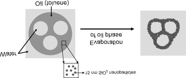
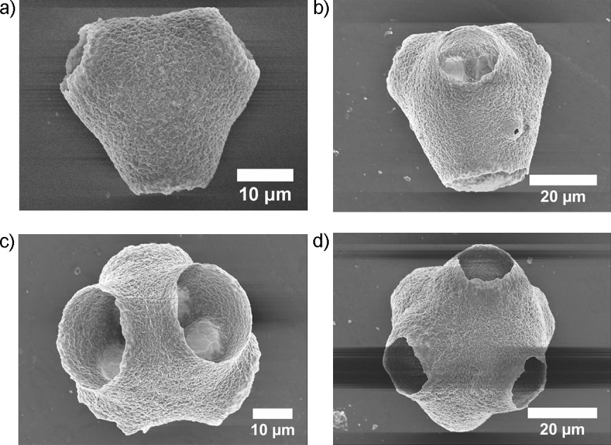
Water-oil-water emulsions are used to construct the multiple compartment non-spherical colloidosomes by glass capillary micro fluidics. There are different numbers of aqueous drop in single oil drop of double emulsions. Hydrophobic SiO2 nanoparticles are suspended in oil phase, polyvinyl alcohol and are dissolved in continuous phase to develop a stabilized double emulsion. Suspended nanoparticles form the shell after the removal of oil. The internal w/o interface remain in spherical shape and outer o/w interface deforms in oil removal process and develops multiple compartment non-spherical colloidosomes. Schematic illustration shown in Figure 7 and SEM images of nonspherical colloidosomes are shown in Figure 8 [51].
Structural Caracterization: Stabilization of emulsions with the help of solid particles was first reported by Ramsden in 1903 [52]. The word Pickering stabilization is derived from the name Pickering, in 1907 on account of a paper published by him [53]. Although many articles have been published related to this field [54, 55, 56] no significant interest in this field was observed until 1990. Velev, et al. reported potential of Pickering stabilization for microencapsulation technique and formation of novel technique [57, 58, 59]. Weitz [60, 61, 62, 63] and Bon [64, 65, 66, 67] with their team reported the use of Pickering emulsions for the synthesis of capsules with mechanical property, size control, permeability properties. The assembly of colloidal particles at liquid-liquid interfaces is influenced by the interfacial free energies γ of the particles and two liquid phases [68]. The interfacial tensions to the equilibrium position of the particle at the interface through the 3-phase contact angle θ, Figure 9 which can be explained by the Young’s equation (Equation 1.1) as given below [69].
γ γ θ γ $$ = \frac {\gamma_ {p o} - \gamma_ {p w}}{v} \dots \dots 1. 1 $$
cos po pw ow In the given equation of 1.1, γpo, γpw, and γow, these are the interfacial tensions of particle/oil, particle/water and oil/ water respectively. When particle is partially wetted by two liquid phases or when 0º < θ < 180º, particles get adsorbed on the oil/water interface shown in Figure 9. Experimental studies reported that the most stable Pickering emulsion can be obtained when intermediate θ values i.e. 60º < θ < 120º and θ determines the types of emulsions [70, 71]. Hydrophilic particles where, θ < 90º project aqueous phase and develops the oil in water (o/w) emulsion and hydrophobic particles where, θ > 90º stabilize water in oil emulsion (w/o). The main characteristic of Pickering emulsion is the large size of the stabilizing entities which causes strong binding energy of particles with the interface shown in Equation 1.2 [72].
![Figure 9: Experimental studies reported that the most stable Pickering emulsion can be obtained when intermediate θ values i.e. 60º < θ < 120º and θ determines the types of emulsions [70,71]. Hydrophilic particles where, θ < 90º project aqueous phase and develops the oil in water (o/w) emulsion and hydrophobic particles where, θ > 90º stabilize water in oil emulsion (w/o). The main characteristic of Pickering emulsion is the large size of the stabilizing entities which causes strong binding energy of particles with the interface shown in Equation 1.2 [72].](/fulltextimages/11308/fig_9.png)
$$ E _ {B} = \pi R ^ {2} \gamma_ {o w} \left(1 - \left| \cos \theta \right|\right) ^ {2} \dots \dots 1. 2 $$ In above equation, EB represents the desorption energy of the particle from liquid interface into the continuous phase of the emulsion. It shows that colloidal particles of radius R having size between 0.01-10 µm and intermediate θ are attached irreversibly to interface which forms highly stable emulsion droplets. Applications of Pickering emulsions found useful in various industrial process such as, cosmetics, food, mineral floatation and crude oil processing [73, 74, 75, 76]. Pickering emulsion droplets are extensively used as scaffolds for the synthesis advanced supracolloidal materials like colloidosomes, colloidal nano-composites, porous solids and foams [77]. Velev, et al. [36] synthesized structures similar to colloidosomes for the first time, but Dinsmore, et al. defined colloidosomes as “selectively permeable capsules that are composed of colloidal particles.” The self-assembly of spherical particles on emulsion droplets resulted in solid shells which were porous (Figure 9b & 9c).
In above figure, (Figure 9a), D ark grey circle is solid particle and light grey area that surrounds the particle is stabilizing group which is soluble in the oil. ‘R’ is the radius of the particle, ‘θ’ is the three phase contact angle. ‘Z’ is the distance from the center of the particle to the interface.
The intrinsic porosity of colloidosomes can be used for controlled release and targeted delivery of drugs. Colloidosomes are most stable structures because of high energy desorption of particles from soft interfaces and it is also possible to dry colloidosomes without collapsing the structure [78, 79, 80]. Recently, there is significant interest increased in packing of particles and defects raised from packing of particle on a curved surface [81, 82]. It is possible to pack same size spherical particles in hexagonal lattice smoothly on flat surface but some surface defect still arises [83, 84]. According to Euler’s theorem, the total disclination charge of any triangulation on a sphere must be 12. The total disclination charge means departure of the coordination number on a planar surface of 6. A total disclination charge of 12 can be obtained in several ways which determine the minimum energy configuration of repulsive particles and is necessary for crystallography on sphere which is very difficult problem. J.J Thomson [85] recognized this problem nearly 100 years ago. He tried, unsuccessfully to explain the periodic table in rigid electron shell. Similar problem occurs in multi-electron bubbles in superfluid helium,88 virus morphology [86, 87, 88, 89, 90, 91, 92, 93, 94, 95, 96, 97, 98] protein s-layers [89, 90] and coding theory [91, 92]. Both the classic Thomson problem related to particle interacting through Coulomb potential and other interaction to potentials remain unsolved even after 100 years [93, 94, 95].
Soccer ball and C60 fullerenes [96, 97] are the familiar examples of this phenomenon, they have 12 pentagonal and 20 hexagonal patches shown in Figure 10. Soccer ball consist of 12 black color pentagonal patches and 20 white color hexagonal patches. As the number of particles on the spheres increases, isolated charge 1 defects induces too much strain, and can be relieved by introducing additional dislocations which consists of tightly bound pairs of 5-7 defects which still satisfy Euler’s theorem as their net disclination charge is zero. Dislocations are the point like topological defects in two dimensions which disrupts the translational order of the crystalline phase but are less disruptive of orientational order [98]. Dislocations play important role in crystallography on spherical surface. When the system size i.e D/4 surpasses approximately the critical value of 5 there is formation of chains of 5-7 dislocations which are called as grain-boundary scars. Where, D is the diameter of spherical and R represent the radius of the particle. Curvature of colloidal film results in formation of grain-boundary scars which is not observed in flat colloidal films. Colloidal particle covered emulsion droplet used as experimental model for general theories on particle configurations with arbitrary repulsive interactions on curved surface such as J.J.Thomson problem or Tammes problem [99].
![Figure 10: Soccer ball consist of 12 black color pentagonal patches and 20 white color hexagonal patches. As the number of particles on the spheres increases, isolated charge 1 defects induces too much strain, and can be relieved by introducing additional dislocations which consists of tightly bound pairs of 5-7 defects which still satisfy Euler’s theorem as their net disclination charge is zero. Dislocations are the point like topological defects in two dimensions which disrupts the translational order of the crystalline phase but are less disruptive of orientational order [98]. Dislocations play important role in crystallography on spherical surface. When the system size i.e D/4 surpasses approximately the critical value of 5 there is formation of chains of 5-7 dislocations which are called as grain-boundary scars. Where, D is the diameter of spherical and R represent the radius of the particle. Curvature of colloidal film results in formation of grain-boundary scars which is not observed in flat colloidal films. Colloidal particle covered emulsion droplet used as experimental model for general theories on particle configurations with arbitrary repulsive interactions on curved surface such as J.J.Thomson problem or Tammes problem [99].](/fulltextimages/11308/fig_10.png)
Last few studies on colloidosomes are mainly related to the large surface coverage of particles which are locally high ordered particles. If particles can interact repulsively then there is formation of locally crystalline particle packing with minimum energy. Non-crystalline packing colloidosomes are found in the literature [100, 101]. Fortuna, et al. reported the assembly of polydispersed particles resulted into non- crystalline packing. S.Jiang studied the transition from low density and disordered packing to crystalline packing depending on the contact angle of paticles [102]. In most of the studies of colloidosomes, structure of particles are characterized by qualitatively by using electron or light microscopy. 3D presentation of the same images of colloidosomes can be used to quantitatively characterize the particle configuration. The parameter which is measured is θ, the droplet diameter and structural organizations of particles on the droplet surface. Three Phase Contact Angle, θ: The three phase contact angle, θ which is related to Pickering emulsion is difficult to measure at liquid interfaces. Advanced technique applied to image as well as to study the wettability of spherical particle at liquid interfaces. In this technique, one of the two liquid consisting particles is gelled to trap the particles and later this gel with trapped particle is allowed to capture image with SEM or AFM [33, 103]. But another method is applied to explore the deformation of particles by heating to temperature above the glass transition. Sintering is essential to strengthen the interactions between the particles on surface of droplet and to achieve stable capsule. It is observed that, during the process particle deforms and reduces particle water contact area shown in Figure 11. This deformation process is carried out by surface tension and θ can be calculated.
In the above mentioned deformation process, it is considered that particle position remain at the same place before and after the deformation. R and w can be determined from SEM images and used to calculate θ by equation 1.3.
$$ \sin \theta = \frac {w}{2 R} \dots \dots \dots 1. 3 $$
In this case, θ ≈ 130, because of the colloidosomes are composed of polystyrene (pS) particles and wide range of values for θ reported i.e 90º < θ < 130º [104, 105] for these type of particles at alkane water interface.
It is necessary to mention that the observed θ is just an indication because possibility of the experimental error in determination of w and R from the SEM image. In glass transition, time of heating is limited and it does not precisely represent the deformation. Extended heating may cause spreading of the particle over the droplet surface and helps to reduce the oil/water interfacial area but measured θ gives useful information related to wettability of particle. Droplet Diameter Distribution (D): Droplet diameter distribution is used to characterize the emulsion as well as Pickering emulsion which is useful to reveal information on total available interface between oil and water phase. The total available interface helpful to determine the maximum number of particles presented at the surface of colloidosomes. In general, estimation of average droplet diameter can be assumed by all available particles covering w/o interface. In this type, attachment of number of particles NA to w/o interface depends on the diameter of the droplet D, as given in below equation.
D
| N .2 | 3.R2 |
|---|
In above equation, VD represents the total volume of dispersed phase [106] and 2√3.R2 is the area of the particle (Ap) which is hexagonally packed, R is the radius of the particle. It is considered that all particles are packed in hexagonal structure. The droplet diameter can be obtained by SEM images and LM (light microscopy). Structural Arrangement of Particles & 3D Structure: Delaunay triangulation (DT) used for characterization highly ordered colloidosomes of 5-,6-, and 7- folded particles but other technique is also needed for non-crystalline system [107]. Laser scanning confocal microscopy (LSCM) is used to characterize any particle in 3D structure which scans the colloidosomes and positions of particles at different height and gives 3D image by combining three different images. Scanning electron image and conventional light microscope images can also be used to construct the 3D image of colloidosomes. It is considered that the colloidosomes are spherical in shape and to obtain 3D coordinates it is necessary to decide the two dimensional (x,y coordinates) i.e 2D image which is subtracted from planar SEM image. The required height z can be calculated from given equation:
2 2 4 pz D p = − ……. 1.5
In above equation, D is the diameter of colloidosome and p is the planar center-to-center distance of the particle to the central axis of the colloidosome (Figure 12) and achieved in similar manner like zp.
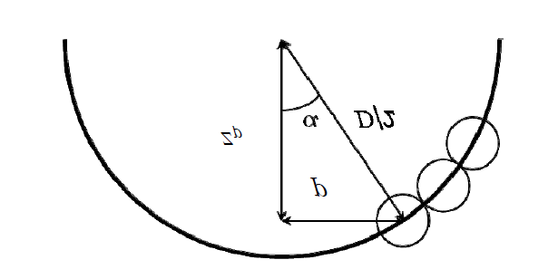
Z is calculated from radius (D/2) of colloidosome and planar distance (p) from the central axis of the colloidosome as explained in equation 1.5 and α is the inclination angle of particle respected to central axis.
If we consider, xc, yc are the planar coordinates of the central axis of the colloidosome and xp, yp are the coordinates of particle then p can be calculated from the following equation ( ) ( ) 2 2 $$ p = \sqrt {\left(x _ {p} - x _ {c}\right) ^ {2} - \left(y _ {p} - y _ {c}\right) ^ {2}} \dots \dots \dots 1. 6 $$ The accuracy of the particle adjacent to colloidosome declines due to error in x,y positions interprets comparatively larger error in z. Voronoi tessellation (VT) and DT are used to check the accuracy of the particle. Delaunay triangulation (DT) method applied to recognize adjacent particles which exhibit defects and grain boundary scars. It can also determine adjacent angle distribution, fraction of defect and interparticle distance. In addition, Voronoi tessellation (VT) provides information of adjacent particle with area occupied by each particle and total surface covered by that particle. Sastry, et al. [108] applied combined DT and VT to conclude area, volume and pores present in particle assembly but not to colloidosome. Grier, et al. explained application of pair correlation function to characterize the degree of order in colloidal films [109]. A single image of the highly ordered colloidosomes are used to exhibit 3D image by Delaunay triangulation (DT) shown in Figure13. Whereas, is complementary method to Delaunay triangulation (DT) to characterize particle configurations and used to recognize coordinate defects shown in Figure 14 [110, 111].
![Figure 13: Whereas, is complementary method to Delaunay triangulation (DT) to characterize particle configurations and used to recognize coordinate defects shown in Figure 14 [110,111].](/fulltextimages/11308/fig_13.png)
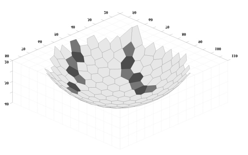
SEM image of the colloidosome and 3D image by Delaunay triangulation (DT). The dot represent pentagonal structure (light gray) and hexagonal (dark grey). At the center of the polygons particles are located. Light-gray polygons and dark-gray polygons indicate pentagonal and heptagonal coordinated particles. Colloidal Cage: Tunable Particle Packing: Colloidosomes are hollow spherical structures which are developed from colloidal particle gathering on the edge of liquid drop. The usage of uniform spherical particle in the formation of colloidosome aids to switch the permeability which forms densely packed particle due to interstitial voids. The size of the particles is mainly depends upon the interstitial voids which also control the permeability and so release kinetics. Porosity and permeability can be tuned by controlling the particle density on colloidal surface. Crystalline particle packing achieved at high densities in which particle size determine the pore size and in case of low densities large pore size with disorder packing achieved. Particle density which control pore size and interaction between particles are the major factor in the formation of colloidosomes. In resemblance with term colloidal armor used for densely packed colloidosome, new type of class is termed as ‘Colloidal cages’. Colloidal particles of repulsive interparticle potential used in most studies of colloidosome. Repulsive particle charge at high and low particle densities exhibit high crystalline particle pattern [112]. The application of such repulsive particle which are packed on droplet surface used as investigational model to study the old Johnson problem related to spherical surface whose structural arrangement exhibit point like charges [113]. In addition this experimental study exposed new problem like grain boundary scars which is only related to spherical surface [114]. These study started new arenas of investigation of organization of matter consist of effect of curvature and topology i.e. studies of shapes and spaces [115]. The colloidal particle interactions at interface are innovative area of research and involvement of particle potential can lead to new class of assemblies.
Colloids with attractive interactions are regularly applied for the study of colloidal gels and glasses [116] but not for colloidosomes. Volume fraction of the particle and inter-particle potential are the major parameter that determine the formation and structure the formation of colloidal gel and glasses. These parameters can be controlled in the development of colloidosome. Factors Affecting Colloidosomes: There are various factors that affect the formation of stable colloidosomes. A system comprises mixture of small and large spherical colloidal particle with opposite electrical charges. Electrostatic attraction attracts the small particle towards large particle. Insufficient concentration of small particle fails to saturate surface of the large particle causes formation of bridging flocculation among the positive and negative large particle. Increases in concentration of small particle saturate the large particle and forms the colloidosomes. Further increase in the concentration of small particle may increases the attraction between the colloidosome that causes flocculation. Here we will discuss the major factors that affect the formation of stable colloidosomes. Critical Saturation Concentration: Volume fraction of small particles essential to saturate the surface of specified large particle concentration can be calculated by geometric consideration. If it is considered that total small spherical particles are monodisperse and incompressible then the required number of small particles to saturate the surface of single large particle of hexagonal packing can be calculated by equation 1.7 [117].
$$n_{S,Sat} = \frac{2\pi}{\sqrt{3}} \left( 1 + \frac{1}{R} \right)^2 \dots 1.7$$
In above equation, $R$ (= dS/dL) which is the ratio of small particles to the large particle.
If total numbers of large particles are also monodisperse then the attraction between small and large particles becomes stronger and binds to the surface of large particles. So following equation (Eq.1.8) is used to achieve the minimum volume fraction that required saturating the surface of large particles.
$$\phi_{S,Sat} = \frac{2\pi}{\sqrt{3}} R \left( 1 + R \right)^2 \phi_L$$
$$\varnothing_S = \varnothing_{Sat}$$
In this, $\varnothing'L$ represent volume fraction of the large particle.
Critical Depletion Concentration: After saturation of large particle surface by small particle, the remaining small particle in continuous phase builds depletion attraction between large particles. Strong depletion attraction than repulsion attraction may cause flocculation. Equation 1.9 expresses the strength of depletion interaction among the two large particles in continuous phase including free small particles [118].
$$\frac{W_{Dep}}{K_B T} = \phi_S \left( 1 + 2\phi_s \right) \left[ \frac{3}{2R} + 1 \right] \dots 1.9$$
In above equation, WDep represent depletion attraction strength when two large particle surfaces come in contact with each other. Depletion attraction strength increases with increase in concentration of small particle and decrease in R.
**Critical Adsorption Concentration**
The stability of colloidosomes also influenced by the time required for saturation (TSat) of large particle surface and time required for collision (Tcol) of small particles with large particle. If the large particle surface are not saturated with small particle (Tcol < Tsat), then the large particle will combined with small particles and act as bridge. If it is considered that the mechanism of large particle collision is Brownian motion, then the average time between collisions can be calculated from equation 2.0 [119].
$$\tau_{Col} = \frac{\pi d_L^3}{8K_B} \frac{n_C}{T\varnothing_L} \dots 2.0$$
Where, $\eta$C represents viscosity of continuous phase.
Effect of Cosolvents: To improve the colloidal particles packing at the interface of water/oil, some cosolvents like ethanol are used in colloidal suspension. Colloidal particle aggregation and coalescence are induced by use of cosolvents like ethanol. Laib, et al. [120] studied and determined the critical concentration of ethanol observed to be about 25 wt % for 11.25 wt % colloidal suspension. The colloidal particle / ethanol (3/1 v/v) suspension air dried and analyzed by SEM to study the effect of ethanol on stability. The SEM images revealed aggregates of fused particle which indicates latex particle are fusing due to plasticization by use of ethanol as cosolvents.
Effect of Surfactant: Aqueous suspension of different colloidal particle concentration is blended with oil and aqueous volume fraction of 1/50 which result in formation of unstable emulsion. This indicates that colloidal particles are not a suitable emulsifier for water in oil droplets. Therefore nonionic surfactant having hydrophobic tail and polar head are added to stabilize water-oil droplets. For the negatively charged colloidal particle cationic surfactant are employed [125].
Applications of Colloidosomes: Colloidosomes are mainly used as encapsulation and controlled release in drug delivery, vitamins, and protein. These are also applied for fragrances and flavors pharmaceutical and cosmetic industries and also useful in food molecule generated by sensitive biomaterial like living cells. Colloidosomes can be used for different particle size which is further used in controlled release drug delivery. Various interesting characteristics like mechanical deformation and rapture are very interesting and used in encapsulation technique. A new technique of formulation of monodisperse stimuli-responsive colloidosomes which consist of stimuli responsive as building block, aqueous droplets as templates and microfluidic devices are used to control the assembly revealed nearly 80% reduction in volume and can be used for targeted-pulsed release [121]. Colloidosomes can be further used in therapeutic and pharmaceutical applications like in tumor therapy, in antimicrobial, antifungal, antiviral therapy, in DNA delivery, in enzyme immobilization, for altered pharmacokinetics and biodistribution [122].
**Conclusion**
NanoColloidosome, a novel class of microcapsules developed with high promises as microencapsulation vehicle. These are the hollow spherical capsule developed from controlled self-assembly of colloidal particles on w/o, o/w, o/w/o, w/o/w emulsion droplets. It is a versatile technique which provides efficient encapsulation to control the size, mechanical strength, compatibility and permeability. Different types of emulsion, water in oil emulsion, oil in water emulsion and water oil water emulsion are used to develop different nanocolloidosome such as aqueous, hairy, nanoparticle, layer by layer and non-spherical colloidosomes [123]. Structural characterization revealed that the colloidosomes which are composed of colloidal particles are most stable structures due to high energy desorption of particles from soft interfaces. Study of packing of particles on curved surface and 3D structure exhibited that the colloidosome consist of hexagonal and pentagonal patches which are similar to structure of soccer ball and C60 fullerenes. Factors which mainly affect colloidosome include critical saturation concentration, critical depletion concentration, critical adsorption concentration, effect of cosolvent and surfactant. Increase in concentration of small spherical particle than the large spherical particle causes flocculation. The stability of colloidosome is also affected by time required for saturation of large particle surface time required for collision of small particle with large particle. The application of colloidosomes is mainly useful in controlled release, as encapsulation agent, in tumor therapy, in nDNA and mtdDNA delivery and altered pharmacokinetics [124]. The flexibility of nanocolloidosomes vaccines will explore the large range of potential applications in future.
References
-
Gupta PN, Vyas SP (2011) Colloidal carrier systems for transcutaneous immunization. Curr Drug Targets 12(4): 579-597.
-
Chanchal D, Swarnlata S (2008) Novel approaches in herbal cosmetics. J Cosmet Dermatol 7(2): 89-95.
-
Rawat M, Saraf S (2008) Liposphere: Emerging carriers in the delivery of proteins and peptides. Int J Pharm Sci Nanotechnol 1: 207-214.
-
Elsayed MMA, Abdullah OY, Naggar VF, Khalafallah NM (2006) Deformable liposome and ethosome: Mechanism of enhanced skin delivery. Int J Pharm 322(1-2): 60-66.
-
Beg S, Samad A, Nazish I, Sultana R, Rahman M, Md Ahmad Z, et al. (2013) Colloidal drug delivery systems in vaccine delivery. Curr Drug Targets 14(1): 123-137.
-
Biju SS, Talegaonkar S, Mishra PR, Khar RK (2006) Vesicular system: An overview. Ind J Pharm Sci 68(2): 141-153.
-
Saraf S (2009) Process optimization for production of nanoparticles for drug delivery applications. Expert Opin Drug Deliv 6(2): 187-196.
-
Jain S, Saraf S (2009) Repaglinide-loaded long-circulating biodegradable nanoparticles: Rational approach for the management of type 2 diabetes mellitus. J Diabetes 1(1): 29-35.
-
Rawat M, Reader SS (2009) Formulation optimization of double emulsification method for preparation of enzyme-loaded Eudragit S100 microspheres. J Microencapsulation 26(4): 306-314.
-
Chanchal D, Swarnlata S (2008) Novel approaches in herbal cosmetics. J Cosmet Dermatol 7(2): 89-95.
-
Ofoegbu O (2003) Force measurements on nanorods- enriched sintered colloidosomes. Gordon Mckay Laboratories: Harvard University Summer, pp: 2-9.
-
Hsu MF, Nikolaides MG, Dinsmore AD, Bausch AR, Gordon VD, et al. (2005) Self-assembled Shells Composed of Colloidal Particles: Fabrication and Characterization. Langmuir 21(7): 2963-2970.
-
Gibbs BF, Kermasha S, Alli I, Mulligan CN (1999) Encapsulation in the food industry: a review. Int J Food Sci Nutr 50(3): 213-224.
-
Hemsley AR, Griffiths PC (2000) Architecture in the microcosm: biocolloids, self-assembly and pattern formation. Philos Trans R Soc Lond Ser A-Math Phys Eng Sci 358(1766): 547-564.
-
Jain RA (2000) The manufacturing techniques of various drug loaded biodegradable poly(lactide-co-glycolide) (PLGA) devices. Biomaterials 21(13): 2475-2490.
-
Rossier Miranda FJ, Schroen C, Boom RM (2009) Colloidosomes: Versatile microcapsules in perspective. Colloids and Surfaces A: Physicochemical and Engineering Aspects 343(1-3): 43-49.
-
Kamen ME, Bernstein P, Rivero RT (1993) Method of Encapsulating Pigment Particles Useful in the Manufacturing of Cosmetic Products and the Products Thereof. USA, 5,234,711.
-
Yow HN, Routh AF (2006) Formation of liquid core- polymer shell microcapsules. Soft Matter 2(11): 940- 949.
-
Peyratout CS, Dahne L (2004) Tailor-made polyelectrolyte microcapsules: From multilayers to smart containers. Angew Chem-Int Edit 43(29): 3762-3783.
-
Hawlader MNA, Uddin MS, Khin MM (2003) Microencapsulated PCM thermal-energy storage system. Applied Energy 74(1-2): 195-202.
-
Chu LY, Park SH, Yamaguchi T, Nakao SI (2002) Preparation of Micron-Sized Monodispersed Thermoresponsive Core-Shell Microcapsules. Langmuir 18(5): 1856-1864.
-
McClements DJ, Decker EA, Weiss J (2007) Emulsion- based delivery systems for lipophilioc bioactive components. Journal of Food Science 72(8): 109-124.
-
Sacanna S, Kegel W, Philipse A (2007) Spontaneous oil- in-water emulsification induced by charge-stabilized dispersions of various inorganic colloids. Langmuir 23(21): 10486-10492.
-
Binks BP, Whitby CP (2005) Nanoparticle silica- stabilised oil-in-water emulsions: improving emulsion stability. Colloids and Surfaces a-Physicochemical and Engineering Aspects 253(1-3): 105-115.
-
Prestidge CA, Simovic S (2006) Nanoparticle encapsulation of emulsion droplets. International Journal of Pharmaceutics 324(1): 92-100.
-
Pickering SU (1907) Cxcvi.—emulsions. Journal of the Chemical Society. Transactions 91(91): 2001-2021.
-
Wang J, Liu G, Wang L, Li C, Xu J, et al. (2010) Synergistic stabilization of emulsions by poly (oxypropylene) diamine and Laponite particles. Colloids and Surfaces A: Physicochemical and Engineering Aspects 353(2): 117- 124.
-
Dinsmore AD, Hsu MF, Nikolaides MG, Marquez M, Bauschet AR, et al. (2002) Colloidosomes: Selectively permeable capsules composed of colloidal particles. Science 298(5595): 1006-1009.
-
Yow HN, Routh AF (2009) Release Profiles of Encapsulated Actives from Colloidosomes Sintered for Various Durations. Langmuir 25(1): 159-166.
-
Kim DG, Jeong YI, Choi C, Roh SH, Kang SK, et al. (2006) Retinol-encapsulated low molecular water- soluble chitosan nanoparticles. International Journal of Pharmaceutics 319(1): 130-138.
-
Hwang YJ, Oh C, Oh SG (2005) Controlled release of retinol from silica particles prepared in O/W/O emulsion: The effects of surfactants and polymers. Journal of Controlled Release 106(3): 339-349.
-
Wang, H, Zhu X, Tsarkova L, Pich A, Möller M (2011) All- Silica Colloidosomes with a Particle-Bilayer Shell. Acs Nano 5(5): 3937-3942.
-
Cayre OJ, Noble PF, Paunov VN (2004) Fabrication of novel colloidosome microcapsules with gelled aqueous cores. J Mater Chem 14(22): 3351-3355.
-
Yi GR, Manoharan VN, Klein S, Brzezinska KR, Pine DJ, et al. (2002) Monodisperse micrometer-scale spherical assemblies of polymer particles. Adv Mater 14(16): 1137-1140.
-
Fang M, Grant P, McShane M, Sukhorukov G, GolubV, et al. (2002) Magnetic Bio/Nanoreactor with Multilayer Shells of Glucose Oxidase and Inorganic Nanoparticles. Langmuir 18(16): 6338-6344.
-
Velev OD, Furusawa K, Nagayama K (1996) Assembly of latex particles by using emulsion droplets as templates. Microstructured hollow spheres. Langmuir 12(10): 2374-2384.
-
Ashby NP, Binks BP, Paunov VN (2004) Formation of giant colloidosomes by transfer of pendant water drops coated with latex particles through an oil-water interface. Phys Chem Chem Phys 6(17): 4223-4225.
-
Yi GR, Manoharan VN, Klein S, Brzezinska KR, Pine DJ, et al. (2002) Monodisperse micrometer-scale spherical assemblies of polymer particles. Adv Matter 14(16): 1137-1140.
-
Caruso FJ, Shi XY, Caruso RA (2001) Fabrication of nanoporous silica nanotubes by inorganic and organic double templates. Adv Matter 13: 740-740.
-
Caruso F, Mohwald H (1999) Preparation and characterization of ordered nanoparticle and polymer composite multilayers on colloids. Langmuir 15(23): 8276-8281.
-
Paunov VN, Noble PF, Cayre OJ, Alargova RG, Velev OD (2005) Fabrication of novel types of colloidosome microcapsules for drug delivery applications. Mater Res Soc Symp Proc 845: 279-283.
-
Kim JW, Utada AS, Fernandez Nieves U, Hu Z, Weitz DA (2007) Fabrication of monodisperse gel shells and functional microgels inmicrofluidic devices, Angewandte Chemie 46(11): 1819-1822.
-
Laib S, Routh AF (2008) Fabrication of colloidosomes at low temperature for the encapsulation of thermally sensitive compounds. J Colloid Interface Sci 317(11): 121-129.
-
Velev OD, Furusawa K, Nagayama K (1997) Assembly of latex particles by using emulsion Droplets. Reverse (water in oil) systems. Langmuir 13(6): 1856-1859.
-
Velev OD, Furusawa K, Nagayama K (1996) Assembly of latex particles by using emulsion droplets as templates. Ball-like and composite aggregates. Langmuir 12(10): 2385-2391.
-
Ashby NP, Binks BP, Paunov VN (2004) Bridging interaction between a water drop stabilized by solid particles and a planar oil/water interface. Chem Comm 2004(4): 436-437.
-
Noble PF, Cayre OJ, Alargova RG, Velev OD, Paunov VN (2004) Fabrication of Hairy colloidosomes with shells of polymeric microrods. J A Chem Soc 126(26): 8092-8093.
-
Lee D, Weitz DA (2008) Double emulsion-templated nanoparticle colloidosomes with selective permeability. Adv Mater 20(18): 3498-3503.
-
Caruso F, Trau D, Mohwald H, Renneberg R (2000) Enzyme Encapsulation in Layer-by-Layer Engineered Polymer Multilayer Capsules. Langmuir 16(4): 1485- 1488.
-
De Geest BG, Sanders NN, Sukhorukov GB, Demeester J, De Smedt SC (2007) Release mechanisms for polyelectrolyte capsules. Chem Soc Rev 36(4): 636-649.
-
Lee D, Weitz DA (2009) Nonspherical colloidisomes with multiple compartments from double emulsions. Small 5(17): 1932-1935.
-
Ramsden W (1903) The Separation of Solid Materials on the Surface of Solutions and Suspensions. Observation Concerning Surface Diagrams, Foam Blisters, Emulsions and Mechanical Coagulation. Proceedings of the Royal Society 72: 156-164.
-
Pickering SU (1907) CXCVI.—Emulsions. J Chem Soc 91(91): 2001-2021.
-
Finkle P, Draper HD, Hildebrand JH (1923) The Theory Ob Emulsification. Journal of the American Chemical Society 45(12): 2780-2788.
-
Wiley RM (1954) Limited coalescence of oil droplets in coarse oil-in-water emulsions. Journal of Colloid Science 9(5): 427-437.
-
Schulman JH, Leja J (1954) Control of contact angles at the oil-water-solid interfaces. Emulsions stabilized by solid particles (BaSO4). Trans Far Soc 50(50): 598-605.
-
Velev OD, Furusawa K, Nagayama K (1996) Assembly of Latex Particles by Using Emulsion Droplets asTemplates. 2. Ball-like and Composite Aggregates. Langmuir 12(10): 2385-2391.
-
Velev OD, Furusawa K, Nagayama K (1996) Assembly of Latex Particles by Using Emulsion Droplets as Templates. 1. Microstructured Hollow Spheres. Langmuir 12(10): 2374-2384.
-
Velev OD, Nagayama K (1997) Assembly of latex particles by using emulsion droplets. 3. Reverse (water in oil) systemLangmuir 13(6): 1856-1859.
-
Dinsmore AD, Hsu MF, Nikolaides MG, Marquez M, Bausch AR, et al. (2002) Colloidosomes: selectively permeable capsules composed of colloidal particles Science 298(5595): 1006-1009.
-
Hsu MF, Nikolaides MG, Dinsmore AD, Bausch AR, Gordon VD, et al. (2005) Self-assembled Shells Composed of Colloidal Particles: Fabrication and Characterization. Langmuir 21(7): 2963-2970.
-
Kim JW, Fernandez Nieves A, Dan N, Utada AS, Marquez M, Weitz DA (2007) Colloidal Assembly Route for Responsive Colloidosomes with Tunable Permeability. Nano Letters 7(9): 2876-2880.
-
Lee D, Weitz DA (2008) Double Emulsion-Templated Nanoparticle Colloidosomes with Selective Permeability. Advanced Materials 20(18): 3498-3503.
-
Bon SAF, Cauvin S, Colver PJ (2007) Colloidosomes as micron-sized polymerisation vessels to create supracolloidal interpenetrating polymer network reinforced capsules. Soft Matter 3(2): 194-199.
-
Bon SAF, Chen T (2007) Pickering Stabilization as a Tool in the Fabrication of Complex Nanopatterned Silica Microcapsules Langmuir 23(19): 9527-9530.
-
Chen T, Colver PJ, Bon SAF (2007) Organic–Inorganic Hybrid Hollow Spheres Prepared from TiO2-Stabilized Pickering Emulsion Polymerization. Advanced Materials 19(17): 2286-2289.
-
Colver PJ, Chen T, Bon SAF (2006) Supracolloidal Structures through Liquid-Liquid Interface Driven Assembly and Polymerization Macromolecular symposia 245-246(1): 34–41.
-
Pieranski P (1980) Two-Dimensional Interfacial Colloidal Crystals. Physical Review Letters 45: (7): 569- 572.
-
Young T (1805) An Essay on the Cohesion of Fluids. Philosophical Transactions of the Royal Society of London 95: 65-87.
-
Aveyard R, Clint JH, Horozov TS (2003) Aspects of the stabilisation of emulsions by solid particles: Effects of line tension and monolayer curvature energy. Physical Chemistry Chemical Physics 5(11): 2398-2409.
-
Kralchevsky PA, Ivanov IB, Ananthapadmanabhan KP, Lips A (2005) On the Thermodynamics of Particle- Stabilized Emulsions: Curvature Effects and Catastrophic Phase Inversion. Langmuir 21(1): 50-63.
-
Levine S, Bowen BD, Partridge SJ (1989) Stabilization of emulsions by fine particles I. Partitioning of particles between continuous phase and oil/water interface. Colloids and Surfaces 38(2): 325-343.
-
Frelichowska J, Bolzinger MA, Pelletier J, Valour JP, Chevalier Y (2009) Topical delivery of lipophilic drugs from o/w Pickering emulsions. International Journal of Pharmaceutics 371(1-2): 56-63.
-
Rousseau D, Ghosh S, Park H (2009) Comparison of the Dispersed Phase Coalescence Mechanisms in Different Tablespreads. Journal of Food Science 74(1): 1-7.
-
Philipsen HJA, Oestreich M, Klumperman B, German AL (1997) Characterization of low-molar-mass polymers by gradient polymer elution chromatography: III. Behaviour of crystalline polyesters under reversed- phase conditions. Journal of Chromatography 775(1-2): 157-177.
-
Dickinson E (2010) Food emulsions and foams: Stabilization by particles. Current Opinion in Colloid & Interface Science 15(1-2): 40-49.
-
Colver PJ, Bon SAF (2007) Cellular Polymer Monoliths Made via Pickering High Internal Phase Emulsions. Chemistry of Materials 19(7): 1537-1539.
-
Arditty S, Schmitt V, Giermanska Kahn J, Leal Calderon F (2004) Materials based on solid-stabilized emulsions. Journal of Colloid and Interface Science 275(2): 659-664.
-
Studart AR, Gonzenbach UT, Akartuna I, Tervoort E, Gauckler L J (2007) Materials from foams and emulsions stabilized by colloidal particles. Journal of Materials Chemistry 17(31): 3283-3289.
-
Tcholakova S, Denkov ND, Lips A (2008) Comparison of solid particles, globular proteins and surfactants as emulsifiers. Physical Chemistry Chemical Physics 10(12): 1608-1627.
-
Bausch AR, Bowick MJ, Cacciuto A, Dinsmore AD, Hsu MF, et al. (2003) Grain boundary scars and spherical crystallography. Science 299(5613): 1716-1718.
-
Fortuna S, Colard CAL, Troisi A, Bon SAF (2009) Packing Patterns of Silica Nanoparticles on Surfaces of Armored Polystyrene Latex Particles. Langmuir 25(21): 12399- 12403.
-
Einert T, Lipowsky P, Schilling J, Bowick MJ, Bausch AR (2005) Grain Boundary Scars on Spherical Crystals. Langmuir 21(26): 12076-12079.
-
Bowick MJ, Nelson DR, Travesset A (2000) Interacting topological defects on frozen topographies Physical Review B 62(13): 8738-8751.
-
Thomson JJ (1904) XXIV. On the structure of the atom: an investigation of the stability and periods of oscillation of a number of corpuscles arranged at equal intervals around the circumference of a circle; with application of the results to the theory of atomic structure. Phil Mag 7(39): 237-265.
-
Leiderer P (1995) Ions at helium interfaces. Z Phys B 98: 303-308.
-
Caspar DLD, Klug A (1962) Physical principles in the construction of regular viruses. Cold Spring Harbor Symposia on Quantitative Biology 27: 1-24.
-
Marzec CJ, Day LA (1993) Pattern formation in icosahedral virus capsids: the papova viruses and Nudaurelia capensis beta virus. Biophys Jour 65(8): 2559-2577.
-
Reddy VJ, Natarajan P, Okerberg B, Li K, Damodaran KV, et al. (2001) Virus Particle Explorer (VIPER), a Website for Virus Capsid Structures and Their Computational Analyses. J Virol 75(24): 11943-11947.
-
Sleytr UB, Sara M, Pum D, Schuster B (2001) Characterization and use of crystalline bacterial cell surface layers. Prog Surf Sci 68(7-8): 231-278.
-
Pum P, Messner P, Sleytr UB (1991) Role of the S layer in morphogenesis and cell division of the archaebacterium Methanocorpusculum sinense. J Bacteriology 173(21): 6865-6873.
-
Sloane NJA (1984) The Packing of Spheres. Sci Am 250(1): 116125.
-
Conway JH, Sloane NJA (1998) Sphere Packings, Lattices and Groups. In: 3rd (Edn.), Springer-Verlag, New York.
-
Smale S (1998) Mathematical problems for the next century. Math Intelligencer 20(2): 7-15.
-
Altschuler EL, Williams TJ, Ratner ER, Tipton R, Stong R, et al. (1997) Possible Global Minimum Lattice Configurations for Thomson’s Problem of Charges on a Sphere. Phys Rev Lett 78: 2681.
-
Erber T, Hockney GM (1997) Complex systems: Equilibrium configurations of N equal charges on a sphere (2 <= N <= 112). In: Prigogine I, Rice SA (Eds.), Advances in Chemical Physics 98: 495-594.
-
Kroto HW, Heath JR, O’Brien SC, Curl RF, Smalley RE (1985) C60: Buckminsterfullerene. Nature 318: 162- 163.
-
Jarrold MF (2000) The smallest fullerene. Nature 407(6800): 26-27.
-
Nelson DR (2002) Defects and Geometry in Condensed Matter Physics, (Cambridge University Press, Cambridge.
-
Tammes PML (1930) On the origin of number and arrangement of the places of exit on pollen grains. Groningen.
-
Binks BP, Kirkland M (2002) Interfacial structure of solid-stabilised emulsions studied by scanning electron microscopy. Physical Chemistry Chemical Physics 4(15): 3727-3733.
-
Gautier F, Destribats M, Perrier Cornet R, Dechezelles JF, Giermanska J, et al. (2007) Pickering emulsions with stimulable particles: from highly- to weakly-covered interfaces. Physical Chemistry Chemical Physics 9(48): 6455-6462.
-
Jiang S, Granick S (2008) Controlling the geometry (Janus balance) of amphiphilic colloidal particles Langmuir 24(6): 2438-2445.
-
Arnaudov LN, Cayre OJ, Stuart MAC, Stoyanov SD, Paunov VN (2010) Measuring the three-phase contact angle of nanoparticles at fluid interfaces. Physical Chemistry Chemical Physics 12(2): 328-331.
-
Paunov VN (2003) Novel Method for Determining the Three-Phase Contact Angle of Colloid Particles Adsorbed at Air−Water and Oil−Water Interfaces. Langmuir 19(19): 7970-7976.
-
Stancik EJ, Fuller GG (2004) Connect the drops: using solids as adhesives for liquids. Langmuir 20(12): 4805-4808.
-
Salari JWO, Van Heck, J, Klumperman B (2010) Steric stabilization of Pickering emulsions for the efficient synthesis of polymeric microcapsules. Langmuir 26(18): 14929-14936.
-
Hansen PHF, Rodner S, Bergstrom L (2001) Structural Characterization of Dense Colloidal Films Using a Modified Pair Distribution Function and Delaunay Triangulation. Langmuir 17(16): 4867-4875.
-
Sastry S, Corti DS, Debenedetti PG, Stillinger FH (1997) Statistical geometry of particle packings. I. Algorithm for exact determination of connectivity, volume, and surface areas of void space in monodisperse and polydisperse sphere packings. Physical Review E 56(5): 5524-5532.
-
Grier DG, Murray CA (1996) Direct imaging of the local dynamics of colloidal phase transitions. VCH Publishers: New York, USA, pp: 69-100.
-
Kraft DJ, Vlug WS, van Kats CM, van Blaaderen A, Imhof A, et al. (2009) Self-Assembly of Colloids with Liquid Protrusions. Journal of the American Chemical Society 131(3): 1182-1186.
-
Wales DJ, McKay H, Altschuler EL (2009) Defect motifs for spherical topologies Physical Review B 79: 22415.
-
Leunissen ME, van Blaaderen A, Hollingsworth AD, Sullivan MT, Chaikin PM (2007) Electrostatics at the oil–water interface, stability, and order in emulsions and colloids. Proceedings of the National Academy of Sciences of the United States of America 104(8): 2585- 2590.
-
Bowick M, Cacciuto A, Nelson DR, Travesset A (2002) Crystalline Order on a Sphere and the Generalized Thomson Problem. Physical Review Letters 89(18).
-
Lipowsky P, Bowick MJ, Meinke JH, Nelson DR, Bausch AR (2005) Direct visualization of dislocation dynamics in grain-boundary scars. Nature Materials 4: 407-411.
-
Bowick MJ, Giomi L (2009) Two-dimensional matter: order, curvature and defects. Advances in Physics 58(5): 449-563.
-
Zaccarelli E (2007) Colloidal gels: equilibrium and non-equilibrium routes. Journal of Physics-Condensed Matter 19: 323101.
-
Yates PD, Franks GV, Biggs S, Jameson GJ (2005) Heteroaggregation with nanoparticles: Effect of particle size ratio on optimum particle dose. Colloids Surf A: Physicochem Eng Aspects 255(1-3): 85-90.
-
McClements DJ (2000) Food Emulsions: Principles, Practice and Techniques. In: 2nd (Edn.), CRC Press, USA, pp: 632.
-
Walstra P (2003) Physical Chemistry of Foods. In: 1st (Edn.), Marcel Decker, New York.
-
Laib S, Routh AF (2008) Fabrication of colloidosomes at low temperature for the encapsulation of thermally sensitive compounds. J Colloid Interface Sci 317(1): 121- 129.
-
Gu YS, Decker EA, McClements DJ (2007) Formation of colloidosomes by adsorption of small charged oil droplets onto the surface of large oppositely charged oil droplets. Food Hydrocolloids 21(4): 516-526.
-
Shah RK, Kim JW, Weitz DA (2010) Monodisperse stimuli-responsive colloidosomes by self-assembly of microgels in droplets. Langmuir 26(3): 1561-1565.
-
Vyas SP, Khar RK (2002) Targeted and Controlled Drug Delivery- Novel Carrier Systems. 1st (Edn.), CBS Publisher, New Delhi.
- Solution-Processed Chiral Perovskites for Biomedical Applications
- Nanotechnology in Health Chemistry and Medicine: Current Challenges and Future Directions
- Human Exposure to Micro- and Nanoplastics: Pathways, Toxicity, and Intervention Strategies
- Exosome Nanomedicine for Cancer Therapy
- Micro and Nanoplastics–Plastisphere, Biotoxicity, Impact on Human Health, and Mitigation Strategies
- Process Validation of Cefixime Powder for Suspension Dosage Form, 50 mL