Quantum Dots' Role in Cancer Diagnosis: An overview on Nano- Fluorescence in Oncology
This paper highlights the significant advantages of utilizing quantum dots (QDs) in cancer detection. The discussion begins with an introduction to Quantum Dots and Their Properties, emphasizing their unique characteristics. Subsequently, the paper explores the utility of Quantum Dots as Imaging Probes, shedding light on their role in enhancing imaging capabilities for cancer diagnosis. The focus then shifts to their pivotal role in cancer targeting, particularly for early detection. Finally, Challenges and Future Prospects in the field of QD-based cancer detection are addressed. This comprehensive overview underscores the potential of quantum dots in revolutionizing cancer diagnosis and opens avenues for further research and development.
Introduction
Quantum dots (QDs) represent a class of nanomaterials renowned for their distinctive chemical and physical characteristics. Their capacity to confine charges, such as electrons and holes, within three dimensions grants us precise control over their energy levels and wave functions, ultimately bestowing upon them remarkable luminescent and absorptive properties. These unique properties have made them a vital nanostructure for a wide variety of applications in various fields such as optoelectronics, biomedical imaging, solar cells, quantum computing, and advanced materials science [1, 2, 3, 4, 5, 6]. Hence, in the year 2023, three pioneers in the field of nanotechnology—namely, Bawendi, Louis E. Brus, and Alexei I. Ekimov—were honored with a Nobel Prize for their groundbreaking contributions. These laureates are celebrated for their pivotal roles in the discovery and development of QDs, semiconductors compressed to such minuscule scales that their electrons find themselves in exceptionally confined carriers, which has unlocked a realm of unprecedented possibilities [7, 8, 9, 10].
Cancer diagnosis hinges on a range of sophisticated imaging techniques, each with its unique advantages and applications [11]. These modalities encompass ultrasound, X-rays, computed tomography (CT) [12], positron emission tomography (PET) [13], magnetic resonance imaging (MRI) [14], and more. Ultrasound, for example, employs high- frequency sound waves to create real-time images and is often employed for the assessment of solid tumors and blood flow evaluation across various cancer types. X-rays are invaluable for detecting abnormalities, particularly in breast and lung cancer screenings. Computed tomography (CT) merges X-ray technology with computer processing to produce detailed cross-sectional images, making it essential for visualizing tumor size and location. Positron emission tomography (PET) offers valuable insights into metabolic activity within tissues, helping identify areas of concern. Magnetic resonance imaging (MRI) harnesses strong magnets and radio waves to produce detailed anatomical images, proving particularly useful in visualizing soft tissues and detecting cancer in organs like the brain, breast, and prostate. These diagnostic tools, in conjunction with emerging technologies, serve as critical assets for early cancer detection and precise characterization, ultimately enhancing treatment strategies and elevating patient outcomes. Targeted treatment’s effectiveness depends on the presence of the specific genetic change or protein that the drug targets within the tumor [15, 16, 17]. However, even when the target is present, the tumor’s response to the drug may vary, and the treatment’s durability over time can be unpredictable.
In recent years, the distinctive characteristics of Quantum Wells (QWs) have garnered significant interest within the biomedical sector [18, 19, 20, 21]. These properties are harnessed for a wide array of applications, including real-time tissue imaging (bioimaging) [22], diagnostics, single molecule probing, drug delivery, and more. Quantum dots offer versatility, as their optical features can be fine- tuned through size and composition adjustments [1]. Their remarkable attributes, such as high brightness, resistance to photobleaching, multiplexing capabilities, and a high surface-to-volume ratio, position them as ideal candidates for intracellular tracking, diagnostics, in vivo imaging, and therapeutic delivery. This study aims to explore the immense potential of colloidal materials as a viable alternative to traditional dyes in various biomedical fields. We investigate their applications and assess their suitability for a wide range of medical and biological purposes, opening new avenues for innovation and improved outcomes in these domains.
Quantum Dots and Their Properties
Nano shells represent plasmonic nanoparticles with tunable diameters ranging from approximately 10 to 300 nm. In contrast, Quantum Dots (QDs) are excitonic nanoparticles with tunable diameters of around 1 to 10 nm [23]. QDs have emerged as a novel class of fluorescent probes for molecular and cellular imaging in the realm of bioimaging. Their distinct optical and electronic properties, including size-tunable light emission, enhanced signal brightness, resistance to photobleaching, and the ability to simultaneously excite multiple fluorescence colors, surpass the capabilities of organic dyes and fluorescent proteins. These unique features hold great promise in significantly enhancing the sensitivity of molecular imaging and quantitative cellular analysis by one to two orders of magnitude [24]. The term “dot” in quantum dots implies an extremely confined region of space, approaching zero dimensions, similar to how “wires” and “wells” in quantum terminology are confined to one and two dimensions, respectively. It is within this nanometer- sized realm that semiconductors undergo a transition from behaving as bulk materials to exhibiting properties predicted for individual or small groups of atoms. This transition gives rise to exceptional and unique phenomena Figure 1 [25].
![Figure 1: Size-Dependent Energy and Frequency Variation of Quantum Dots and Their Diverse Applications in Biomedical and Bioconjugation Method of synthetized are also mentioned. Adopted from Wagner AM, et al. [1].](/fulltextimages/11449/fig_1.png)
In the physics of semiconductors and materials science, when a photon with energy exceeding the semiconductor’s band gap energy is absorbed, it generates an electron-hole pair, commonly known as an exciton. If the semiconductor nanocrystal has a diameter smaller than its exciton Bohr radius, typically just a few nanometers, the electrons and holes become confined, giving rise to the quantum confinement effect [26]. In this state, energy levels become quantized and directly dependent on the nanocrystal’s size. As the quantum dots’ size decreases, typically to less than 10nm, the quantum confinement effects become more prominent. Quantum dot photoluminescence (PL) occurs when the excited electron returns to the ground state and recombines with the hole, releasing electromagnetic energy within a narrow and symmetrical energy band (frequency) [2]. This emission falls within the UV to near-infrared range. QW’s are nanostructures that are typically composed of elements from groups II-VI or III-V in the periodic table, these nanocrystals are defined by their physical size, which is smaller than the exciton Bohr radius [27, 28]. For instance, in the case of spherical CdSe particles, this size threshold is around 10 nm. While extensive research over the past two decades has predominantly concentrated on the photophysics of nanostructures and their applications in microelectronics and optoelectronics [29, 30, 31], recent advancements indicate that the first practical utility of QDs is unfolding in the fields of biology and medicine [32, 33]. A pivotal factor enabling these applications is the ability to synthesize large quantities of highly luminescent QDs.
Quantum Dots as Imaging Probes
In direct photoluminescence (PL) assays using quantum dots (QDs), when an excitation light source generates excitons, they can recombine and emit light energy. However, sometimes this energy can be transferred to nearby molecules without the QD emitting light. To make use of this energy transfer, a well-designed system incorporates fluorescent molecules as energy acceptors. In such a system, the transferred energy can excite an outer shell electron in the fluorescent molecule, causing it to release light (fluoresce) when the electron returns to its ground state. This energy transfer mechanism, known as Förster resonance energy transfer (FRET), was first described by Förster in 1948 [34]. Remarkably, it was not until 2001 that QD-based FRET was demonstrated in a bioassay. It is essential to note that FRET is a highly distance-dependent phenomenon, with the efficiency of energy transfer decreasing as the sixth power of the distance between the donor and acceptor.
The FRET mechanism enhances QD-based assays, allowing for the development of highly sensitive tests. FRET occurs when the donor and acceptor molecules are very close. This property enables the detection of nearby fluorophores on the QD surface. By attaching fluorophores to specific molecules, like complementary DNA sequences, QD-based FRET can identify the presence of target molecules and DNA sequences. For example, we used biotin-conjugated DNA capture probes and Cy5 fluorophore-conjugated reporter probes that hybridized with target DNA. When streptavidin-coated QDs were added, they formed complexes (QD-[capture probe]-[target DNA]-[reporter probe]). It is explained schematically in the Figures 2A-2C.
In the presence of target DNA, excited QDs emitted light and transferred energy to the reporter probes. The emitted light from both QDs and reporter probes was simultaneously detected using a custom confocal spectroscopic setup. The nanosensor assay demonstrated sensitivity over 100 times better than conventional molecular beacon DNA detection methods.
![Figure 2: Nanoscale DNA Sensing with QD-Fluorophore Nanosensors: (A) Streptavidin-coated quantum dots (QDs) selectively bind to target DNA molecules that have been hybridized with pairs of complementary probes, each labeled with either a fluorophore or biotin. (B) When illuminated, both the quantum dots (QDs) and the fluorophores emit light simultaneously through photoluminescence and Förster resonance energy transfer (FRET), respectively. (C) A custom microcapillary-based detection system is employed to detect the emitted light, enabling the precise analysis of individual nanocomplexes. Adapted from the original work of the authors Zhang CY, et al. [35].](/fulltextimages/11449/fig_2.png)
Figure 2: Nanoscale DNA Sensing with QD-Fluorophore Nanosensors: (A) Streptavidin-coated quantum dots (QDs) selectively bind to target DNA molecules that have been hybridized with pairs of complementary probes, each labeled with either a fluorophore or biotin. (B) When illuminated, both the quantum dots (QDs) and the fluorophores emit light simultaneously through photoluminescence and Förster resonance energy transfer (FRET), respectively. (C) A custom microcapillary-based detection system is employed to detect the emitted light, enabling the precise analysis of individual nanocomplexes. Adapted from the original work of the authors Zhang CY, et al. [35].
QD-Based Charge Transfer Multiplexing: QD-based fluorescence multiplexing is widely used in biological assays. Beyond common approaches, multiplexing signals can also be achieved by transferring charge (electrons) between QDs and nearby redox complexes. For instance, Medintz IL, et al. [36] demonstrated controlled quenching of QD photoluminescence using charge-transfer interactions between QDs and ruthenium phenanthroline (Ru-phen) complexes as illustrated in Figure 3. They connected Ru- phen complexes to QDs via peptide bridges, serving as electron conduits. The transfer of charge from the metal complex to the QD surface efficiently quenched QD PL. The extent of quenching was directly related to the number of Ru- phen complexes near each QD, with smaller QDs exhibiting higher quenching efficiencies due to their greater surface state density. This approach enabled multiplex quenching across a wide optical spectrum, with at least eight optical channels resolved. Applications include biosensors for proteins, maltose, fatty acids, DNA hybridization, and monitoring enzymatic activity. This interaction may find uses in multicolor fluorescence barcodes and multiplex small molecule detection, such as DNA.
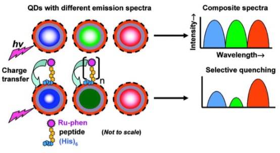
Figure 3: Selective Quenching of Quantum Dot Emissions by Ru-phen Complexes via Peptide Linkage: Quantum dots (QDs) with distinct emission spectra are specifically linked to Ru-phen complexes through a peptide bridge, resulting in the precise quenching of their photoluminescence emissions via electron transfer from the complex to the QD surface. The degree of quenching in each QD’s emission spectrum can be finely controlled by adjusting the number of complexes attached to the QD. This content is reprinted with permission from Medintz IL, et al. [36], ©2009 American Chemical Society.
Fluorescence intensity is determined by two factors: the extinction coefficient (ε) and the quantum yield (Φ). The extinction coefficient (ε) is linked to the amount of incident light absorbed per unit concentration of the dye and per unit length of the path traveled. Quantum dots (QDs) have an exceptionally high ε due to quantum confinement, making them absorb light very efficiently, with ε values as high as 3.5×〖10〗^6 at low excitation wavelengths of 400 nm, far surpassing values for organic dyes like Alexa 594, with ε values as high as 1.3×〖10〗^5 [37]. Quantum yield (Φ) measures the number of emitted photons compared to the number of absorbed photons and is crucial for assessing fluorophores’ efficiency and signal intensity. High-quality QDs can have quantum yields as high as 85%, and their conjugation with proteins has minimal impact on yields. In contrast, organic dyes often see a significant reduction in quantum yield when bound to biomolecules. Therefore, QDs have significantly higher absorbance and comparable quantum yield, making them exceptionally bright for visualizing even a single dot. This brightness is particularly advantageous when marking antigens with low copy numbers per cell.
QDs have demonstrated significant potential in cancer detection, enabling precise and sensitive diagnostics. These nanoparticles offer several advantages over traditional methods using organic dyes and fluorescent proteins, making them valuable tools in immuno-histochemical analysis, single-molecule tracking, and in vivo imaging for cancer-related research and diagnosis. Their unique spectral properties empower the creation of multiplexed systems for simultaneous detection of multiple targets through multicolor imaging, Figure 4. Additionally, QDs can be conjugated with drug molecules or incorporated into QD-based drug delivery particles, allowing real-time drug tracking and image-guided therapy in cancer treatment [38]. Furthermore, QDs with tunable photophysical properties, particularly those emitting in the near-infrared range, have become vital for deep-tissue mono- and multiphoton in vivo imaging. This capability is especially advantageous in cancer research, where visualization of tumor tissues at greater depths is essential. Overall, QDs are emerging as a versatile and powerful tool in cancer detection, aiding in more effective research and treatment strategies.
![Figure 4: Additionally, QDs can be conjugated with drug molecules or incorporated into QD-based drug delivery particles, allowing real-time drug tracking and image-guided therapy in cancer treatment [38]. Furthermore, QDs with tunable photophysical properties, particularly those emitting in the near-infrared range, have become vital for deep-tissue mono- and multiphoton in vivo imaging. This capability is especially advantageous in cancer research, where visualization of tumor tissues at greater depths is essential. Overall, QDs are emerging as a versatile and powerful tool in cancer detection, aiding in more effective research and treatment strategies.](/fulltextimages/11449/fig_4.png)
Early Cancer Detection
Sentinel lymph node (SLN) diagnosis plays a crucial role in cancer surgery, especially in detecting metastatic cancer cells in SLNs. Near-infrared (NIR) QDs emitting at 850 nm have demonstrated superiority in SLN mapping, offering high sensitivity and minimal background during surgeries. Real-time intra-operative SLN mapping using NIR QDs has been reported for gastrointestinal tract cases, enabling accurate tumor border definition and reduced dissection size. QDs, especially self-illuminating QDs, have shown promise in tracking cancer cells and studying metastasis. Targeted delivery of QDs can be achieved by attaching specific molecules to their surface (Figure 5).
![Figure 5: In vivo Targeting and Imaging of a Lung Metastasis Model Using QD-Based Nanotechnology: A Live Animal Imaging System. Reproduced from Chen C, et al. [39].](/fulltextimages/11449/fig_5.png)
QDs have been used successfully in mouse models for cancer imaging, showing potential for precise diagnosis and real-time monitoring, such as in prostate cancer and glioblastoma cases. Furthermore, a standardized protocol for liver cancer imaging in animal models was developed using QD probes, aiding in the early monitoring of liver cancer metastasis. These findings represent significant advancements in preclinical studies, with potential for clinical application. In addition, a group has developed a standard protocol for in vivo imaging of liver cancer xenograft animal models. They successfully achieved animal imaging by injecting human hepatocellular carcinoma cell lines (HCCLM6) that overexpress alpha-fetoprotein (AFP) with antiAFP monoclonal antibody and QD-IgG probes. HCCLM6 has an increased potential for lung metastasis, making it a valuable tool for constructing a platform for the early monitoring of liver cancer metastasis.
Targeted Cancer Therapies
Quantum Dots (QDs) offer a versatile approach to engineer drug delivery systems for potential cancer treatment enhancement. The success of delivering QD/drug formulations to tumor cells hinges on certain principles:
Functionalized Surface: The nanoparticle’s surface must incorporate targeting ligands for specific tumor cell delivery and allow simultaneous drug delivery.
Size Optimization: The nanoparticle size must be minimized for eventual body excretion.
Drug Confinement: The drug must be securely confined within the nanoparticle system to prevent harm to normal tissues. Still, it should release at tumor cells upon external triggers or local environmental cues.
Biocompatible Surface: The QDs’ surface should be coated with a long-lasting biocompatible polymer to safeguard against degradation in biological environments.
Two Strategies Exist to Combine Qds and Drug Molecules into a Nanoparticle System
Direct Conjugation: Here, drug molecules link to the QD surface, which subsequently delivers drug-conjugated QDs to target locations. Drug release occurs in response to local biological conditions, like pH or enzyme presence.
Polymer Encapsulation: In this approach, the drug resides within a polymer nanoparticle along with either hydrophobic or hydrophilic QDs, depending on the polymer type. The entire QD/drug nanoparticle system is transported to the target site, and drug molecules are either released as the polymer degrades in low pH conditions or diffuse out from the polymer particle.
One study involved QD-Aptamer(Dox) conjugates, where the QD surface was tailored with an RNA aptamer recognizing prostate cancer cells [40] see Figure 6. The anticancer drug, Dox, intercalated with the RNA aptamer, was gradually released from the QD system. Another study employed nanoconjugates of CdSe/CdS/ZnS QD and doxorubicin [41], targeting alveolar macrophages for the treatment of pulmonary disease. In both cases, the released drug maintained its bioactivity. These examples illustrate the potential of nanoparticle platforms in targeted therapy and sensing applications. This makes QDs able to revolutionize drug delivery systems, enhancing their precision and efficacy, particularly in the context of cancer treatment and other medical applications.
![Figure 6: The anticancer drug, Dox, intercalated with the RNA aptamer, was gradually released from the QD system. Another study employed nanoconjugates of CdSe/CdS/ZnS QD and doxorubicin [41], targeting alveolar macrophages for the treatment of pulmonary disease. In both cases, the released drug maintained its bioactivity. These examples illustrate the potential of nanoparticle platforms in targeted therapy and sensing applications. This makes QDs able to revolutionize drug delivery systems, enhancing their precision and efficacy, particularly in the context of cancer treatment and other medical applications.](/fulltextimages/11449/fig_6.png)
Figure 6: (a) Schematic of QD−Apt(Dox) Bi-FRET system: QDs are surface-functionalized with A10 PSMA aptamer. Dox intercalation into the A10 PSMA aptamer on QD surface results in QD−Apt(Dox) formation and quenching of both QD and Dox fluorescence through Bi-FRET. (b) Schematic of QD−Apt(Dox) uptake into cancer cells: QD− Apt(Dox) conjugates are specifically taken up by target cancer cells through PSMA-mediated endocytosis. Dox release from QD−Apt(Dox) induces fluorescence recovery from both QD and Dox, allowing intracellular Dox delivery sensing and synchronized cancer cell localization and treatment. Adopted from Yong KT, et al. [41].
![Figure 7: Schematic illustration of nanocarrier-based active and passive targeting drug delivery systems. Adopted from Li J, et al. [42].](/fulltextimages/11449/fig_7.png)
Nanocarriers are popular for precise drug delivery, enhancing drug bioavailability, targeting tumor cells, and reducing toxic effects. They can be modified through controlled chemosynthesis, carry multiple drugs, and optimize drug release. Nanocarriers interact with biomolecules, improving stability and circulation time.
Various strategies involve targeting molecules and delivery vehicles. However, challenges like tumor complexity and drug metabolism persist (Figure 7).
Although significant progress has been made in targeted drug delivery in oncotherapy, many challenges and limitations, such as the complexity of tumor structure, the ambiguous biosafety of delivery systems, as well as the rapid metabolism and clearance of drugs, still remain. Therefore, further research is still necessary to promote the development and clinical application of targeted drug delivery technologies in efficient oncotherapy [43, 44]. In light of the above considerations, as shown in Figure 7.
Cadmium-based QDs are widely used for in vitro and in vivo studies, despite their known toxicity due to cadmium and selenium content. Cadmium, a potential carcinogen, can distribute throughout the body, with liver and kidneys as major target organs. Recent studies have shown increasing cadmium concentration in these organs after intravenous injection of cadmium-based QDs in small animals, suggesting QD degradation in vivo. Researchers are now working on cadmium-free QDs, like InP QDs, which offer advantages such as robust covalent bonds, reduced toxicity, and ease of dispersion in aqueous systems. These cadmium-free QDs have potential for targeted delivery to tumor cells. However, comprehensive research on biocompatible cadmium-free QDs for in vitro and in vivo applications is still limited, presenting both opportunities and challenges in the QDs community [45].
Challenges and Future Prospects
Nanotoxicology, while quantum dots (QDs) show great promise in biomedical imaging and detection, concerns about their toxicological and pharmacological effects, mainly related to heavy metal content and colloidal instability, present significant hurdles to their application in cancer diagnosis and therapy [46]. These concerns may not be as problematic for in vitro applications but pose substantial barriers to their safe use in human in vivo cancer imaging. Efforts have been made to develop novel QDs with reduced toxicity and enhanced detection efficiency, considering their components, sizes, surface coatings, and valences. However, challenges like coating shell degradation due to QD modifications must be addressed. Moreover, non- specific accumulation by the reticuloendothelial system (RES), including the liver, spleen, and lymphatic system, as well as immune responses and genotoxic effects, have been reported [46, 47]. Some studies have indicated that QDs smaller than 5 nm can be excreted by the kidneys, offering potential biosafety for in vivo applications. To ensure safety, comprehensive long-term studies are required to investigate QD degradation, excretion, persistence, and immune responses systematically. To mitigate the toxicity concerns associated with elements like Cd, Se, Zn, Te, Hg, and Pb, researchers have developed low-toxicity QDs, for instance, by substituting Cd with Zn [48]. These doped QDs exhibit reduced sensitivity to environmental factors and offer good quantum efficiency, making them promising candidates to reduce QD cytotoxicity. They also have narrow emission spectra and can cover most of the visible spectral range. In the future, doped QDs emitting in the near-infrared (NIR) region may be developed. However, extensive research and scrutiny are necessary to understand the toxicity profiles and clearance mechanisms of these QDs from living systems. Design and Generation of Biocompatible and Biodegradable Nanoparticles: The application of QDs in in vivo imaging and targeting is constrained by nonspecific organ uptake and RES clearance. This is primarily due to the relatively large size (15 nm to 30 nm) and short circulation half-life in the bloodstream. To address this limitation, current research focuses on extending QDs’ circulation time by attaching passivating molecules like PEG and controlling particle charges to prevent plasma protein adsorption. Additionally, achieving clearance from the body is essential for clinical use. Recent findings suggest a size threshold (5 nm to 6 nm in diameter) below which QDs cannot escape the liver and are cleared through the kidneys [49]. Reproducibility, Reliability, and Comparability of QDs: The clinical application of QDs faces limitations in terms of data on reproducibility, comparability, and quantifiability. QDs with different surface functionalizations from various sources may exhibit varying fluorescence quantum yields due to differences in materials and surface chemistries. Establishing quality criteria for differently functionalized QDs is an essential first step [50, 51, 52, 53, 54, 55].
Conclusions
To summarize, QDs are promising technological innovations with the potential to revolutionize cancer diagnosis and treatment. They are currently widely used in vitro for tasks like detecting cancer biomarkers, studying cancer invasion, exploring the tumor microenvironment, and improving our understanding of tumor heterogeneity, diagnosis, classification, and treatment. However, complex in vivo studies still face challenges in identifying dominant and compensatory mechanisms in tumor invasion and the microenvironment. In clinical settings, optical imaging is suitable for tissues near the skin’s surface, accessible via endoscopy or intraoperative visualization. The future of nanomedicine lies in multifunctional nanoplatforms that combine therapeutic components and multimodal imaging. The ultimate goal is to develop nanoplatform-based agents that enable efficient and specific in vivo targeted drug delivery without systemic toxicity. This should also allow accurate noninvasive measurement of delivered doses and therapeutic efficacy over time. However, challenges like inefficient delivery, potential toxicity, and a lack of quantification remain significant obstacles to the clinical translation of QDs.
References
-
Wagner AM, Knipe JM, Orive G, Peppas NA (2019) Quantum Dots in Biomedical Applications. Acta Biomater 94: 44-63.
-
Yoffe AD (2001) Semiconductor quantum dots and related systems: Electronic, optical, luminescence and related properties of low dimensional systems. Adv Phys 50(1): 1-208.
-
En-nadir R, El ghazi H, Leontie L Tihtih M, Zaki SE, et al. (2023) Tailoring optoelectronic properties of InGaN-based quantum wells through electric field, indium content, and confinement shape: A theoretical investigation. Phys B Condens Matter 663: 414976.
-
En-nadir R, Haddou EG, Mohammed T, Zaki SE, Walid B, et al. (2023) Exploring the electronic properties of shallow donor impurities in modified ∩-shaped potential: effects of applied electric field, parabolicity, compositions, and thickness. Eur Phys J B 96(6): 78.
-
En-nadir R, Mohamed A, Kabatas BM, Tihtih M, Belaid W, et al. (2023) Enhancing Emission via Radiative Lifetime Manipulation in Ultrathin InGaN/GaN Quantum Wells: The Effects of Simultaneous Electric and Magnetic Fields, Thickness, and Impurity. Nanomaterials 13(21): 2817.
-
Maouhoubi I, En-nadir R, Bekkari KE, Zorkani I, Hassani AOT, et al. (2022) Effects of applied magnetic field and pressure on the diamagnetic susceptibility and binding energy of donor impurity in GaAs quantum dot considering the non-parabolicity model’s influence. Philos Mag 103(3): 286-303.
-
Efros AL, Brus LE (2021) Nanocrystal Quantum Dots: From Discovery to Modern Development. ACS Nano 15(4): 6192-6210.
-
Murray CB (2023) Delving into quantum dots. Penn Today.
-
Bawendi MG, Brus LE, Yekimov A (2023) Press Release. The Nobel Prize in Chemistry.
-
Fischman J (2023) Nobel Prize in Chemistry Goes to Tiny Quantum Dots with Huge Effects. Sci Am.
-
Wang L (2017) Early diagnosis of breast cancer. Sensors 17(7): 1572.
-
Withers PJ, Bouman C, Carmignato S, Cnudde V, Grimaldi D, et al. (2021) X-ray computed tomography. Nat Rev Methods Primer 1(1): 18.
-
Gallamini A, Zwarthoed C, Borra A (2014) Positron emission tomography (PET) in oncology. Cancers 6(4): 1821-1889.
-
Katti G, Ara SA, Shireen A (2011) Magnetic resonance imaging (MRI)–A review. Int J Dent Clin 3(1): 65-70.
-
Lee YT, Tan YJ, Oon CE (2018) Molecular targeted therapy: Treating cancer with specificity. Eur J Pharmacol 834: 188-196.
-
Baudino TA (2015) Targeted cancer therapy: the next generation of cancer treatment. Curr Drug Discov Technol 12(1): 3-20.
-
Al-Lazikani B, Banerji U, Workman P (2012) Combinatorial drug therapy for cancer in the post- genomic era. Nat Biotechnol 30(7): 679-692.
-
Grady R (2018) Design of an Infrared LED for use in Medical Applications.
-
Ahn D, JEON S, SUH H, WOO S, CHU RJ, et al. (2023) High- responsivity InAs quantum well photo-FET integrated on Si substrates for extended-range short-wave infrared photodetector applications. Photonics Res 11(8): 1465- 1473.
-
Coldren LA, Corzine SW, Mashanovitch ML (2012) Diode lasers and photonic integrated circuits. John Wiley & Sons.
-
Rasool R Ur, Ahmad HF, Rafique W, Qayyum A, Qadir J, et al. (2023) Quantum Computing for Healthcare: A review. Future Internet 15(3): 94.
-
Eichenholz JM, Barnett N, Juang Y, Fish D, Spano S, et al. (2010) Real-time megapixel multispectral bioimaging. SPIE, Imaging, Manipulation, and Analysis of Biomolecules, Cells, and Tissues VIII 7568: 337-346.
-
Sasi S, Francis SM, Jacob J, Thomas VI (2021) A Tunable Plasmonic Refractive Index Sensor with Ultrabroad Sensing Range for Cancer Detection. Plasmonics 16(5): 1705-1717.
-
Murray CB, Norris DJ, Bawendi MG (1993) Synthesis and characterization of nearly monodisperse CdE (E = sulfur, selenium, tellurium) semiconductor nanocrystallites. J Am Chem Soc 115(19): 8706-8715.
-
Pisanic TR, Zhang Y, Wang TH (2014) Quantum Dots in Diagnostics and Detection: Principles and Paradigms. The Analyst 139(12): 2968-2981.
-
Takagahara T, Takeda K (1992) Theory of the quantum confinement effect on excitons in quantum dots of indirect-gap materials. Phys Rev B 46(23): 15578-15581.
-
Charra F, Gota-Goldmann S, Warlimont H (2018) Nanostructured Materials. Springer Handbook of Materials Data. In: Warlimont H, Martienssen W (Eds.), Springer Handbooks. Cham: Springer International Publishing pp: 1041-1080.
-
Sanguinetti S, Guzzi M, Gurioli M (2008) 6-Accessing structural and electronic properties of semiconductor nanostructures via photoluminescence. Characterization of Semiconductor Heterostructures and Nanostructures. Elsevier pp: 175-208.
-
Kim FS, Ren G, Jenekhe SA (2010) One-Dimensional Nanostructures of π-Conjugated Molecular Systems: Assembly, Properties, and Applications from Photovoltaics, Sensors, and Nanophotonics to Nanoelectronics. Chem Mater 23(3): 682-732.
-
Soavi G, Scotognella F, Lanzani G, Cerullo G (2016) Ultrafast Photophysics of Single‐Walled Carbon Nanotubes. Adv Opt Mater 4(11): 1670-1688.
-
Kianinia M, Xu ZQ, Toth M, Aharonovich I (2022) Quantum emitters in 2D materials: Emitter engineering, photophysics, and integration in photonic nanostructures. Appl Phys Rev 9(1): 011306.
-
Ghosh SK, Pal T (2009) Photophysical aspects of molecular probes near nanostructured gold surfaces. Phys Chem Chem Phys 11(20): 3831-3844.
-
Ma Z, Zhang Y, Han B, Chen Q, Sun H (2018) Femtosecond‐ Laser Direct Writing of Metallic Micro/Nanostructures: From Fabrication Strategies to Future Applications. Small Methods 2(7): 1700413.
-
T. Forster, ‘Intermolecular energy migration and fluorescence (Ger.)’, Ann. Phys., vol. 2, pp. 55–75, 1948.
-
Zhang CY, Yeh HC, Kuroki MT, Wang TH (2005) Single- quantum-dot-based DNA nanosensor. Nat Mater 4(11): 826-831.
-
Medintz IL, Farrell D, Susumu K, Trammell SA, Mattoussi H, et al. (2009) Multiplex Charge-Transfer Interactions between Quantum Dots and Peptide-Bridged Ruthenium Complexes. Anal Chem 81(12): 4831-4839.
-
Arya H, Kaul Z, Wadhwa R, Taira K, Hirano T, et al. (2005) Quantum dots in bio-imaging: Revolution by the small. Biochem Biophys Res Commun 329(4): 1173-1177.
-
Chen C, Peng J, Xia HS, Yang GF, Wu QS, et al. (2009) Quantum dots-based immunofluorescence technology for the quantitative determination of HER2 expression in breast cancer. Biomaterials 30(15): 2912-2918.
-
Bilan R, Nabiev I, Sukhanova A (2016) Quantum Dot- Based Nanotools for Bioimaging, Diagnostics, and Drug Delivery. ChemBioChem 17(22): 2103-2114.
-
Bagalkot V, Zhang L, Levy-Nissenbaum E, Jon S, Kantoff PW, et al. (2007) Quantum Dot−Aptamer Conjugates for Synchronous Cancer Imaging, Therapy, and Sensing of Drug Delivery Based on Bi-Fluorescence Resonance Energy Transfer. Nano Lett 7(10): 3065-3070.
-
Chakravarthy KV, Davidson BA, Helinski JD, Ding H, Law W, et al. (2011) Doxorubicin conjugated quantum dots to target alveolar macrophages/inflammation. Nanomed 7(1): 88-96.
-
Yong KT, Wang Y, Roy I, Rui H, Swihart MT, et al. (2012) Preparation of Quantum Dot/Drug Nanoparticle Formulations for Traceable Targeted Delivery and Therapy. Theranostics 2(7): 681-694.
-
Li J, Wang Q, Xia G, Adilijiang N, Li Y, et al. (2023) Recent Advances in Targeted Drug Delivery Strategy for Enhancing Oncotherapy. Pharmaceutics 15(9): 2233.
-
Shah A, Aftab S, Nisar J, Ashiq MN, Iftikhar FJ (2021) Nanocarriers for targeted drug delivery. J Drug Deliv Sci Technol 62: 102426.
-
Erogbogbo F, Yong KT, Roy I, Hu R, Law WC, et al. (2011) In Vivo Targeted Cancer Imaging, Sentinel Lymph Node Mapping and Multi-Channel Imaging with Biocompatible Silicon Nanocrystals. ACS Nano 5(1): 413-423.
-
Rzigalinski BA, Strobl JS (2009) Cadmium-containing nanoparticles: Perspectives on pharmacology and toxicology of quantum dots. Toxicol Appl Pharmacol 238(3): 280-288.
-
Smith AM, Duan H, Mohs AM, Nie S (2008) Bioconjugated quantum dots for in vivo molecular and cellular imaging. Adv Drug Deliv Rev 60(11): 1226-1240.
-
Thoai DT, Zimmermann R, Grundmann M, Bimberg D (1990) Image charges in semiconductor quantum wells: Effect on exciton binding energy. Phys Rev B Condens Matter 42(9): 5906-5909.
-
Koole R, Van Schooneveld MM, Hilhorst J, Castermans K, Cormode DP, et al. (2008) Paramagnetic Lipid-Coated Silica Nanoparticles with a Fluorescent Quantum Dot Core: A New Contrast Agent Platform for Multimodality Imaging. Bioconjug Chem 19(12): 2471-2479.
-
Mohammadi S, Park JW, Pavlidis D, Guyaux JL, Garcia JC (2000) Design optimization and characterization of high-gain GaInP/GaAs HBT distributed amplifiers for high-bit-rate telecommunication. IEEE Trans Microw Theory Tech 48(6): 1038-1044.
-
Kuznetsov D, Dezhurov S, Krylsky D, Neschisliaev V (2022) Fluorescent nanosensors for molecular visualization of the c-Met tumor marker. Nano-Struct Nano-Objects 31: 100890.
-
Gidwani B, Sahu V, Shukla SS, Pandey R, Joshi V, et al. (2021) Quantum dots: Prospectives, toxicity, advances and applications. J Drug Deliv Sci Technol 61: 102308.
-
Freitas M, Neves MM, Nouws HP, Delerue-Matos C (2023) Quantum dots as nanolabels for breast cancer biomarker HER2-ECD analysis in human serum. Talanta 208: 120430.
-
Tade RS, Patil PO (2022) Biofabricated functionalized graphene quantum dots (fGQDs): unraveling its fluorescence sensing mechanism of human telomerase reverse transcriptase (hTERT) antigen and in vitro bioimaging application. Biomed Mater 17(5): 055010.
-
Randive DS, Bhutkar MA, Bhinge SD, Wadkar GH, Pattekari SN (2023) Theranostic Applications of Quantum Dots. In: Pardeshi CV (Ed.), Nanomaterial-Based Drug Delivery Systems, Cham: Springer International Publishing, pp: 209-238.
- Solution-Processed Chiral Perovskites for Biomedical Applications
- Nanotechnology in Health Chemistry and Medicine: Current Challenges and Future Directions
- Human Exposure to Micro- and Nanoplastics: Pathways, Toxicity, and Intervention Strategies
- Exosome Nanomedicine for Cancer Therapy
- Micro and Nanoplastics–Plastisphere, Biotoxicity, Impact on Human Health, and Mitigation Strategies
- Process Validation of Cefixime Powder for Suspension Dosage Form, 50 mL