Role of Irvingia gabonensis Seed Extract in Streptozotocin- Induced Hyperglycemia Memory Deficit in Rodents
The study evaluated the role of Irvingia gabonensis as an antidote for memory loss in streptozotocin (STZ) hyperglycemia. Forty-two rats were grouped into six groups of seven rats. A single dose of STZ (60mg/kg) given intraperitoneally was used to induce hyperglycemia and confirmed after 72 hours with over 200mg/dL of fasting blood glucose. Three doses of extract (100, 200, and 300mg/kg) were used to treat groups C, D, and E, while F received 500mg/kg of the standard drug (metformin). Spatial memory was evaluated using Morris Water Maze, while blood was used to estimate biomarkers. The result showed that hyperglycemia significantly reduced memory with decreased latency compared to control (β) in both the acquisition and reversal memory phases (p≤0.05). The extract improved memory in C, D, and E compared to B (ϒ) and in the standard drug (α) at p≤0.05. Group A showed distinct hippocampal layers with CA 1-4 vital for spatial memory, while group B showed cell vacuolations and hypertrophied neurons with large nuclei. Groups C, D, E, and F showed healthier cells than B. A significant reduction was observed in GST, GOT, and GPX in B compared to the treated groups ((P≤0.05). In conclusion, Irvingia gabonensis seed extract may ameliorate hippocampal alterations and improve learning and memory.
Introduction
Diabetes is one of the debilitating neurodegenerative diseases, as well as a group of metabolic diseases with high blood glucose levels [1]. Patients with high blood sugar typically experience polyuria, polydipsia, and polyphagia [2]. Diabetes impairs the metabolism of carbohydrate, fat, and protein metabolisms either due to the deficiency of insulin or cells insensitivity to insulin leading to either chronic hypoglycemia or hyperglycemia [3]. It is associated with chronic complications such as nephropathy, angiopathy, retinopathy, and peripheral neuropathy [4, 5]. Many organs are affected by diabetes, including the brain, which undergoes changes that can increase the risk of cognitive decline, as reported by Revsin Y [6]. Streptozotocin (STZ)
diabetic models help elucidate the mechanisms of diabetic pathogenesis and pharmacological agents capable of lowering blood glucose levels which are implicated in impairing brain functionalities by Jinzi, et al. [7]. It was also reported that learning and memory capacities are reduced in type 1 diabetes mellitus patients [8]. One of the crucial roles of the hippocampus is in learning and memory, and it is also the center for alterations associated with diabetes in the central nervous system [9]. Rats treated with STZ showed hyperglycemia with increased corticosterone and impaired hippocampus synaptic plasticity and learning [10]. STZ- induced diabetic rats showed learning deficits and impaired long-term potentiation of the hippocampus [11]. According to Joghataie_, et al. [12] diabetes induced a reduction in the spine density of apical dendrites of medial prefrontal cortex neurons only in two-month diabetic rats. Studies revealed that STZ-induced diabetes significantly reduced the population of proliferating neurons in the dentate gyrus by altering the hippocampal synaptic plasticity, according to Jackson-Guilford J, et al. [13] _Irvingia gabonensis is an edible tree commonly known as the African mango, dika nut, or bush mango [14]. The seeds are known in three major Nigerian languages: Ogbuno in Igbo, Aponin Yoruba, and Kwayan Dika in Hausa. The seeds of the fruits are used for soup thickening and the leaves in the treatment of dysentery and wound dressing. In a previous study by Ngondi_, et al. [15] they reported that the seeds of _I. gabonensis reduced fasting blood glucose levels in obese subjects. In the present study, the effort was focused on the effect of I. gabonensis in restoring learning and memory loss due to alterations in the hippocampal structure induced by streptozotocin- induced diabetes in Wistar rat models.
Materials and Methods
Collection, Identification, and Extraction
Sun-dried seeds of I. gabonensis were collected from the University of Nigeria Nsukka, herbarium, and identified with herbarium number UNN221a. The extraction was done by cold extraction method. The sun-dried seeds of I. gabonensis (1.2kg) were pulverized, transferred into a conical flask, and extracted by cold maceration with distilled water at room temperature for 48 hours with intermittent shaking. Then the sludge was filtered, and the filtrate was concentrated to dryness using a water bath set at 40 0C. The aqueous extract in powdered form was stored in a refrigerator at 40c until required [16].
Chemicals
The streptozotocin with the CAS number 18883-66-4 and Batch number T-8361536 was used for induction. The buffer solution of P.H. 4.5 was prepared by dissolving 24.096g of sodium citrate and 3.471g of citric acid in 800 ml of distilled water. The P.H. of the buffer was adjusted using HCL with the help of a P.H. monitor. Metformin hydrochloride with Batch No: 2024 and NAFDAC No: A46597 were purchased from Octavia Pharmacy, Abakaliki.
Ethical Clearance
This study was conducted strictly to the guidelines approved by the National Guide for care and Use of Laboratory Animals. The ethical clearance was obtained from Alex Ekwueme Federal University Ndufu-Alike Nigeria (AE-FUNAI) Animal Use and Research Ethical Committee with Reference Number AE-FUNAI-2021/0022341. The animals’ suffering was minimized by euthanizing them with chloroform before cervical dislocation.
Experimental Protocol
Thirty (30) male Wistar rats weighing 160-180g were purchased from the animal house of Alex Ekwueme Federal University Ndufu Alike, Nigeria, and housed in netted cages under a standard condition of equal light and dark as well as temperature and all freely accessed fed and water ad libitum. After one week of acclimatization, the rats fasting blood glucose levels were measured using Accu- Chek Glucose Monitor. Then the rats was randomly divided into six (6) groups of seven rats per group. Groups B, C, D, E, and F were induced with diabetes using STZ. Diabetes was confirmed using Accu-Chek Glucose Monitor after 48 hours, and rats with fasting blood glucose levels greater than 200mg/dl were considered diabetic. The glucose levels were monitored for fluctuations at an interval of five days. After that, the glucose levels were maintained for 14 days, and by checking glucose levels at an interval of 4 days before treatment commenced. The groupings and treatments were as follows; group A (control) received regular rat feed and water. Groups B (diabetes untreated), C, D, E, and F received a single intra-peritoneal dose of streptozotocin (60mg/kg body weight) Olawayemi, et al. [16] Hosan, et al. [17] Group C (low dose) received 100mg/kg of Irvingia gabonensis extract, group D (medium dose) received 200mg/kg of Irvingia gabonensis extract, and group E (high dose) received 300mg/kg of Irvingia gabonensis extract. In contrast, group F (metformin) received metformin. These treatments were administered orally using oral gavage, once daily every morning for seven days.
Induction of Diabetes
The animals were fasted for 12hrs and weighed prior to induction of diabetes. Experimental diabetes was induced with a single intra-peritoneal dose of 60mg/kg of STZ freshly prepared in 0.1M of citrate buffer (PH4.5) and administered intra-peritoneally. Thirty minutes after the induction, the rats were allowed free access to food and water. After 6hrs STZ injections, rats were given 5% dextrose solution for 24hrs. Diabetes was confirmed after 72hrs and rats with fasting blood glucose levels greater than 200mg/dl were considered diabetic.
Morris Water Maze
The animals were trained before the task to acquaint them with the Morris water maze (MWM) apparatus. The apparatus was employed to assess rodents’ learning, spatial memory, recall ability, and navigation [18]. This MWM apparatus was made up of four quadrants, and the rats were placed on one quadrant at a time and allowed to find the hidden platform for 60 seconds. Then, the rat navigates from the start location around the perimeter of an open swimming area to locate a submerged escape platform The apparatus included a pool of water with a diameter of 120 cm2 and a depth of 150cm. The escape platform was made of a diameter of 10cm2 and 130cm. The rats were placed in the pool with an escape platform hidden a few millimeters away and 1cm below the water surface [19]. During the training, the rats were released and allowed to swim around the pool for an exit for 60 seconds. Subsequently, the tests were performed to know if the rats could locate the platform in a shorter time than during the training. The tests were performed on days 26, 27, and 28 of the experiment before the animals were sacrificed.
Estimation of Biomarkers
The activity of glutathione peroxidase was determined according to [17]. GPx, in the presence of H2O2, oxidizes and reduces glutathione (GSH) to form H2O. The amount of GSH consumed is directly proportional to the activity of GPx and was expressed as U/ml. The remaining GSH reacted with dithiobis-2-nitrobenzoic acid (DTNB) to form a yellow complex that absorbs maximally at 412 nm. The reaction mixture contained 0.4 ml of phosphate buffer (pH 7.0), 0.1 ml sodium azide and 0.2 ml of the plasma or standard, 0.2 ml of glutathione, and 0.1 ml of H2O2. The solution was thoroughly mixed and allowed to incubate at 37°C for 10 minutes and, after that, was arrested by adding 0.4 ml of 10% trichloroacetic acid. The tubes were centrifuged at a speed of 4000 rpm for 5 minutes. After that, about 0.5 ml of the supernatant was added into a cleaned test tube, followed by 2 ml of phosphate buffer (pH 7.0) and 0.5 ml of 40 mM DTNB. The solution was thoroughly mixed, and the yellow color was read at 412 nm. A blank was treated the same way, except it contained 0.2 ml of Distilled water instead of the sample. 20 mg/100 ml of GSH standard (0.651µmol/ml) was also used. The activity of glutathione peroxidase was expressed as U/ mL of plasma.
Animal Sacrifice
After the experiment, the animals were sacrificed through cervical dislocation after 24 hours of fasting, and blood samples were collected from the apex of the heart for biochemical analysis. Then, the animals were decapitated and skinned, and the skull was fixed in Bouin’s fluid. Finally, after 48 hours, the skull was excised, and the hippocampus was harvested and further re-fixed in 10% formal saline for histological studies.
Statistical Analysis
Data generated were analyzed with Statistical Package of Social Sciences (SPSS) version 20, and the results expressed as mean ± S.E. and the presence of a significant difference between means of groups were determined by making use of one-way analysis of variance (ANOVA) at a P-value less than or equal to 0.05 was considered to be statistically significant (Table 1).
| Groups | Initial | Final weight | Weight change |
|---|---|---|---|
| weight (gm) | |||
| A | 140.70±1.30 | 194.80±0.20 | 54.10±1.17 |
| B | 140.20±0.80 | 152.49±0.51 | 12.29±0.29* |
| C | 129.10±0.90 | 143.50±0.50 | 14.40±0.40*r |
| D | 138.30±0.70 | 131.73±1.37 | 06.57±0.67*r |
| E | 142.60±0.40 | 163.75±0.45 | 21.15±0.05** |
| F | 126.40±0.60 | 146.25±0.45 | 19.85±0.15*y |
Table 1: ** Bodyweight changes of the rats during the period of the experiment.
Values represent Mean ± SEM; n = 7 (Number of animals). *Significant decrease compared to A at P≤0.05, *rSignificant decrease compared to B at P≤0.05, Significant increase compared to B at P≤0.05, *y Significant increase compared to B, C, and D at P≤0.05 Table 1:** Bodyweight changes of the rats during the period of the experiment.
Results
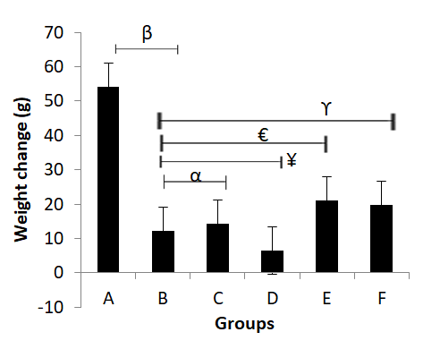
Change in Weight
Weight change was calculated by subtracting the initial weight from the final weight. The results showed that the untreated diabetic (Group B) had a significant weight change less than group A which improved in group C. Groups E and F had significantly increased body weight change compared to group B; however, there was a significant decrease in group D.
Spatial Memory and Learning
The spatial memory and Learning ability of control (group A), diabetic untreated (group B), medium and high doses, as well as standard drug groups, were measured using Morris Water Maze (Table 2, Figure 2). Diabetes induction hampered the spatial memory and learning ability by significantly decreasing the latency in group B compared to the control group (β)(p<0.01). In the present experiment, there was a noticeable improvement in the memory and learning of the rats in the medium and high doses of the extract as compared to diabetic untreated Group B (ϒ) (p<0.01) while group F (α) showed a significant improvement in learning compared diabetic untreated (p<0.01), (Figure 2).
| Acquisition phase | Reverse Phase | |||||
|---|---|---|---|---|---|---|
| Groups | Day 1 (s) | Day 2 (s) | Day 3 (s) | Day 1 | Day 2 | Day 3 |
| A | 22.69±7.71 | 25.39±11.07 | 16.78±6.89 | 19.68±1.16 | 24.38±2.52 | 24.28±2.10 |
| B | 47.06±10.70* | 48.81±8.11* | 56.87±2.10* | 16.18±0.05* | 48.57±2.72*y | 33.43±3.89*y |
| C | 15.57±1.28** | 28.65±3.16** | 25.43±6.05** | 25.52±6.81** | 49.88±3.29 | 37.60±4.22** |
| D | 19.89±3.91** | 36.53±2.76** | 27.20±7.92** | 24.32±6.39** | 53.27±0.48** | 40.70±3.45** |
| E | 29.67±6.06** | 36.58±0.09** | 29.03±9.25** | 29.69±6.06** | 36.58±0.09a | 29.03±9.25a |
| F | 21.34±8.88** | 31.52±7.70** | 25.75±7.17** | 21.34±8.88** | 31.52±7.70a | 25.75±7.17a |
Table 2: ** The effect of aqueous extract of _Invirngia gabonensis_ (Ig) on the spatial acquisition memory level in Streptozotoci
Groups; N= no of trials = 4; *Significant decrease in time compared to A at P≤0.01; Significant increase in time compared to B at P≤0.01; *y Significant increase in time compared to A at P≤0.01; a Significant decrease in time compared to B at P≤0.01 Table 2:** The effect of aqueous extract of Invirngia gabonensis (Ig) on the spatial acquisition memory level in Streptozotocin- induced diabetic rats.
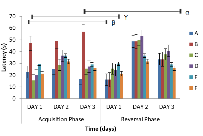
Figure 2: Change in the spatial memory during the experiment. ϒ-Day 1 of group A acquisition compared to reversal of B; β-Day 1 of group B acquisition compared to reversal C and α- Day 3 of group B acquisition compared to F reversal phase. Estimation of Biomarkers The results of some biomarkers estimated are represented in Table 3 and figure 3 below. In the present research, TBARS significantly decreased in the diabetic group before progressively decreasing in Groups C and D, after which it increased significantly in groups E and F. GPX showed a significant increase in the diabetic group compared to the control (p≤0.01) before an increase in groups C and D. There was a significant decrease in GST value of Group B compared to group A (p≤0.01), while group C had a significant increase in GST value compared to group B. However, groups D, E, and F significantly decreased GST levels compared to Group B (p≤0.01). Group B significantly increased GOT value compared to group A at p≤0.01, while groups C, D, and E significantly decreased GOT level compared to group B (p≤0.01). However, group F significantly increased compared to group B (p≤0.01).
| Groups | T-BARS | GPX | GST | GOT |
|---|---|---|---|---|
| A | 3.59±0.009 | 0.69±0.045 | 6.25±0.044 | 41.25±0.48 |
| B | 1.96±0.023* | 1.11±0.042a | 5.41±0.009* | 66.50±0.65a |
| C | 1.95±0.09 | 1.01±0.032 | 6.53±0.013*y | 39.00±0.41** |
| D | 1.69±0.09 | 1.06±0.031 | 4.54±0.013** | 36.00±0.41** |
| E | 2.50±0.023*y | 0.86±0.023** | 4.51±0.055** | 23.00±0.41** |
| F | 2.30±0.037*y | 0.80±0.048** | 4.82±0.014** | 75.00±0.41*y |
Table 3: ** The effect of aqueous extract of _Invirngia gabonensis_ (Ig) on the biochemical brain markers in Streptozotocin-induc
*Significant decrease compared to A at P≤0.01; Significant decrease compared to B at P≤0.01; *y Significant increase compared to B at P≤0.01; a Significant increase compared to A at P≤0.01 Table 3:** The effect of aqueous extract of Invirngia gabonensis (Ig) on the biochemical brain markers in Streptozotocin-induced diabetic rats.
Microscopic Examination
After microscopically examining the hippocampus, group A presented normal hippocampal histoarchitecture of the pyramidal, polymorphic, and molecular layers (Plate 1). It clearly showed CA1, CA2, CA3, and CA4 that play a vital role in spatial learning with several blood vessels and normal dentate gyrate. Group B (diabetes untreated) showed some degeneration, such as vacuolation of the pyramidal cell, hypertrophied neuron, cells with large nuclei, and loss of nuclei (Plate 2). Group C showed moderate regeneration with a distinct pyramidal cell layer and blood vessels with normal nuclei, as shown in Plate 3. Group D showed mild regeneration with a pyknotic pyramidal neuron (Plate 4). Group E showed moderate regeneration with mild vacuolation within the plexiform layer; however, the pyramidal layer appeared distinct.
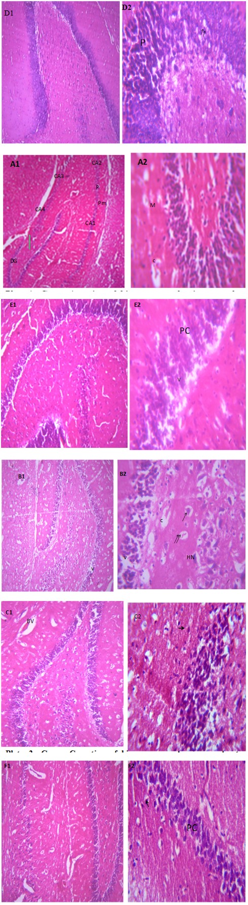
Plate 1: Group A section of hippocampus showing normal three layers: pyramidal layer (p), polymorphic layer (PM), molecular layer (M). It shows CA1, CA2, CA3, widened blood vessel and dentategyrate (DG), X100 and 400, H&E.
Plate 2: Group B section of hippocampus showing moderate to severe degeneration with severe vacuolation (V) of the pyramidal cell with moderate hypertrophied neuron (HN), cell with large nuclei (arrow), cell with lost nuclei (double arrow) and wid ending of vessels (c), (X400)(H/E)
Plate 3: Group C section of hippocampus showing moderate regeneration with distinct pyramidal cell layer, blood vessels (BV), normalnuclei (arrow)(X100 & 400) (H/E).
Plate 4: Group D section of hippocampus showing mild regeneration with moderate pyknotic (p) appearance of pyramidal neuron (double arrows) (X100 & 400)(H/E).
Plate 5: Group E section of hippocampus showing (X400) (H/E) shows moderate regeneration with mild vacuolation (V) within the plexiform layer however the pyramidal cell distinct.
Plate 6: Group F section of hippocampus showing moderate regeneration with the pyramidal layer having healthy prominent pyramidal cell (PC), normal nuclei (arrow) (X100 & 400)(H/E).
Discussion
According to Kumar, et al. [20] diabetes has been reported to lower patients’ weight as the cells cannot effectively utilize the available nutrients in the body, which agrees with the present study. Therefore, upon introduction of the I. gabonensis, weight improvement might be a pointer to the extract’s ability.
Examination of the animal’s behavior on the third day of training, no significant differences in the meantime taken by the rats to locate the platform were observed, which indicated a reduction in the latency period it took them to find the hiding platform. This research showed an increase in the latency period it took diabetic untreated compared to the control group. The increase in time for diabetic untreated to discover the hidden platform may be attributed to degenerations seen in the hippocampus, such as vacuolations and loss of the basic histological form of the pyramidal cells. The degeneration of pyramidal cells implies that the activities projected into the pyramidal layers of the hippocampus will also be lost, such as memory and learning ability [20]. Our result agrees with the previous study by Ryan [20, 21]. which reported that learning and memory capacities are reduced in diabetic patients. The present study showed that groups treated with a low, medium, and high dose of the extract significantly decreased the latency period it took to locate the platform compared to diabetic untreated, which may be a result of the ameliorative role of aqueous seed extract of I. gabonensis. This present work agreed with the previous work of when they stated that ethanol extract of I. gabonensis has an anti-hypoglycemic effect. However, in the group treated with a low, medium, and high dose of the extract, the reversal testing period showed that there was a significant increase in time to complete the Morris Water Maze task on some days compared to diabetic untreated, which could be as a result of certain factors that can influence the performance including sex, the environment in which there were raised, exposure to the drug 21 [23].
Hyperglycemia has been implicated in promoting increased oxidative stress with harmful effects on the central nervous system Chilelli, et al. [24] and, on the other hand, increases the concentration of TBARS. Oxidative stress, a common pathology, occurs due to an imbalance during the production and detoxification of reactive oxygen species (ROS) and has been implicated in many neurodegenerative diseases [25]. In this study, there is a significant decrease in the level of TBARS in diabetic untreated in contrast with the previous work of El-Bahr, 2013. TBARS measures lipid peroxidation in tissue, and its increase in the blood could lead to neuronal damage. There was a further decrease in the level of TBARS in low, medium, and high doses as well as the standard drug. Diabetic untreated GOT levels significantly increased [26]. All animals that received the extract had a decreased GOT activity level while increasing the standard. According to diabetes increases the level of GPX, but in contrast to the present study, GPX in the person with diabetes was reduced while all other animals showed a decrease in GPX.
Previous studies have revealed that Sustained hyperglycemia leads to hippocampal degeneration, neurological damage, and behavioral disruptions [27]. The control showed normal histoarchitecture of the hippocampus with the pyramidal layer, polymorphic layer, and molecular layer, as well as the CA1, CA2, CA3, and CA4, which play a role in spatial learning and cognition [27, 28]. In addition, it has widened blood vessels and normal dentate gyrate. Diabetic untreated showed moderate degeneration, vacuolation of the pyramidal cell, hypertrophied nuclei, and some with lost nuclei, in agreement with Mohammed and [29, 30, 31]. Extract low dose showed moderate regeneration with distinct pyramidal cell layer and blood vessels with normal nuclei, while medium dose showed a moderate pyknotic appearance of the neuron. The high dose appeared very distinct from others, and the standard drug proved healthy with prominent pyramidal cells. Thus, the aqueous extract of I. gabonensis may have a dose-dependent ameliorative effect on the diabetic histoarchitecture of the hippocampus.
Conclusion
I. gabonensis had an effect in ameliorating diabetes in adult Wistar rats; it also showed regenerations in the hippocampus of the treated diabetic rat. Therefore, early management of diabetes with I. gabonensis should be encouraged to avoid complications in diabetes that can lead to neuronal damage.
Conflict of Interest
The authors of this research declare that there is neither any conflict of interest in this experiment nor will it ever arise.
References
-
American Diabetes Association (2012) Diagnosis and classification of diabetes mellitus. Diabetes Care 33(1): S62-S69.
-
Selvarajah D, Wilkinson ID, Davies J, Gandhi R, Tesfaye S (2011) Central nervous system involvement in diabetic neuropathy. Curr Diab Rep 11(4): 310-322.
-
Ye L, Wang F, Yang RH (2011) Diabetes impairs learning performance and affects the mitochondrial function of hippocampal pyramidal neurons. Brain Res 9(1411): 57- 64.
-
Biessels GJ, Gispen WH (2005) The impact of diabetes on cognition: what can be learned from rodent models?. Neurobiol Aging 26(1): 36-41.
-
Watson NA, Dyer KN, Buckley JD, Brinkworth GD, Coates AM (2015) A randomized trial comparing low-fat diets differing in carbohydrate and protein ratio, combined with regular moderate intensity exercise, on glycaemic control, cardiometabolic risk factors, food cravings, cognitive function and psychological wellbeing in adults with type 2 diabetes: Study protocol. Contemp Clin Trials 45: 217-225.
-
Revsin Y, Saravia F, Roig P, Lima A, Kloet ER, et al. (2005) Neuronal and Astroglial Alterations in the hippocampus of the mouse model for type 1 diabetes_._ Brain Res 1038(1): 22-31.
-
Jinzi Wu, Liang-Jun Y (2015) Streptozotocin-induced type 1 diabetes in rodents as a model for studying mitochondrial mechanisms of diabetic β cell glucotoxicity. Dove Press Journal 8: 181-188_._
-
Xiong Y, Chen X, Zhao X, Fan Y, Zhang Q, et al. (2020) Altered regional homogeneity and functional brain networks in Type-2-diabetes with and without mild cognitive impairments. Sci Rep 109(1): 21254.
-
Amin SN, Younan SM, Youssef FM, Rashed LA, Mohamady I, et al. (2013) A histological and functional study on the hippocampal formation of normal and diabetic rats. F1000Res 2: 151-173.
-
Sibiya N, Mabandla M (2017) The Application of Pectin- Insulin Patch on Streptozotocin-Induced Diabetic Rats: Implications in the Hippocampal Function. J Diabetes Metab 8: 779.
-
Duan W, Sehrawat P, Balachandrasekaran A, Bhumkar BA, Boraste PB, et al. (2020) Cerebral blood flow Associated with Diagnostic Class and Cognitive decline in Alzheimer’s. J Alzheimers Dis 76(3): 1103-20.
-
Joghataie MT, Roghani M, Jalali MR, Baluchnejadmojarad T, Sharayeli M (2007) Dendritic Spine Changes in Medial Prefrontal Cortex of Male diabetic rats using Golgi- impregnation method. Arch Iran Med 10(1): 54-58.
-
Jackson-Guilford J, Leander JD, Nisenbaum LK (2000) The effect of streptozotocin-induced diabetes on the cell proliferation in the rats’ dentate gyrus. Neurosci Lett 293(2): 91-94.
-
Obianime AW, Uche FI (2010) The Phytoconstituents and comparative effects of aqueous extract of _Irvingia_ _gabonensis_ seeds and Proviron on the biochemical parameters of male guinea pigs. Asian Pac Journal of Tropical Medicine; 3(2): 101-105.
-
Ngondi JL, Oben JE, Minka SR (2005) The effect of Irvingian gabonensis seed on the body weight and blood lipids of obese subjects in Cameroon. Lipids Health Disease 4: 12.
-
Olawayemi AO, Basiru OO, Babatunji EO, Adebola BO (2014) Hematological properties of _Irvingia gabonensis_ in male adult rats J. Sci Innov 3(5): 434-436.
-
Hossan E, Jameel AAM, Ahmed A, Kairy Z, Jamaa A, et al. (2015) Effect of STZ-induced diabetes on the camel whey proteins. Pakistan Journal of zoology. 47(4): 1109-1116.
-
Morris R (1984) Development of Water-Maze procedure for studying spatial learning in rats. J Neurosci Methods; 11(1): 47-60.
-
Liu Li, Jiong D, Charles M, Junying G, Gang Hu M, et al. (2011) Pretraining affects Morris water maze performance with different patterns between control and ovariectomized plus d-galactose-injected mice. Behavioral Brain Research. 217(1): 244-247.
-
Kumar S, Vasudeva N, Sharma S (2012) GC-MS analysis and screening of antidiabetic, antioxidant, and hypolipidemic potential of Cinnamomum Tamala oil in streptozotocin-induced diabetes mellitus in rats. Cardiovasc Diabetol 11: 95.
-
Wolf U, Rapoport MJ, Schweizer TA (2009) Evaluating the affective component of the cerebellar cognitive and affective syndrome. Journal Neuropsychiatry Clinical Neuroscience 21(3): 245-253.
-
Erus G, Battapady H, Zhang T, Lovato J, Miller ME, et al. (2015) Spatial patterns of structural brain changes in type 2 diabetes and the longitudinal progression with the intensive blood glucose control. Diabetes Care. 38(1): 97-104.
-
Nagayach A, Patro N, Patro I (2014) Experimentally induced diabetes causes glial activation, glutamate toxicity, and cellular damage leading to changes in motor function. Front Cell Neurosci 8.
-
Chilelli N, Burlina S, Lapolla A (2013) Age, rather than hyperglycemia, is responsible for microvascular complication in diabetes: “glycoxidation-centric” point of view. Nutr Metab Cardiovasc Dis 23(10): 913-919.
-
Borra SK, Gurumurthy P, Mahendra M, Jayamathi KM, Cherian CN, et al. (2013) Antioxidant and free radical scavenging activities of curcumin determined by different in vitro and ex vivo models. J Med Plants Res 7(36): 2680-2690.
-
Pintana H, Apaijai N, Pratchayasakul W, Chattipakorn N, Chattipakorn SC (2012) Effects of metformin on learning and memory behavior and mitochondrial brain functions in high-fat diet-induced insulin-resistant rats. Life Sci 91(11,12): 409-414.
-
El-Bahr SM (2013) Curcumin regulates insulin-like growth factor gene expression, B-cell CLL/lymphoma 2, and antioxidant enzymes in streptozotocin-induced diabetic rats. BMC Complement Altern Med 13: 368.
-
Sun LN, Liu XC, Chen XJ, Guan GJ, Liu G, et al. (2016) Curcumin attenuates high glucose-induced podocyte apoptosis by regulating functional connections between caveolin-1 phosphorylation and ROS. Acta Pharmacol Sin 37: 645-655.
-
Mohammed Faheem N, Askary AE (2017) Neuroprotective role of curcumin on the hippocampus against the structural and serological alterations of streptozotocin-induced diabetes in Sprague Dawley rats. Iran J Basic Med Sci 20(6): 690-699.
-
Quirino CJ, Medrado Faria MA, Fraguas R (2012) Depression, insomnia, and memory loss in a patient with chronic intoxication by inorganic mercury. Journal of Neuropsychiatry and Clinical Neurosciences 15(4): 457-458.
-
Matough FA, Budin SB, Hamid ZA, Alwahaibi N, Mohamed J (2012) The role of oxidative stress and antioxidants in diabetic complications. Sultan Qaboos Univ Med J 12(1): 5-18.
- A Review of Gene Therapy for Parkinson's Disease to Control Dopaminergic Neurons
- Late-Onset Myasthenia Gravis in a Patient with Recurrent Breast Cancer: A Case Report
- Covid-Induced Dystonia and Opsoclonus: A Case Report
- Generalized Tonic-Clonic Seizure in a Pediatric Patient with Sunflower Syndrome: A Case Report
- Comparison of Doppler Guided Seldinger Technique Versus Classic Palpatory Seldinger Technique for Radial Artery Cannulation-an Open Label Randomized Controlled Trial
- Brown Sequard Syndrome: Understanding the Complexities of Spinal Cord Injury