Intentional Replantation of Endodontically Treated Tooth- A Case Report
Conventional and surgical endodontic therapy is accepted as foreseen clinical procedures that save teeth with pulpal disease which would otherwise be extracted. When a tooth's pulp is irreversibly damaged or infected due to caries, cracks, trauma, or leaky restorations, endodontic therapy or extraction are the only workable options [1]. Teeth undergoing initial endodontic therapy have a very high survival rate. Some teeth that continue to show signs of pathosis after the initial therapy will require nonsurgical (orthograde) retreatment [2]. Intentional replantation is valid and inevitable for cases in which nonsurgical endodontic retreatment is unsuccessful or is impractical and endodontic surgery is hindered because of anatomic restrictions [3].
Case Report
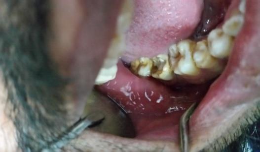
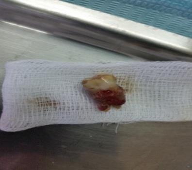
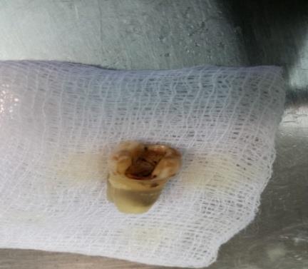
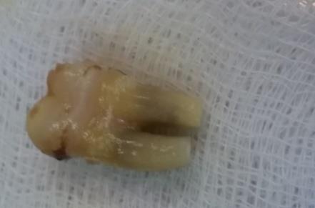
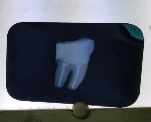
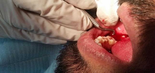
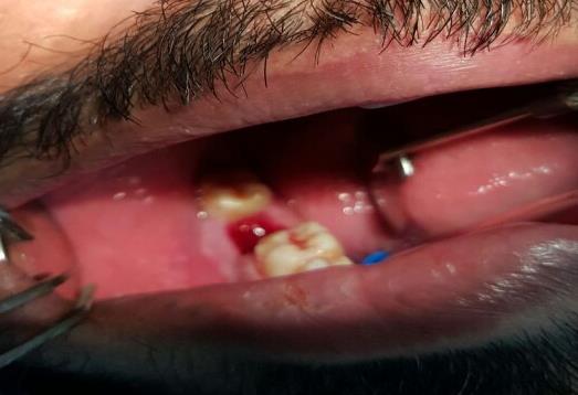
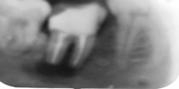
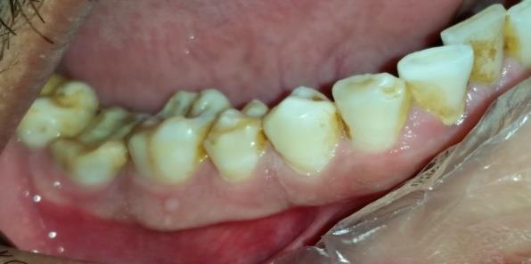
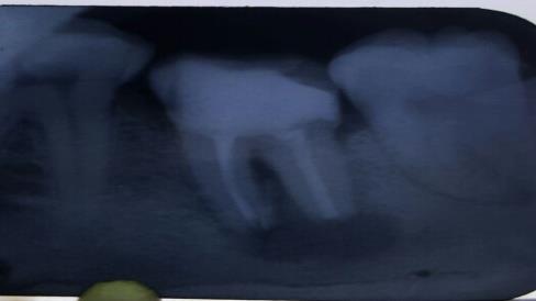
A 35-year-old male patient presented in outdoor patient department had a complain of pain in lower right molar area when chewing. Upon examination, lower right second molar was found tender to percussion. History revealed that the patient underwent root canal treatment for this tooth and was informed about root perforation. Further clinical and radiographic examination showed root perforation just apical to Cementoenamel junction in distal root and signs of chronic periapical periodontitis. All possible treatment options were explained to the patient including i) tooth extraction with/without replacement, ii) retreatment, hemi section and coronal restoration, iii) peri radicular surgery, and iv) Intentional Replantation (IR). The risks and benefits of each option were thoroughly explained and an informed consent was taken. The clinical procedure for IR was as follows: A Chlorhexidine rinse was carried out to control the oral microflora prior to procedure. After achieving complete local anesthesia, the tooth was intentionally and gently extracted using a suitable periotome and forceps. Extracted tooth was carefully root planed extra-orally. First removal of necrotic cementum, granulation tissues and all irritants from the root surfaces was carried out using periodontal curettes. Root conditioning was done using EDTA and doxycycline rinses. Extra oral root canal treatment was performed and the perforation was repaired by using Mineral trioxide aggregate (MTA). The 2-3 mm root-end was resected using diamond bur. Granulation tissue in the extraction socket was removed by gentle curettage and the socket was rinsed with sterile saline. The tooth was then gently replanted into the socket followed by gently pressing the buccal and lingual plates. The patient was asked to bite gently on a wooden stick afterwards which helped in stabilizing the tooth. The accurate repositioning was confirmed by radiography. The tooth did not require stabilization with splints as it was slightly cut-off from occlusion. Tab. Augumentin 625mg in combination with metronidazole 400mg was prescribed in BD dose for 5 days. Patient was recalled after 7 days for suture removal and every month thereafter for 3 months (Figures 1-11).

Discussion
When nonsurgical and surgical endodontic treatment has been considered unreasonable and the patient wishes to make every attempt to save his tooth, intentional replantation can be deemed lastly [4]. Intentional dental reimplantation is a treatment option that should not be underestimated, as its treatment outcomes are excellent.
In order to be successful with extraction and reimplantation cases, the dentist must have the right selection of patient and the right rapport with that patient. The practitioner also must be able to assess the tooth and be affluent with the fact that it can be extracted without breakage. Moreover, being able to identify tooth morphologies that can cause difficulty in extraction is must. This is a skill that is perfected through experience. Reimplantation is a predictable and acceptable method of treatment when patients present with root canals that require retreatment due to failure or those that cannot be completed due to sclerosing of the canals [5].
Indications
a) When routine RCT is impossible or impractical, as with some patients with limited mouth opening or cannot open for the necessary time. b) When a previous RCT has been unsuccessful but orthograde retreatment or apical surgery is impractical. c) Obstruction of canal due to instrument separation or calcification. d) When surgical access would be inadequate, such as a shallow buccal vestibule or short roots. e) When an iatrogenic perforation or a perforating internal or external resorption is present, but surgery is impractical. f) When a foreign body is present in periapical (PA) tissues or PDL, but surgery is impractical. g) If PA radiographs show a large region of rarefaction or a large cyst. h) When the root canal is furcated as it approaches the apex. i) For RCT of immature teeth. j) For primary teeth, as an alternative to extraction and placement of a space maintainer. k) Management of vertical root fractures by reconstructing with dentin bonding agents- management of complicated crown-root fractures. l) For RCT of teeth with certain anatomical malformations such as radicular groove or double teeth. m) Progressive destruction of periodontal tissues and management of teeth with advanced endo-perio lesions. n) Management of maxillary sinusitis [6]. Contraindications: IR is not recommended in the following situations:
a) Patient’s medically compromised history may prevent PA surgery or extraction. b) Extensive caries or no restorability of the tooth. c) Severe periodontally compromised tooth. d) Curved roots or the possibility of fracture during extraction or during the replantation. e) Broken or fractured teeth. f) When labial/buccal plate has been damaged and gingival inflammation and the involvement of furcation exist. g) In ankylosed teeth, IR may be indicated only when the ankylosis is diagnosed at the early stages [6].
Conclusion
Conventional apicoectomy or non-surgical retreatment is a first choice for cases with persistent and recurrent periapical symptoms after root canal therapy. However, in some cases, due to anatomic difficulties or contraindications for surgery or retreatment, an intentional replantation procedure can be performed as an alternative to complete extraction. Case selection is very important for the success; the most significant selection criteria include non-fractured teeth with conical roots, lack of periodontal pathology, and intact, well- sealing coronal restorations. Atraumatic extraction and extraoral times less than 10 minutes will give intentional replantation procedure a better prognosis.
References
-
Nasseh AA (2015) Intentional Replantation: Does It Work? A technique to consider in rare cases when conventional surgery is impossible. Inside dentistry 11(5).
-
Salehrabi R, Rotstein I (2010) Epidemiologic evaluation of the outcomes of orthograde endodontic retreatment. J Endod 36(5): 790-792.
-
Grzanich D, Rizzo G, Silva RM (2017) Saving Natural Teeth: Intentional Replantation-Protocol and Case Series. J Endod 43(12): 2119-2124.
-
Majd NM, Arvin A, Darvish A, Aflaki S, Homayouni H (2014) Treatment of Necrotic Calcified Tooth Using Intentional Replantation Procedure. Case Report in Dentistry 2014.
-
McFarland (2005) Intentional Extraction and Reimplantation of the Same Tooth. Dentistry today.
-
Rouhani A, Javidi B, Habibi M, Jafarzadeh H (2011) Intentional Replantation: A Procedure as a Last Resort. J Contemp Decnt Pract 12(6): 486-492.
- Diagnosis and Management of Mental Nerve Paresthesia Secondary to Apical Periodontitis of Mandibular Second Premolar: A CBCT Based Case Report
- A Randomized, Double Blinded Clinical Trial to Compare the Effect of Oral Premedication (Diclofenac Potassium or Dexamethasone) on Post-Operative Pain Following Pulpectomy
- Modified Lip Repositioning Technique for the Management of Excessive Gingival Display
- Integral Role of Non-Dental Providers and Fluoride Dissemination
- Root Canal Treatment Rate in Deciduous Teeth Among 6-Year- Olds in the Era of Discontinuing Water Fluoridation - Historical Cohort Study
- The Impact of the Notch1 on the Migratory Capacity and the Expression of E-Cadherin and CyclinD1 in Ameloblastoma Cells