Molar-Incisor Hypomineralization–A Review
Molar incisor hypomineralization (MIH) describes the hypomineralization of systemic origin affecting one or more first permanent molars (FPMs) and are associated with affected incisors. Etiological associations with systemic conditions or environmental insults during the prenatal, natal and postnatal times have been implicated. The comprehensive care involved in treating affected children must address their behavior and anxiety, aiming at early diagnosis, remineralization and desensitization, prevention of caries and posteruption breakdown; restorations and extractions under pain-free conditions and maintenance. The challenges include adequate anaesthesia, suitable cavity design, and choice of restorative materials. Restorations in hypomineralized molars appear to fail frequently; there is little evidence-based literature to facilitate clinical decisions on cavity design and material choice. The high prevalence of MIH indicates the need for research to clarify etiological factors and improve the durability of restorations in affected teeth. The purpose of this paper was to describe the prevalence, etiological factors, and clinical features of hypomineralized enamel in molar incisor hypomineralization and to present a sequential approach to management.
Introduction
Developmental defects of tooth enamel are not uncommon, both in the primary and permanent dentitions, and can be divided into hypomineralisation and hypoplasia. Enamel hypomineralisation can be observed visually because of a different translucence that is known as opaque enamel. The opacity may be diffuse or sharply defined, whereas in cases of hypoplasia parts of the enamel are absent or very thin with smooth borders adjacent to normal tissue.
The causes of developmental enamel defects may be congenital, acquired or unknown. Congenital defects, such as amelogenesis imperfecta, have a genetic basis. In the case of acquired defects, the aetiology is very often unknown. Examples of an acquired defect with a known cause are trauma or an excessive use of fluoride. Molar- Incisor Hypomineralisation (MIH) is an example of an acquired defect of unknown aetiology. Molar incisor hypomineralization is defined as the developmentally derived dental defect that involves hypomineralization of one to four permanent first molars that is frequently associated with similarly affected permanent incisors.
Hypomineralisation of permanent teeth has been described in the literature since 1970s but it was in 2001 Weerheijm, et al. suggested the term molar incisor hypomineralisation for this developmental disorder teeth. It was first noted in Sweden in 1970 [1].
Nomenclature
Differential terms were given to MIH before 2001 when Weerheijm KL, et al. suggested the term MIH [2, 3, 4]. The other terms given were: a) Cheese molar in Netherlands b) Non- flupride enamel opacities c) Internal enamel hypoplasia d) Non-endemic mottling of enamel e) Idioathic enamel opacities f) Opaque spots
Prevalence
Etiology
The etiology of MIH is considered to be a combination of factors. The mineralization of first permanent molar begin before birth [32nd week] and is completed around 4year of age development of upper central incisor closely follows, hence the cause of abnormal enamel formation must be present in this particular period. The length of the time period from the beginning of mineralization to eruption of tooth is very similar is molar and incisors [7, 8].
Maxillary lateral incisor, mandibular central and lateral incisor are less frequently affected, difference in developmental timetable partly explain this.
An early insult of the ameloblast may influence the cell in three different ways: First: The disturbance of the ameloblasts ability to produce the correct deposition of proteins. If the protein deposition is incorrect, subsequent accurate maturation is impossible. An increase of 3-15 times more protein is hypomineralised enamel has been found. Most of the amelogenin was resorbed. However, other proteins such as albumin which is a protein known to inhibit tooth mineralization persisted. Secondly: Ameloblasts are affected during late secondary phase or early maturation stage. These stages are very sensitive process. In secretory phase partially mineralised enamel is deposited to the whole enamel thickness. Later organic material and water in the enamel are removed to allow an additional influx of mineral. An insult of ameloblast during this phase is considered to result in hypomineralisation. If the protein resorption does not occur, calcium and phosphate will not be able to enhance the prism development, crystal hydroxyapatite does not form. Thirdly: Ameloblast in a transistional stage during enamel mineralization are the most vulnerable type of ameloblast. An insult of ameloblast at a specific time of this sensitive transition may affect the ameloblasts and cause a disturbed function [7].
The enamel mineralization front is subject to periodic changes and regular variations in ameloblast activity. These variations produce short and long period incremental lines in the enamel. The long period growth lines, or Retzius lines, mark the layers of enamel produced by the secretory ameloblasts, which occur every 6 to 12 days in modern humans. Short-period growth lines, or cross-striations, represent a daily circadian rhythm in secretory ameloblast activity. Both short- and/or long- period growth lines have been used to calculate rates of enamel secretion and cusp formation time. Even if the estimation of the timing of disturbance of the ameloblasts is crude, a possible time range covers a period up to around 200 days after the start of the enamel mineralization. The location of a Retzius line at the enamel surface of the hypomineralized enamel would correspond to 300-400 days, which cannot be realistic since the hypomineralized zone has a cervical limitation along a prism.
Some researchers speculate that prevalence of developmental enamel defects is highest at locations where the enamel is thickest, it has been suggested that increased metabolic demand of ameloblasts in the thicker areas and hence they are more vulnerable to insult [7]. Classification of etiologies of molar-incisor hypomineralisation: Prenatal medical conditions [1, 5, 8, 9] a) High episodes of fever , common cold b) Maternal diabetes {hypocalcemia} c) Hypertension d) Prolonged use of medication [Myometrium spasmoytics] e) Malnutrition f) Vomiting upto last month [imbalance in fluid and eiectrolyte balance, hypoxia]
g) Chicken pox h) Renal deficiency Perinatal a) Caesarean section b) Prolonged / Complicated delivery[ Respiratory distress and birth asyphxia] c) Twins d) Premature birth – low birth weight [Hypocalcemia] e) Haemorrhage –[ detachment during delivery] f) Cyanosis Postnatal a) Repeated episodes more than 5 episodes of high fever b) Common cold/Coryza c) Otitis media d) Bronchitis/ Bronchiolitis e) Asthma f) Laryngitis/Tonsilitis g) Seizures – afebrile/febrile h) Urinary infection i) Encephalitis j) Gastroenteritis k) Exanthematous diseases[Incubator] l) Dioxin present in Mothers milk Prolonged uses of antibiotics were initially considered to cause hypomineralisation but subsequently it was understood that it was the underlying disease which was causing hypomineralisation. All the conditions are known to cause hypoxia and hypocalcemia which has detrimental influence on amelogenesis, ranging from ameloblastic dysfunction to complete cellular degeneration [2].
Clinical Features
MIH can be visualized as abnormality in the translucency of tooth. It varies from creamy white to yellowish brown in color. It shows a clear border between affected and sound enamel. The enamel of MIH molar looks soft and porous and has the appearance of discoloured chalk or old dutch cheese. MIH tooth shows smooth and rounded margins. Some opacities have significant subsurface porosity leading to breakdown of the surface after leading to breakdown of the surface after eruption. Hypomineralised porous enamel can chip off easily, leading to unprotected dentine and also an unexpectedly rapid caries development. The loss of enamel can occur immediately after eruption as the result of masticatory forces on the fragile enamel. The borders to the normal enamel is irregular incase of posteruptive breakdown. The teeth are brittle and there is increased porosity and hence more susceptible to fracture. There is increased dentinal sensitivity [3].
Histological Features
The yellowish brown defects are more porous than the creamy white to cover the entire thickness of the enamel. The creamy white defects are located in the internal part of the enamel [2]. Diagnostic criteria [European academy of pediatric dentistry 2009] [3, 4, 10] Demarcated opacity: A demarcated defect involving an alteration in the translucency of the enamel, variable in degree. The defective enamel is of normal thickness with a smooth surface and can be white, yellow or brown in colour. Posteruptive enamel breakdown: A defect that indicates deficiency of the surface after eruption of the tooth. Loss of initially formed surface enamel after tooth eruption. The loss is often associated with a pre-existing demarcated opacity. Atyptical restoration: The size and shape of restoration conform the temporary caries picture. In most cases restoration will extend to buccal or palatinal smooth surface. At the border of the restorations frequently an opacity can be noticed. In incisors restoration can be noticed not related to a trauma. Extracted molar: Absence of molar in a sound dentition in combination with demarcated opacities or atypical restoration in the other first permanent molar combined with absence of molars. Unerupted: The first permanent molar or incisor to be examined are not yet erupted. On the basis of a time table of human dentition.
Clinical Implications
If molars show signs of opacities and/or post-eruptive breakdown, a child should be seen every three months until the time when the six year molars have completely erupted. In order to minimise the loss of enamel and any damage due to caries, both preventive and interceptive treatment is required. The presence of MIH not only requires the dentist to identify problems at the earliest opportunity, but also to thoroughly explain the problems to the parent and child. As only the permanent first molars (and sometimes the incisors) are affected by the developmental enamel defect, the parents can be reassured with respect to the quality of the remaining teeth that have not yet erupted [2].
Clinically MIH molar can create discomfort to the child. The affected teeth can be very sensitive to current of air, cold and warmth. Even with enamel that has not disintegrated, mechanical stimuli for instance tooth brushing may instigate toothache. It is important to stress that the restorative treatment can be painful due to difficulties in getting a good anesthetic effect probably there is a subclinical inflammation in the pulp cells caused by porosity of the enamel, thus making the management of the child difficult. The children try to avoid the sensitive molars when brushing leading to increased stagnation of food and plaque. The fast caries progression clinically mask the actual reason of hypomineralisation. First year after eruption they are more fragile and brittle and susceptible to posteruptive enamel breakdown [6].
Severity of MIH can be classified as
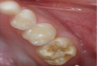
a) Degree 1 [Mild]: Isolated white and cream to yellowish – brown discolouration on the chewing surface and upper part of the crown [3, 9] (Figure 1,2). b) Degree 2 [Moderate]: Hypomineralisation yellowish brown enamel affecting more or less all the on the top of the crown, but with slight loss of substance (Figure 3). c) Degree 3 [severe]: Large scale mineral deficiency with distinct yellowish brown discolorations and defect in crown morphology resulting from extensive loss of enamel (Figure 4).
![Figure 1: Mild [Demarcated opacities on incisors].](/fulltextimages/3331/fig_1.jpeg)
![Figure 2: Mild defects [Demarcated opacities on PFMs].](/fulltextimages/3331/fig_2.jpeg)
![Figure 4: Severe defect [Posteruptive enamel breakdown].](/fulltextimages/3331/fig_4.jpeg)
Management
a) A very useful 6-step management approach for MIH has been proposed by William, et al. [11], b) Risk identification, c) Early diagnosis, d) Remineralisation (a better term may be mineralisation;
the tooth was never ‘completely’ mineralised during development although there may also be an element of demineralisation from enamel caries, superimposed upon the hypomineralised areas) and desensitisation, e) Prevention of dental caries and post eruptive enamel breakdown, f) Restorations or extractions g) Maintenance.
Risk Identification and Early Diagnois
With increased awareness it is possible to trace patients at risk much earlier and initiate preventive treatment more rapidly. Children at risk for MIH should be identified prior to PFM eruption. In the studies it is seen that children with MIH fell ill more frequently in the first four years of life than children in the same age group with normal molars. It appears that diseases concerning the head and neck area were relatively frequent.
Therefore, based upon a relevant history, in children with repeated illnesses in the first four years it seems useful to increase the frequency of dental check-up’s during the period when the permanent first molars erupt. This would be a means to detect the clinical symptoms as early as possible and from careful study under magnification of the unerupted molar crowns on any available radiographs
Preventive Management
The initial treatment is the re-assurance to parents and the children. They should be reassured that the teeth can be restored and it is confined mainly to molars It is very important to start approaching the affected children and their parents with the appropriate dietary and preventive advice. The poor oral hygiene due to increased sensitivity and fragility of the affected enamel were prevalent for the rapid and extensive tooth destruction [11].
During PFM eruption, the hypomineralized surface is very susceptible to caries and erosion. The cariogenicity and erosivity of the child’s diet should be assessed and appropriate recommendations made for dietary modification. Thorough oral hygiene should be instituted; this could include a desensitizing toothpaste. If the child is still using a low-fluoride children’s toothpaste then the parents should be encouraged to change to one with a higher fluoride level of at least 1,000 ppm F.
Remineralization
Topical fluoride, delivered as concentrated varnishes or gels, can remineralize enamel, reduce sensitivity, and enhance resistance to demineralization by providing a reservoir of fluoride ions for redeposition as fluorapatite during remineralization. Anecdotal reports ascribe considerable clinical benefit to topical application of fluoride on hypomineralized molars, resulting in surface hardening of demineralized enamel prior to restoration. Although there is no research at present to evaluate their efficacy in MIH patients, all these products may help to reduce sensitivity and enhance mineralisation of the hypomineralised areas [5, 12].
Remineralization therapy should commence as soon as the defective surface is accessible, aiming to produce a hypermineralized surface layer and to desensitize the tooth. Remineralization and desensitization may be accomplished with casein phosphopeptide-amorphous calcium phosphate (CPP-ACP) oral care products. The CPP-ACP can interact with fluoride ions, producing an amorphous calcium phosphate stabilized by CPP at the tooth surface and providing soluble calcium, fluoride, and phosphate ions to promote remineralization with fluorapatite that is more acid resistant. These products enhance remineralization by creating a state of super saturation followed by deposition of calcium and phosphate ions at the enamel surface. While clinical protocols for CPP-ACP oral care products await development, anecdotal reports describe surface hardening and reduction in tooth sensitivity from daily home use [13].
As remineralization and desensitization of the affected molars occurs, regular oral hygiene strategies can be instituted. For partially erupted PFMs where moisture control is suboptimal, glass ionomer cement sealants can provide caries protection and reduce surface permeability. Retention is poor, however, and such sealants may need rebuilding later with a resin-based sealant when optimal moisture control is possible [13].
Treatment of MIH Divided According to the Severity
Mild cases
Fissure sealants may be useful in mild cases which are not sensitive and without breakdown. They should be regularly monitored and replaced when lost. Opaque or yellow brown fissure can be treated for 60sec pre- treatment with 5% sodium hypochlorite which remove the intrinsic enamel proteins. Fissure sealants (FS) may also be useful for FPM with mild defects, not sensitive and without breakdown, particularly when they are regularly monitored and replaced when lost William, et al. 2006 [11]. Mathu and Wright, suggested that if the fissures appeared opaque or yellow-brown then a 60 second pre- treatment with 5% sodium hypochlorite might be beneficial in that it might remove intrinsic enamel proteins. For partially erupted FPM with MIH, glass ionomer cements (GIC) can be used as FS, providing temporarily, caries and sensitivity protection and minimizing break-downs; as retention of such materials is poor, these should be replaced as soon as the tooth is fully erupted with resin-based sealants [1].
Moderate Cases
Restoring hypomineralized first permanent molars Restoring affected PFMs is complicated frequently by: a) difficulties in achieving anesthesia; b) managing the child’s behaviour; c) determining how much affected enamel to remove; and d) selecting a suitable restorative material.
The porous exposed subsurface enamel or dentin may promote chronic inflammation of the pulp, complicating anaesthesia. The adjunctive use of nitrous oxide-oxygen analgesia may alleviate anxiety and reduce dental pain, or general anaesthesia may be required for restorative treatment.
Commonly used Restorative Materials for the Treatment of MIH
Glass ionomer cements: provides temporary caries and sensitivity protection and minimizing breakdown, but the retention is poor and should be replaced with resin based sealant. It is used as a transitional stage to promote remineralisation of the surrounding structures. It is biocompatible moisture tolerant and enables functional recovery of the patient. GIC are not recommended in stress bearing areas of first permanent molars. GIC can be used as intermediate layer with resin based sealants. When the presence of MIH requires a restoration, the GIC should be considered at least as a transitional stage to promote remineralization of the surrounding structures. It is important to stress that the restorative treatments in hypomineralized teeth can be painful due to the difficulties in getting a good anesthetic effect, probably because there is a subclinical inflammation in the pulp cells caused by the porosity of the enamel; thus making the management of the child difficult. In young children that are difficult to handle, the GIC can be maintained until the complete tooth eruption occurs and the most appropriate behavior is displayed, allowing the use of more resistant restorative materials as it is biocompatible, moisture tolerant, and effective in controlling the sensitivity, enabling a functional recovery of the patient. However, a lower adhesion of these restorative materials can occur because of the changes of the hypoplastic enamel of the affected teeth, early loss, and fractured edges of the restorations and recurrent caries are common. Repeated treatments of teeth with GIC in small intervals are common and may occur 10 times more than in children who do not present this alteration. Adhesive materials are usually chosen due to the atypical cavity outlines following removal of hypomineralized enamel. For dentin replacement or as an interim restoration, GIC provides: (1) placement ease; (2) fluoride release; and (3) chemical bonding [13]. Resin modified GIC: The incorporation of resin monomers in GIC compositions give rise to resin-modified glass ionomer cements (RMGICs). They are material of choice as they have improved mechanical properties, greater working time, Command set on use of visible light, good adaptation and adhesion, Acceptable fluoride release as well as aesthetics. . The RMGICs offer similar advantages to GIC; the incorporation of resin and photo initiators improves: (1) handling; (2) wear resistance; (3) fracture toughness; and (4) fracture resistance.60 Restorations of GIC or RMGIC are not recommended in stress-bearing areas, such as occlusal surfaces of hypomineralized molars, but may suffice until a definitive restoration is achievable. Polyacid modified composite resin: Compomer is a combination of the word ‘comp’ for composite “omer” for ionomer. Though introduced a type of GIC, it became apparent that terms in of clinical use and performance it is best considered as a composite. They have superior working characteristics to RMGIC. Ease of use, easily adapts to the tooth and good esthetics Composite restorations: They are increasingly used nowadays with the advent of newer adhesives. Self- etching enamel etchants have superior bond strength to hypomineralised enamel than single bottle alcohol containing adhesives. This is because of vomiting the step of rinsing hence eliminating the contamination of residual water. In single etching adhesive both micromechanical and chemical bonding to hydroxyapatite is seen as compared with micromechanical seen in single bottle adhesive [13, 14].
Adhesion to Hypomineralised Enamel
The use of various adhesive resin systems has certain limitations in MIH teeth as a result of defective enamel. Study by William, et al. [11] demonstrated that adhesion to MIH enamel is possible, but the enamel-adhesive interface of defective enamel was porous with cracks, had decreased bond strength, and a higher likelihood of cohesive failure compared with sound enamel. A number of studies dealing with the ultrastructure and biochemical make-up of MIH enamel and dentine indicated that the ‘full thickness enamel’ surrounding the clinically defective lesions is less affected and the underlying dentine has no major structural changes. These findings may explain the acceptable results for adhesive composite restorations in molars with MIH, if all apparently defective enamel is removed [13]. With physical properties superior to GIC and RMGIC, the composite are esthetic materials with high wear resistance and adhesion when used with resin- based adhesives; they can be used solely or in a sandwich technique following previous temporization with GIC.
Amalgam is least retentive due to very poor adhesion to hypoplastic teeth. Amalgam is a non-adhesive material and its use in these atypically shaped cavities is not indicated; the inability to protect the remaining structures usually results in further enamel breakdown. The few existing clinical studies of amalgam restorations in MIH molars support this view as they report lower success rates when compared with composite resins. From all available restorative materials, many reviews William, et al. [11], agree on the superior properties of composites, combined with the new adhesive materials. Amalgam is the least durable due to: a) Poor retention in shallow cavity preparations; and b) The inability to protect remaining tooth structure, which is likely to result in restoration failure [13, 14].
Stainless steel crown are best recommended treatment option. They prevent further tooth loss, control sensitivity, establish correct interproximal and proper occlusal contacts. When PFMs have moderate to severe PEB, preformed SSCs are the treatment of choice. These crowns: a) Prevent further tooth deterioration; b) Control tooth sensitivity; c) Establish correct interproximal contacts and proper occlusal relationships; d) Are not as technique sensitive or costly as cast restorations; and e) Require little time to prepare and insert.
Restoring Hypomineralized Permanent Incisors
Hypomineralized incisors in MIH may present esthetic concerns to children and their parents. Microabrasion can be an effective treatment in shallow defects, but the defects usually extend through the full enamel thickness. A conservative approach in managing yellow-brown hypo-mineralized enamel involves: a) Etching the lesion with 37% phosphoric acid; b) Bleaching with 5% sodium hypochlorite; and then c) Re-etching the enamel prior to placing a sealant over the surface to occlude porosities and prevent restaining.
One report of this approach described clinical success with little or no staining up to 5 years post-treatment. Others report little improvement with acid/pumice microabrasion used alone, but esthetic improvement was achieved when the enamel reduction was combined with opaque resins then direct RC veneering. Porcelain veneers are typically delayed until late adolescence when the teeth have fully erupted and the gingival architecture has stabilized.
Severe Cases
Although both the profession and the public believe nowadays in a more conservative treatment plan, some thought might still be given for such a radical approach, resulting usually in the extractions of several permanent teeth. Variables affecting this decision include the child’s age, orthodontic considerations, presence of other dental anomalies, degree of severity of MIH, pulp involvement, presence of third molar germ(s), restorability of the tooth/teeth and expected long term treatment cost. The FPM is not an orthodontist’s first choice for extraction, because later orthodontic treatment may be complicated. Therefore, the decision to extract any of the FPM should be seriously evaluated and discussed with an orthodontist as early as possible if a good result is to be anticipated [14, 15] (Table 1).
| Mild defects | Moderate/ Severe defects | ||||||
|---|---|---|---|---|---|---|---|
| Molars | Incisors | Molars | Incisors | ||||
| Fluoride varnish in partially erupted teeth | In brownish yellow defects, etch bleach sea approach in younger children or chair side bleaching with 10% carbamide peroxide in order. | Consider extractions | Wait until the defect get better, since a degree of enamel mineralisation may occur in the salivary environment. | ||||
| When fully erupted, sealants with prior adhesives | In whitish defects, microabrasion followed if needed by composite restorations | Fluoride varnish or GIC in partially erupted teeth. | Composite restorations or veneers following micro-abrasion or enamel reduction and intermediate opaque resins. | ||||
| Composite restorations if breakdown or caries occur | Composite restorations following enamel reduction | Composite restorations for upto 3 surfaces. | Porcelain veneers if needed | ||||
| Full coverage crown if needed in adulthood | Full coverage restorations for more than 3 surfaces |
Table 1: Treatment summary for patients with MIH [14].
References
-
Weerheijm KL, Duggal M, Mejàre I, Papagiannoulis L, Koch G, et al. (2003) Judgement criteria for Molar Incisor Hypomineralisation (MIH) in epidemiologic studies: a summary of the European meeting on MIH held in Athens, 2003. Eur J Paediatr Dent 4: 110-113.
-
Beentjes VE, Weerheijm KL, Groen HJ (2002) Factors involved in the aetiology of molar-incisor hypomineralisation (MIH). Eur J Paediatr Dent 3(1): 9-13.
-
Bhaskar SA, Hegde S (2014) Molar-incisor hypomineralization: Prevalence, severity and clinical characteristics in 8-to13-year-old children of Udaipur, India. J Indian Soc Pedod Prev Dent 32(4): 322-329.
-
de Oliveira DC, Favretto CO, Cunha RF (2015) Molar incisor hypomineralization: considerations about treatment in a controlled longitudinal case. J Indian Soc Pedod Prev Dent 33(2): 152-155.
-
Kirthiga M, Poornima P, Praveen R, Gayathri P, Manju M, et al. (2015) Prevalence and severity of molar incisor hypomineralization in children aged 11-16 years of a city in Karnataka, Davangere. J Indian Soc Pedod Prev Dent 33(3): 213-217.
-
Weerheijm KL (2003) Molar incisor hypomineralisation (MIH). Eur J Paediatr Dent 4: 115- 120.
-
Fagrell TG, Salmon P, Melin L, Norén JG (2013) Onset of molar incisor hypomineralization (MIH). Swed Dent J 37(2): 61-70.
-
Lygidakis NA, Dimou G, Marinou D (2008) Molar- incisor-hypomineralisation (MIH), A retrospective clinical study in Greek children II, Possible medical aetiological factors. Eur Arch Paediatr Dent 9(4): 207- 217.
-
Lygidakis NA, Dimou G, Briseniou E (2008) Molar- incisor-hypomineralisation (MIH). Retrospective clinical study in Greek children. I. Prevalence and defect characteristics. Eur Arch Paediatr Dent 9(4): 200-206.
-
Weerheijm KL (2004) Molar incisor hypomineralization (MIH): clinical presentation, aetiology and management. Dental Update 31(1): 9- 12.
-
William V, Messer LB, Burrow MF (2006) Molar incisor hypomineralization: review and recommendations for clinical management. Pediatr Dent 28(3): 224-232.
-
Kotsanos N, Kaklamanos EG, Arapostathis K (2005) Treatment management of first permanent molars in children with Molar-Incisor Hypomineralisation. Eur J Paediatr Dent 6(4): 179-184.
-
Lygidakis NA (2010) Treatment modalities in children with teeth affected by molar-incisor enamel hypomineralisation (MIH): a systematic review. Eur Arch Paediatr Dent 11(2): 65-74
-
Calderara PC, Gerthoux PM, Mocarelli P, Lukinmaa P, Tramacere PL, et al. (2005) The prevalence of Molar Incisor Hypomineralisation (MIH) in a group of Italian school children. Eur J Paediatr Dent 6(2): 79- 83.
-
Jasulaityte L, Veerkamp JS, Weerheijm KL (2007) Molar incisor hypomineralization: review and prevalence data from the study of primary school children in Kaunas (Lithuania). Eur Arch Paediatr Dent 8(2): 87-94.
- Diagnosis and Management of Mental Nerve Paresthesia Secondary to Apical Periodontitis of Mandibular Second Premolar: A CBCT Based Case Report
- A Randomized, Double Blinded Clinical Trial to Compare the Effect of Oral Premedication (Diclofenac Potassium or Dexamethasone) on Post-Operative Pain Following Pulpectomy
- Modified Lip Repositioning Technique for the Management of Excessive Gingival Display
- Integral Role of Non-Dental Providers and Fluoride Dissemination
- Root Canal Treatment Rate in Deciduous Teeth Among 6-Year- Olds in the Era of Discontinuing Water Fluoridation - Historical Cohort Study
- The Impact of the Notch1 on the Migratory Capacity and the Expression of E-Cadherin and CyclinD1 in Ameloblastoma Cells