Effect of 9.3μm CO2 Laser and Fluoride on Shear Bond Strength, Microleakage, Acid Resistance and Microhardness of Sealants
Objectives: This study aimed to evaluate the effects of a 9.3 µm microsecond short-pulsed CO2 laser and acidulated phosphate fluoride (APF) 1.23% gel on enamel acid resistance, microleakage, failure mode, and shear bond strength of dental sealants. Materials and Methods: 120 extracted posterior teeth were randomly divided into 3 groups (n=40): Group A, resin-based sealant; Group B, moisture-tolerant resin-based sealant; and Group C, glass-ionomer cement. Each group were divided into 4 subgroups (n= 10) treated with: 1) sealant alone, 2) APF 1.23% fluoride gel + sealant, 3) CO2 laser + sealant, and 4) CO2 laser + APF 1.23% fluoride gel + sealant. Sealants were applied on the buccal and occlusal surfaces. Shear bond strength (SBS) test was performed on the buccal surface sealants using the Instron® 5566A, followed by failure mode assessment under a digital microscope. After the SBS test, the teeth were sequentially thermocycled, immersed in a 2% methylene blue solution, and sectioned longitudinally in a buccal-lingual direction. The sectioned mesial halves were examined under a digital microscope to assess dye penetration. The sectioned mesial halves were assessed for baseline Vickers microhardness at four standardized points on mesial surface, and for the microhardness after treatment at four standardized points located 50 micrometers on enamel adjacent to the enamel-sealant interface on the cut surface. The mesial halves were treated to 9 days of pH-cycling of remineralization and demineralization solutions. After acid challenge, the microhardness measurement was performed at four standardized points located 50 micrometers next to the four original points. Results: For the three sealant materials, CO2 laser + sealant provided higher SBS than other subgroups: 15.5 ± 8.2 MPa for Clinpro, 16.3 ± 7.8 MPa for Embrace, and 4.9 ± 4.7 for Fuji. CO2 laser + sealant also had lower percentage of adhesive failure than other subgroups for Clinpro and Embrace. For Clinpro, microhardness values of subsurface enamel adjacent to the enamel-sealant interface after pH cycling were 202.8 ± 30.1 for sealant, 186.7 ± 39 for fluoride + sealant, 231.4 ± 34.1 for laser + sealant, and 258.3 ± 28.2 for laser + fluoride + sealant. In Clinpro group, subgroup sealant experienced 40% at microleakage score 0, 40% at score 1 and 20% at score 2. Fluoride + sealant subgroup experienced only 20% at score 1 and 80% at score 2. Laser + sealant subgroup experienced 70% of score 1 and laser + fluoride + sealant subgroup experienced 30% at score 1. Conclusion: The 9.3μm CO2 laser had an overall beneficial effect to sealants on the enamel. It increased shear bond strength of dental sealants and reduced adhesive failure for the three sealant materials. For Clinpro, the microhardness of subsurface enamel adjacent to the enamel/sealant interface pre-treated with 9.3μm CO2 laser was higher than those pre-treated with APF only or no treatment after pH cycling. CO2 laser groups had less microleakage than APF groups.
Introduction
Although pit and fissure sealants are effective for preventing occlusal caries of permanent molars [1], their highest retention rate was reported around 60% after 5-7 years [2]. Sealants’ retention remarkably depends on materials and sensitivity to moisture. Conventional and resin-reinforced glass ionomer cements are less sensitive to moisture but have lower retention rates than resin based sealants [2]. Keeping a dry environment in the oral cavity is difficult, especially in uncooperative children, when the isolation with rubber dam or cotton roll is impossible. The moist environment significantly affects the bonding of resin-based sealants to enamel, increasing microleakage and the risk of dental caries. Given these major drawbacks of sealant materials, it is necessary to accompany sealants with additional strategies to enhance enamel resistance to cariogenic acids to control caries progression.
Studies have been conducted to assess the ability of different lasers in increasing enamel resistance to acid challenge [3, 4, 5, 6]. CO2 lasers have been long known for their ability to prevent dental caries by altering enamel surfaces and inhibiting demineralization both in vitro [7, 8, 9, 10] and in vivo [11, 12]. Further studies have explored the ability of these lasers to prevent such demineralization at varying wavelengths [9, 13, 14]. Since this finding, others have re- enforced the ability of 9.3μm CO2 laser to prevent enamel demineralization and analyzed its protective features in connection with fluoride use [6, 8, 15, 16]. When enamel was irradiated with a CO2 laser, the carbonated hydroxyapatite was converted to a much more acid resistant purer phase hydroxyapatite (HAP) [9, 17]. In the presence of fluoride, CO2 laser possibly converts HAP to fluoroapatite (FAP), which is even more resistant against acid dissolution [18]. Chin-Ying CSH, et al. reported that CO2 laser increased the fluoride uptake into enamel by forming calcium fluoride (CaF2) both superficially and in its crystalline structure [19]. CaF2 inhibits demineralization and activates enamel remineralization. Studies of the effects of lasers in combination with fluoride were either for CO2 lasers of different wavelengths (10,600- nm laser [20], 10.6 μm [6, 21], 9.4 μm [16], 9.3 μm [15]) or for other lasers such as Er: YAG laser [3], diode laser [22], and Argon laser [4, 23]. Rechmann, et al. reported an increased resistance to caries around orthodontic brackets, and molar fissures with the presence of fluoride varnish [12, 24] when using a new microsecond pulsed CO2 9.6-μm laser, which deposits lower energy depositions to avoid harm to the pulpal tissue. He also showed that a new 9.3 µm microsecond short-pulsed CO2 laser appeared to provide higher bond strength values to pit and fissure sealants [8].
Little information is available on the caries prevention effect, microleakage, and shear bond strength when using a 9.3 µm microsecond short-pulsed CO2 laser and acidulated phosphate fluoride 1.23% (APF) gel as an additional resource for various dental sealants. A study has shown that dental lasers in combination with topical fluoride treatment increased the acid resistance of the enamel against the cariogenic challenge. Previous studies [12, 15] tested the 9.3 µm microsecond short-pulsed CO2 laser and APF gel on enamel under acid challenge but did not consider the effect of various type of sealants and acid etching with 35% phosphoric acid or conditioner prior to sealant placement. It is unknown to what extent the 35% phosphoric acid or conditioner can neutralize the positive effect of CO2 laser and APF gel. This study aimed to evaluate the effects of the 9.3 µm microsecond short-pulsed CO2 laser (Solea, Convergent Dental, Inc., Needham, MA) and acidulated phosphate fluoride (APF) 1.23% gel on enamel acid resistance, microleakage, failure mode, and shear bond strength of dental sealants. We hypothesized that the treatment of the enamel surface with the 9.3 µm CO2 laser and the APF gel before sealant placement will increase the enamel microhardness and shear bond strength, reduce microleakage and adhesive failure, resulting in reducing the risk of dental caries compared to sealants alone.
Material and Methods
One hundred and twenty extracted posterior teeth were randomly divided into three groups each with a different sealant (n=40) (Table 1). Each group was divided into four subgroups (n=10) (Table 1). Subgroup 1 was the control group with just sealant alone. Subgroup 2 samples were exposed to APF 1.23% fluoride gel (Acclean Fluoride Gel, 1.23% APF, Henry Schein) and sealant. Subgroup 3 samples were exposed to CO2 laser and sealant. Lastly, subgroup 4 samples were exposed to CO2 laser, APF 1.23% fluoride gel and sealant. The CO2 laser was used to irradiate the buccal surface of samples in subgroups 3 and 4 with the following parameters: pulse fluence of 0.7J/cm2, pulse energy 5.5mJ, pulse repetition rate 750 Hz, 1 mm diameter beam scanned in a pattern of an area 7.3mm2. The APF gel was applied to the buccal surface in subgroups 2 and 4 for four minutes then rinsed thoroughly. In subgroup 4, the sample was exposed to the CO2 laser fluences before the application of fluoride.
Shear Bond Strength and Failure Mode
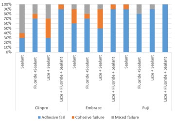
For each sample, roots were embedded in acrylic resin as support base exposing the occlusal-buccal surface above the CEJ. One hundred and twenty teeth were divided and labelled into three sealant groups, then divided into four subgroups (Table 1). On the buccal surface a button of sealant with the dimensions of 2.38mm in diameter by 2.00mm in height was applied for each material per manufacturer’s instructions. Each sample were stored for 24 hours after application in relative humidity at 37°C. The shear bond strength (SBS) test was performed using the Instron material testing machine (Instron 5566A, Norwood, MA). The crosshead speed moving at 1.0 mm/min at 500N. Each value was recorded and analyzed for the failure mode under a digital stereomicroscope (Olympus® SZX16). Each failure mode was recorded based on adhesive failure (A): debonding occurred at the interface between the treated buccal surfaces and fissure sealant; cohesive failure (B): failure in sealant/ enamel; mixed failure (C): occurrence of both adhesive and cohesive failures.
| Groups | Brand/Type | ||
|---|---|---|---|
| Group A | Clinpro™ Sealant, Syringe Introductory Kit - 3M Resin-based sealant | ||
| Group B | Embrace Wetbond Pit & Fissure Sealant Pulpdent Moisture-tolerant resin-based sealant | ||
| Group C | Fuji TRIAGE Pit & Fissure Sealant GC America Glass-ionomer cement. | ||
| Subgroups | Group A | Group B | Group C |
| 1: Sealant Only | A1 | B1 | C1 |
| 2: Fluoride + Sealant | A2 | B2 | C2 |
| 3: Laser + Sealant | A3 | B3 | C3 |
| 4: Laser + Sealant + Fluoride | A4 | B4 | C4 |
Table 1: List of groups and subgroups.
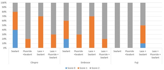
Microleakage
Each sample was thermocycled for 6,000 cycles between temperatures of 4-5°C and 55-60°C with 15 second dwell time. Samples were painted with nail varnish and immersed in 2% methylene blue for 24 hours. The samples were sectioned buccal-lingually using a slow speed diamond saw (Isomet 1000, Buehler). Each sectioned mesial half was examined under a digital microscope (Olympus SZX16) to asses dye penetration. Microleakage was evaluated based on the following scores described in Table 2.
| Score | Type of Stain |
|---|---|
| Score 0 | No stain is present at enamel-fissure sealant interface |
| Score 1 | Stain is present in middle-third of enamel-fissure sealant interface |
| Score 2 | Stain is present at the base of enamel-fissure sealant interface |
Table 2: Microleakage evaluation scores.
Microhardness and pH Cycling
The sectioned mesial halves were then assessed for baseline Vickers microhardness using the Micromet 2104 (Buehler) at four standardized points on the mesial surface and at four standardized points located 15 micrometers on the enamel, adjacent to the enamel-sealant interface on the cut surface. The sample was treated to 9 days of pH-cycling of remineralization and demineralization solutions. The pH-cycling process consisted of a 6-hour demineralization period in acetate/calcium/phosphate buffer solution at of pH 4.5-5 followed by an 18-hour remineralization period in a calcium phosphate buffer solution at pH 7.0 [15]. After pH- cycling, the microhardness measurement was performed at four standardized points located 15 micrometers next to the four original points.
Sample Size
We calculated sample size using results from a published study that examined in-vitro effect of CO2 laser and topical fluoride gel application on enamel microhardness after acid challenge [20]. The authors reported that the microhardness was 238.4 for the teeth treated with CO2 laser and 268.3 for the teeth treated with acidulated phosphate fluoride (APF) gel and CO2 laser. Based on the paper, standard deviations were assumed to be 21 and 16.5, respectively. To detect the difference in microhardness between the two groups, a sample of 20 teeth (n=10 per group) would achieve a power of 92% and significance level of 0.05. A total of 120 teeth were included for 12 groups in the proposed study.
Data Analysis
Number and percentage of sealants with adhesive failure, cohesive failure, and mixed failure were calculated. Means and standard deviations were calculated for Vickers microhardness, microleakage, and shear bond strength. The one-way Analysis of Variance (ANOVA) was used to compare the SBS, the microleakage, and the microhardness at baseline, after treatment, and after acid challenge between the four groups of each material. Bonferroni correction was used for post-hoc pairwise comparisons. The chi-square test was used to compare the failure modes between the four groups of each material. Statistically significant level was set at 0.05. Data was analyzed using Stata 15.
Results
There was a significant difference in SBS between sealant groups and between subgroups. CO2 laser + Sealant subgroup for Clinpro and Embrace had higher SBS than other groups (Figure 1 & Table 3). CO2 laser + Sealant group also had significant lower percentage of adhesive fail than other groups for Clinpro (Figure 2). Subgroup Clinpro sealant and Laser + Clinpro sealant had SBS of 13.1 ± 5.9 MPa and 15.5 ± 8.2 MPa, respectively. SBS of subgroup Embrace sealant was 11.9 ± 9.6 MPa and of laser + Embrace sealant was 16.3 ± 7.8 MPa. Group Fuji performed significantly lower than Groups Clinpro and Embrace. The SBS of subgroup laser + fluoride + Fuji sealant (0.5 ± 1.7 MPa) and laser + Fuji sealant (4.9 ± 4.7 MPa) were significantly different.

| Clinpro (MPa) | Embrace (MPa) | Fuji (MPa) | P value* | |
|---|---|---|---|---|
| Sealant | 13.1 ± 5.9a,b,A | 11.9 ± 9.6a,B | 1.5 ± 2.5a,B | <0.001 |
| Fluoride + Sealant | 4.5 ± 3.3a,c,A | 10.7 ± 7.3A,B | 3.7 ± 2.4B | 0.006 |
| Laser + Sealant | 15.5 ± 8.2c,d,A | 16.3 ± 7.8a,b,B | 4.9 ± 4.7a,A,B | 0.002 |
| Laser+ Fluoride + Sealant | 4 ± 3.3b,d,A | 1.5 ± 2.7a,b | 0.5 ± 1.7a,A | 0.02 |
| P value** | <0.001 | <0.001 | 0.01 |
Table 3: Shear bond strength (SBS) of dental sealants.
*: p value for one-way ANOVA test of means SBS values of groups in the same row : p value for one-way ANOVA test of means SBS values of groups in the same column In each column, mean SBS values of two groups with the same lowercase letter were significantly different. In each row, mean SBS values of two groups with the same uppercase letter were significantly different. Table 3:** Shear bond strength (SBS) of dental sealants.
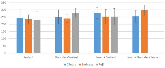
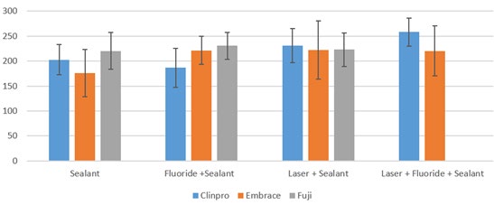
Group Clinpro had less microleakage overall compared to Group Embrace and Fuji (Figure 3). In group Clinpro, subgroup sealant experienced 40% at score 0, 40% at score 1 and 20% at score 2. Fluoride + sealant experienced only 20% at score 1 and 80% at score 2. Laser + sealant experienced 70% of score 1 and laser + fluoride + sealant experienced 30% at score 1. In group Embrace, subgroup sealant and laser +sealant experienced score 1 microleakage at 30% and 20% respectively. In group Fuji, experienced excessive leakage, however subgroup laser + sealant experienced about 10% of score 0 and 40% of score 1. There was no significant difference in microhardness of mesial surface enamel unrelated to the occlusal sealant before pH-cycling (Table 4). Group Clinpro showed a significant difference in microhardness from prior pH cycling values to after pH cycling values within the subgroups (Tables 5 & 6). Prior pH cycling hardness values for the four subgroups were 244.5 ± 54.2, 252.1 ± 48, 279.5 ± 39.2 and 255.4 ± 42.1. After pH cycling, hardness values were 202.8 ± 30.1, 186.7 ± 39, 231.4 ± 34.1 and 258.3 ± 28.2 respectively. There was a significant difference in the microhardness of subsurface enamel adjacent to the enamel/sealant interface after pH-cycling between subgroups of Clinpro sealant. Values for subgroup laser + Fuji sealant were not recorded because sealants fell off after thermocycling (Figures 4 & 5, Tables 5 & 6).
| Clinpro | Embrace | Fuji | P value* | |
|---|---|---|---|---|
| Sealant | 239.2 ± 33.5 | 243.8 ± 35 | 249.8 ± 30.9 | 0.78 |
| Fluoride + Sealant | 253.3 ± 80.6 | 261.1 ± 47.3 | 285.5 ± 40.5 | 0.45 |
| Laser + Sealant | 267.6 ± 28.8 | 263.4 ± 43.7 | 246.6 ± 61.9 | 0.58 |
| Laser + Fluoride + Sealant | 267 ± 48.3 | 265.9 ± 40.2 | 239.5 ± 35.7 | 0.27 |
| P value** | 0.58 | 0.64 | 0.11 |
Table 4: Microhardness of mesial surface enamel unrelated to the occlusal sealant before pH-cycling.
*: p value for one-way ANOVA test of means SBS values of groups in the same row **: p value for one-way ANOVA test of average microhardness of groups in the same column Table 4: Microhardness of mesial surface enamel unrelated to the occlusal sealant before pH-cycling.
| Clinpro | Embrace | Fuji | P value* | |
|---|---|---|---|---|
| Sealant | 244.5 ± 54.2 | 236.4 ± 28.5a | 231 ± 56.2 | 0.8 |
| Fluoride + Sealant | 252.1 ± 48.5 | 239.7 ± 25.7b | 278.8 ± 29.8 | 0.06 |
| Laser + Sealant | 279.5 ± 39.2 | 253.2 ± 50.9 | 252.3 ± 56.5 | 0.39 |
| Laser + Fluoride + Sealant | 255.4 ± 42.1A | 298.1 ± 35.5a,b,A | n/a | 0.04 |
| P value** | 0.38 | 0.005 | 0.11 |
Table 5: Microhardness of subsurface enamel adjacent to the enamel/sealant interface before pH-cycling.
*: p value for one-way ANOVA test of means SBS values of groups in the same row : p value for one-way ANOVA test of average microhardness of groups in the same column In each column, average microhardness of two groups with the same lowercase letter were significantly different. In each row, mean microhardness of two groups with the same uppercase letter were significantly different. n/a: sealants fell off after thermal cycling Table 5:** Microhardness of subsurface enamel adjacent to the enamel/sealant interface before pH-cycling.
| Clinpro | Embrace | Fuji | P value* | |
|---|---|---|---|---|
| Sealant | 202.8 ± 30.1a | 176.1 ± 47.3 | 220.2 ± 36.8 | 0.052 |
| Fluoride + Sealant | 186.7 ± 39b,c,A | 221.7 ± 27.7 | 230.9 ± 27A | 0.01 |
| Laser + Sealant | 231.4 ± 34.1b | 222.1 ± 58.7 | 222.9 ± 33.7 | 0.87 |
| Laser + Fluoride + Sealant | 258.3 ± 28.2a,c | 220.6 ± 49.9 | n/a | 0.06 |
| P value** | <0.001 | 0.1 | 0.8 |
Table 6: Microhardness of subsurface enamel adjacent to the enamel/sealant interface after pH-cycling.
*: p value for one-way ANOVA test of means SBS values of groups in the same row : p value for one-way ANOVA test of average microhardness of groups in the same column In each column, average microhardness of two groups with the same lowercase letter were significantly different. In each row, mean microhardness of two groups with the same uppercase letter were significantly different. n/a: sealants fell off after thermal cycling. Table 6:** Microhardness of subsurface enamel adjacent to the enamel/sealant interface after pH-cycling.
Discussion
9.3um CO2 laser irradiation was demonstrated effective in reducing demineralization and inhibits caries prevention [5, 12, 15]. Many theories have suggested the mechanism of treating dental caries with laser irradiation can increase enamel resistance to caries [5]. Laser converts the carbonated hydroxyapatite to a much more acid resistant purer phase hydroxyapatite (HAP) [9, 17]. The combination of CO2 laser and fluoride has been studied and shows that it enhances the prevention of caries. CO2 laser irradiation on enamel surface with fluoride application may produce fluorohydroxyapatite and calcium fluoride (CaF2) which increase enamel resistance to acid attacks [25].
Bond strength was measured to compare to the bonding ability of pit and fissure dental materials. Our results corroborate with other studies showing a significant improvement in SBS values when irradiating enamel surfaces with low power CO2 laser before placing sealants [26]. The increased SBS with CO2 laser irradiation resulted in a reduction in the enamel-sealant interface de-bonding. In addition, the parameters of the CO2 laser in our study caused a very minor change in the enamel surface. Although other studies showed an increase in enamel microhardness after applying APF gel, it is still unknown how the APF gel affect the SBS. In our study, we found that the APF gel applied after CO2 laser irradiation might have interfered with the true bond that worked between the material and enamel surface. In subgroups fluoride + sealant of the 3 materials where APF was used, values were much lower compared to the Groups where APF wasn’t used. An alternative explanation could be the difficulty of removing APF properly. There should be an extra effort to make sure the bonding site is clean and properly prepped after APF application.
Marginal microleakage is to mainly measure the amount of dye penetration which directly represents bacteria penetration that can lead to caries. Our study results and a previous study [27] examining microleakage between restorative materials and the walls of preparations irradiated with a 9.3um CO2 laser did not show a significant improvement in microleakage. The data obtained showed less microleakage without APF. The resin-based cement used in Group Clinpro, performed better than the moisture- tolerant resin-based sealant and the glass-ionomer cement. Fluoride releasing dental cements can be tested to further analyze how fluoride directly effects of 9.3um CO2 irradiation on pits and fissures to reduce microleakage.
Microhardness values represents the resistance of a material or a surface against challenges to the enamel surface [28]. It expresses the mechanical strength characteristics if the material and the challenges to the tooth by the increase in microhardness [28]. It is important to test for mineral content and enamel resistance to demineralization. After pH cycling, subgroup fluoride + Clinpro sealant and laser + fluoride + sealant showed that the 9.3um CO2 laser had an increasing effect on microhardness. The APF enhanced the mineral content and will be beneficial to improve caries prevention. There are several limitations with this study. First, the effect of moisture environment on the bonding of sealant to enamel cannot be assessed in this in vitro study. Second, although increased SBS, microhardness, and decreased microleakage will be expected with CO2 laser treatment, dental practitioners will be more interested in clinical outcomes such as retention rate and the reduction in the development of new dental caries when they decide which additional methods should be used with sealant placement. Therefore, this work provided an avenue for further testing to see what combination of methods will help to prevent caries and will lead to further in-vivo studies.
Conclusion
Under the limitations of the current study, the 9.3μm CO2 laser had an overall beneficial effect to the enamel. It increased shear bond strength of dental sealants and reduced adhesive failure for the three sealant materials. For Clinpro, the microhardness of subsurface enamel adjacent to the enamel/sealant interface pre-treated with 9.3μm CO2 laser was higher than those pre-treated with APF only or no treatment after pH cycling. The 9.3μm CO2 laser reduced microleakage of Clinpro sealant more than APF.
References
-
Ahovuo Saloranta A, Forss H, Walsh T, Nordblad A, Makela M, et al. (2017) Pit and fissure sealants for preventing dental decay in permanent teeth. Cochrane Database Syst Rev 7(7): Cd001830.
-
Locker D, Jokovic A, Kay EJ (2003) Prevention. Part 8: The use of pit and fissure sealants in preventing caries in the permanent dentition of children. Br Dent J 195(7): 375-378.
-
Allam GG, Abdel Aziz AF (2018) Comparing topical fluoride application, laser irradiation and their combined effect on remineralisation of enamel. Future Dental Journal 4(2): 318-323.
-
Anderson JR, Ellis RW, Blankenau RJ, Beiraghi SM, Westerman GH (2000) Caries resistance in enamel by laser irradiation and topical fluoride treatment. J Clin Laser Med Surg 18(1): 33-36.
-
Bahrololoomi Z, Lotfian M (2015) Effect of Diode Laser Irradiation Combined with Topical Fluoride on Enamel Microhardness of Primary Teeth. J Dent 12(2): 85-89.
-
Mahmoudzadeh M, Rezaei Soufi L, Farhadian N, Jamalian SF, Akbarzadeh M, et al. (2018) Effect of CO2 Laser and Fluoride Varnish Application on Microhardness of Enamel Surface Around Orthodontic Brackets. J Lasers in Med Sci 9(1): 43-49.
-
Stern RH, Vahl J, Sognnaes RF (1972) Lased enamel: ultrastructural observations of pulsed carbon dioxide laser effects. J Dent Res 51(2): 455-460.
-
Rechmann P, Sherathiya K, Kinsel R, Vaderhobli R, Rechmann BM (2017) Influence of irradiation by a novel CO2 9.3-mum short-pulsed laser on sealant bond strength. Lasers Med Sci 32(3): 609-620.
-
Kim JW, Lee R, Chan KH, Jew JM, Fried D (2017) Influence of a pulsed CO2 laser operating at 9.4 mum on the surface morphology, reflectivity, and acid resistance of dental enamel below the threshold for melting. J Biomed Opt 22(2): 28001.
-
Esteves Oliveira M, Wollgarten S, Liebegall S, Jansen P, Bilandzic M, et al. (2017) A New Laser-Processing Strategy for Improving Enamel Erosion Resistance. J Dent Res 96(10): 1168-1175.
-
Rechmann P, Fried D, Le CQ, Nelson G, Hilo MR, et al. (2011) Caries inhibition in vital teeth using 9.6-mum CO2-laser irradiation. J Biomed Opt 16(7): 071405.
-
Rechmann P, Charland DA, Rechmann BM, Le CQ, Featherstone JD (2013) In-vivo occlusal caries prevention by pulsed CO2 -laser and fluoride varnish treatment--a clinical pilot study. Lasers Surg Med 45(5): 302-310.
-
Featherstone JD, Barrett-Vespone NA, Fried D, Kantorowitz Z, Seka W (1998) CO2 laser inhibitor of artificial caries-like lesion progression in dental enamel. J Dent Res 77(6): 1397-1403.
-
Rodrigues LK, Nobre dos Santos M, Pereira D, Assaf AV, Pardi V (2004) Carbon dioxide laser in dental caries prevention. J Dent 32(7): 531-540.
-
Rechmann P, Rechmann BM, Groves WH, LE QC, Hilo MLR, et al. (2016) Caries inhibition with a CO2 9.3 mum laser: An in vitro study. Lasers Surg Med 48(5): 546-554.
-
Lee R, Chan KH, Jew J, Simon JC, Fried D (2017) Synergistic effect of fluoride and laser irradiation for the inhibition of the demineralization of dental enamel. Proc SPIE Int Soc Opt Eng 2017: 10044.
-
Fox JL, Yu D, Otsuka M, Higuchi WI, Wong J, et al. (1992) Combined effects of laser irradiation and chemical inhibitors on the dissolution of dental enamel. Caries Res 26(5): 333-339.
-
Meurman JH, Hemmerle J, Voegel JC, Rauhamaa-Makinen R, Luomanen M (1997) Transformation of hydroxyapatite to fluorapatite by irradiation with high-energy CO2 laser. Caries Res 31(5): 397-400.
-
Chin Ying SH, Xiaoli G, Jisheng P, Wefel JS (2004) Effects of CO2 laser on fluoride uptake in enamel. J Dent 32(2): 161-167.
-
Belcheva A, El Feghali R, Nihtianova T, Parker S (2018) Effect of the carbon dioxide 10,600-nm laser and topical fluoride gel application on enamel microstructure and microhardness after acid challenge: an in vitro study. Lasers Med Sci 33(5): 1009-1017.
-
Valério RA, Rocha CT, Galo R, Borsatto MC, Saraiva MCP, et al. (2015) CO2 laser and topical fluoride therapy in the control of caries lesions on demineralized primary enamel. The Scientific World Journal 2015: 547569.
-
Santaella MR, Braun A, Matson E, Frentzen M (2004) Effect of diode laser and fluoride varnish on initial surface demineralization of primary dentition enamel: an in vitro study. Int J Pediatr Dent 14(3): 199-203.
-
Westerman GH, Hicks MJ, Flaitz CM, Ellis RW, Powell GL (2004) Argon laser irradiation and fluoride treatment effects on caries-like enamel lesion formation in primary teeth: an in vitro study. AM J Dent 17(4): 241-244.
-
Gorton J, Featherstone JD (2003) In vivo inhibition of demineralization around orthodontic brackets. Am J Orthodont Dentofac Orthoped 123(1): 10-14.
-
Kim JW, Chan KH, Fried D (2016) Evaluation of enamel surface modification using PS-OCT after laser treatment to increase resistance to demineralization. Proc SPIE Int Soc Opt Eng 2016: 9692.
-
Sherathiya K (2016) In Vitro Study of Enamel Bond Strength Following 9.3 μm CO2 LASER Treatment. UCSF.
-
Kotin A, Afutu R, Tran D, Kugel G (2019) Does Tooth Preparation with a CO2-Laser Prevent Demineralization Around Restorations?. J Dent Res 98: 1809.
-
Aziznezhad M, Alaghemand H, Shahande Z, Pasdar N, Bijani A, et al. (2017) Comparison of the effect of resin infiltrant, fluoride varnish, and nano-hydroxy apatite paste on surface hardness and streptococcus mutans adhesion to artificial enamel lesions. Electron Physician 9(3): 3934-3942.
- Diagnosis and Management of Mental Nerve Paresthesia Secondary to Apical Periodontitis of Mandibular Second Premolar: A CBCT Based Case Report
- A Randomized, Double Blinded Clinical Trial to Compare the Effect of Oral Premedication (Diclofenac Potassium or Dexamethasone) on Post-Operative Pain Following Pulpectomy
- Modified Lip Repositioning Technique for the Management of Excessive Gingival Display
- Integral Role of Non-Dental Providers and Fluoride Dissemination
- Root Canal Treatment Rate in Deciduous Teeth Among 6-Year- Olds in the Era of Discontinuing Water Fluoridation - Historical Cohort Study
- The Impact of the Notch1 on the Migratory Capacity and the Expression of E-Cadherin and CyclinD1 in Ameloblastoma Cells