Enhanced Enamel Whitening, Surface Hardness and Stain Resistance by 9.3-µm CO2 Laser Irradiation: An In-Vitro Pilot Study
Objectives: In vitro studies were performed to investigate the effects of irradiating incisor enamel with a 9.3 µm CO2 laser in enhancement of whitening, stain resistance and surface hardness. Methods: A 9.3 µm CO2 laser beam was used to irradiate the labial surface of incisors with 0.8 J/cm2 pulse fluence at 333 Hz automatically scanning a 5.8 mm2 area for 0.381 seconds repeated to cover the whole teeth surface. The teeth were divided into three groups of n=10 each: a control group that received 40% H2 O2 gel for 20-minutes application time, an irradiated group followed with two 10-minute gel applications, and another irradiated group followed with two 20-minute gel applications. A spectroscopy device was used to analyze the overall color change (delta E) and degree of whitening. A 24-hour tea stain protocol was used to investigate the uptake of stain after the treatment procedure. Additionally, a small flat polished area on each sample was used to investigate microhardness before and after an acid challenge with pH 3.6 citric acid buffer. Result: Both irradiated groups showed an enhanced whitening effect, with a delta E that was 1-2 higher than the H2 O2 gel control group. The 10-minute application time was sufficient to reach the full benefit from irradiation. The irradiated groups showed increased resistance to acid solubility and reduced the absorption of tea stain. Conclusion: Irradiation with a 9.3 µm CO2 laser before application of whitening gel on incisor enamel increased the effect of whitening in half the time. It also provided an increased resistance to acid attack and staining.
Introduction
A substantial amount of work has been done in the development of tooth whitening techniques [1, 2, 3]. These methods generally involve the use of hydrogen peroxide or carbomide peroxide to oxidize and remove organic chromophores that produce discoloration [4, 5]. However, there are many known side effects caused by the use of these compounds, especially at concentrations commonly used in the clinic [5]. Such effects include damage to the oral mucosa, digestive issues after swallowing, and pulp and gum sensitivity. Generally, hypersensitivity peaks soon after the procedure and diminishes within a week [6]. An additional problem is that traditional treatments often require multiple applications of bleach and repeated visits.
In an effort to improve on the side effects of whitening with bleaching gels, new technologies have been developed with a goal of speeding up the procedure, reducing the active ingredient concentrations, or improving sensitivity, and reducing the number of treatments needed [7]. Among these technologies, power bleaching through light-activated chromophores has been a primary focus showing an improvement on whitening in less time and fewer treatments [8, 9]. Despite their success, additional side effects may occur. Since this method requires heating the bleach and enamel through light absorption, undesired heating of the pulp and discomfort to the patient may occur [9, 10].
In this study, a novel approach was taken using a 9.3 µm CO2 to safely modify the tooth surface before application with a hydrogen peroxide bleach gel in order to improve the outcome of whitening and speed up the overall treatment. This type of sub-ablative laser irradiation at a 9.3 µm wavelength has been demonstrated to reduce demineralization of enamel from acid erosion [11, 12, 13].and carious lesion formation [14, 15, 16].
The high absorbance at this wavelength allows for a rapid and controlled superficial heating to the necessary temperatures to remove carbonate groups without additional damage to the enamel structure [11, 12, 13, 14, 15, 16, 17]. The changes to the surface caused by this irradiation are not visible to the naked eye and are considered safe. Furthermore, the enamel will remineralize in saliva, and particularly when combined with fluoride, will provide a highly acid-resistant layer [15, 16, 17, 18]. The benefit of this enhanced acid resistance from laser irradiation was studied here through a staining protocol after completion of the whitening treatment in vitro.
Materials and Methods
Study Design and Sample Preparation
The study design is outlined in Figure 1. A total of 30 human incisors in sound condition and with no signs of fluorosis or caries were obtained, cleaned, disinfected and transported in 0.1% thymol to prevent microbial growth before use (Therametric Technologies, Inc., Nobelsville, IN). These teeth were selected from a batch to ensure that all had a starting classical Vita Shadeguide (Vita, Zahnfabrik, Germany) value of 8 or higher on a scale of 1-16. The roots were removed by a diamond saw (Isomet, Buehler, Lake Bluff, IL) and the samples were mounted in Durabase hard chairside reline (Reliance Dental Mfg, Alsip, IL) with the labial surface up (and all other surfaces covered). The samples were then cleaned gently with a standard rotary polisher and pumice paste. To prepare for microhardness measurements, a ~3 mm diameter area toward the bottom of the enamel surface was formed by serially polishing up to a 1 µm diamond grit finish (Forcipol 1V with Forcimet, Metkon Instruments, Inc., Bursa, Turkey).
Samples were left in a 1:3 slurry of toothpaste (Crest cavity protection, Proctor and Gamble, Inc., Cincinnati, OH) to distilled water for 1 minute. The samples were then placed into a CaPO4 Tris-HCl pH 7.1 remineralization buffer solution overnight.
The samples were randomized into 3 groups of n=10, and baseline spectroscopy and microhardness measurements were obtained. Group 1 was a control group with 40% whitening bleach (Opalescence Boost, Opalescence, South Jordan, UT) for 2 applications of 20 minutes; group 2 samples were irradiated with the laser, followed by two applications of the same whitening bleach for 10 minutes each; group 3 were irradiated with the laser, followed by two applications of the same whitening bleach for 20 minutes each. The whitening gel was placed on the entire tooth surface to a thickness of about 1 mm.
Spectroscopy measurements were taken between each application of bleach. After the 2nd application, samples were stored in remineralization solution, and microhardness measurements were taken. Samples were gently mixed in the fluoride slurry for 1 minute, and then placed in remineralization buffer for 7 days at room temperature. Spectroscopy and microhardness measurements were obtained again, after which the samples were subject to an acid challenge by exposing them to a citric acid buffer of pH 3.6 for 6 minutes. Measurements were then taken, and the samples were then placed in a fresh remineralization solution for one week. Spectroscopy measurements were then obtained. The samples were placed in a tea stain for 24 hours, and a final spectroscopy measurement of each sample was obtained.
Laser Settings
Laser irradiation was performed using a 9.3 µm CO2 laser (Solea, Convergent Dental, Inc., Needham, MA). A beam diameter of 1 mm was scanned over a ~7 mm area at a pulse repetition rate of 750 Hz. Pulses had a fluence of about 0.8 J/cm2 on this system by setting the optical pulse duration of the laser to 20 µs, which provides the median fluence used in a previous study with similar parameters 15. The distance from the tip of the handpiece output to the sample surface was maintained at a set position of 10 mm. A continuous airflow out of the handpiece was maintained at about 9 L/ min for cooling the tooth.
Spectroscopy Measurements of Whitening
The effect of tooth whitening was measured by a spectroscopy method. Spectroscopy was done using a color/ shade measurement device (MHT Spectroshade Micro, Oxnard CA), with tooth surfaces wet and the mounted samples placed in a black box. The device outputs brightness (L), red-green axis (a), and blue-yellow axis (b) values for each measurement. These values are combined into one value, ΔE, 19,20 which represents the overall color change between a pair of measurements, calculated by:
Higher ΔE values indicate a greater effect of whitening, and should be associated with an increase in L, and a decrease in a and b. These values were calculated at each step of the experiment for the 3 groups. A standard ~5 mm circular region close to the center of the incisors was used, while avoiding the flattened area that was used for microhardness measurements. To reduce variations in measurements, a black box was used to encase the samples and device opening and remove any interference caused by ambient lighting.
Microhardness Measurements
A microhardness tester Matsuzawa Seiki DMH-2, (Matsuzawa, Akita Pref, Japan) was used with a Knoop diamond tip and a load of 50 g, with a 10 sec dwell time. 5 indents were taken on each sample at each experiment step and averaged. Indent lengths were measured in a digital microscope (RH-2000, Hirox-USA, Inc., Hackensack, NJ) at 1000x magnification. Indent lengths were converted to Knoop hardness (KHN) using the following formula: KHN=g/(0.0703*L^2 ) where g is the load in grams, and L is the indent length in millimeters.
Staining Protocol
Two bags of Lipton black tea were placed in 500 ml of boiled water for 10 minutes, and then cooled to room temperature. The samples were removed from the remineralization solution and gently cleaned with a toothbrush for 30 seconds. The samples were then left in the tea for 24 hours.
Statistical Analysis
Data were analyzed in Minitab 18 (Minitab, Inc. State College, PA) using one-way ANOVA and post-hoc Tukey tests for pairwise comparisons.
Results
Irradiated samples in both groups 2 and 3 produced an overall larger increase in brightness and delta E after both bleach applications, and this increase persisted after remineralization of the outer enamel. Example images showing the different in whitening are shown in Figure 1. The data sets for delta E and delta L are plotted in Figure 2. The overall change in delta E and delta L were 1-2 values higher for the irradiated groups than the bleach control group, with a significant difference (p < 0.05). This difference was observed after both bleach applications and remineralization.
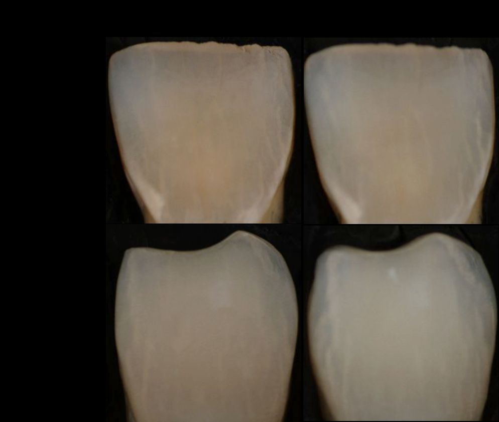
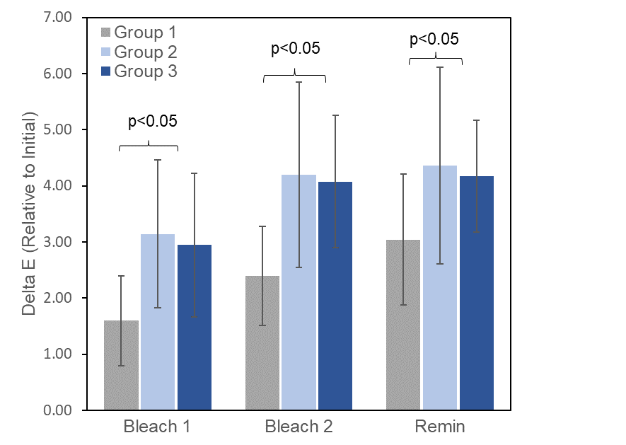
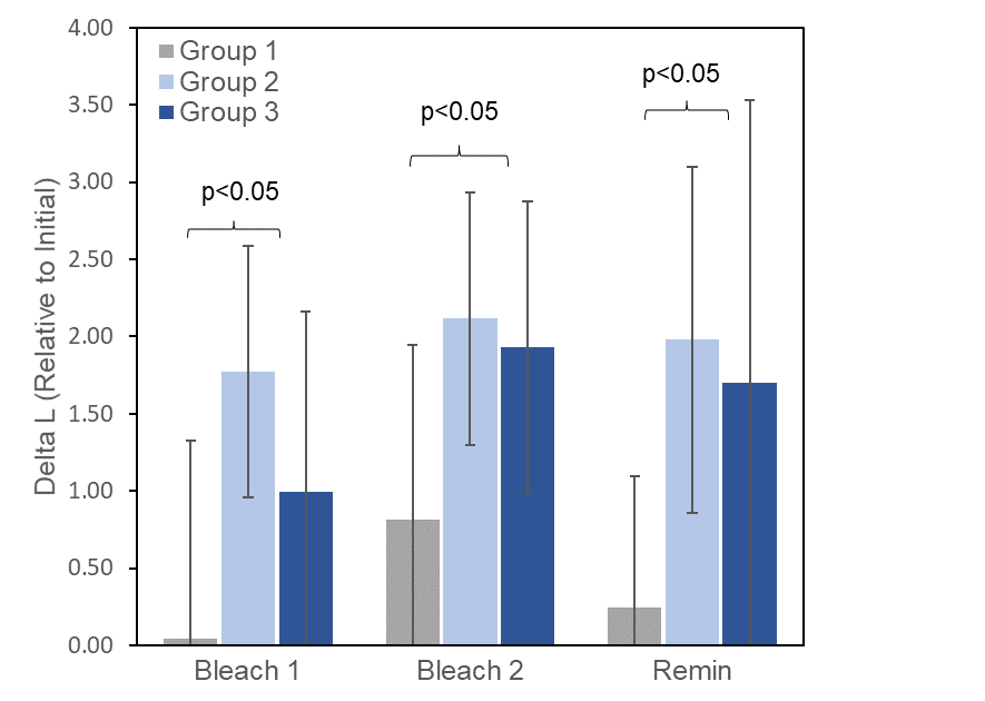
Figure 2: A) Delta E measurements of whitening (shown with standard deviation bars) after two applications of bleach and one week in remineralization solution. The laser treated groups (2 and 3) produced a significantly higher delta E compared to the bleach control group. B) Delta L measurements of the change in brightness for the same time points and groups.
Figure 3 shows the Knoop microhardness measurements for the groups. The initial Knoop hardness values for the three groups prior to the experiment averaged 280 and were not significantly different. Groups 1 and 2 were not different after the bleach applications, but group 3 was smaller by around 7% (p < 0.05). After remineralization, groups 2 and 3 were both significantly harder than the bleach control group 1. After acid challenge, all three groups exhibited a decrease in hardness, but the bleach control group was around 40% softer than the irradiated groups.
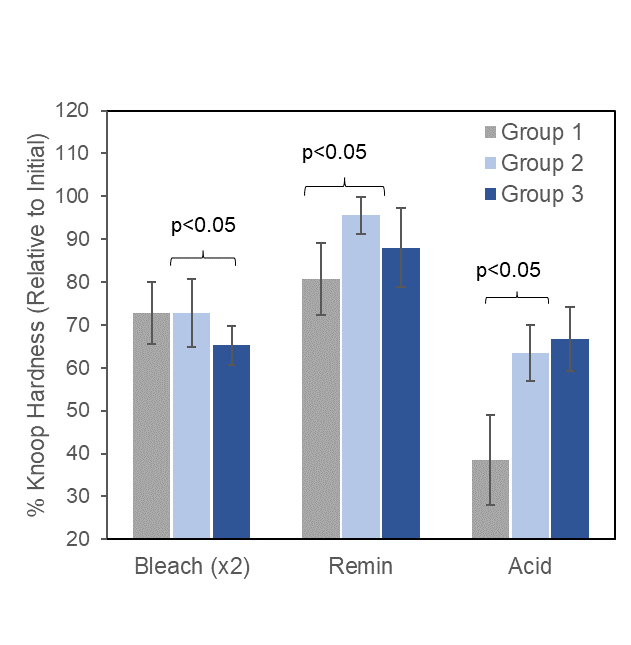
Figure 3: Knoop microhardness measurements for the three groups after two applications of bleach each for 20 minutes (groups 1 and 3) or 10 minutes (group 2). Both groups of laser irradiated samples (groups 2 and 3) showed an enhanced hardness after remineralization relative to the bleach controls (group 1), and a higher retention of mineral content after the acid challenge.
Images of the incisors before and after the 24 hour tea stain are shown in Figure 4A. The stain appeared to be more strongly absorbed into the non-irradiated incisors. Delta E values for the three groups are shown in Figure 4B. These values were inverted since the changes in L, a and b
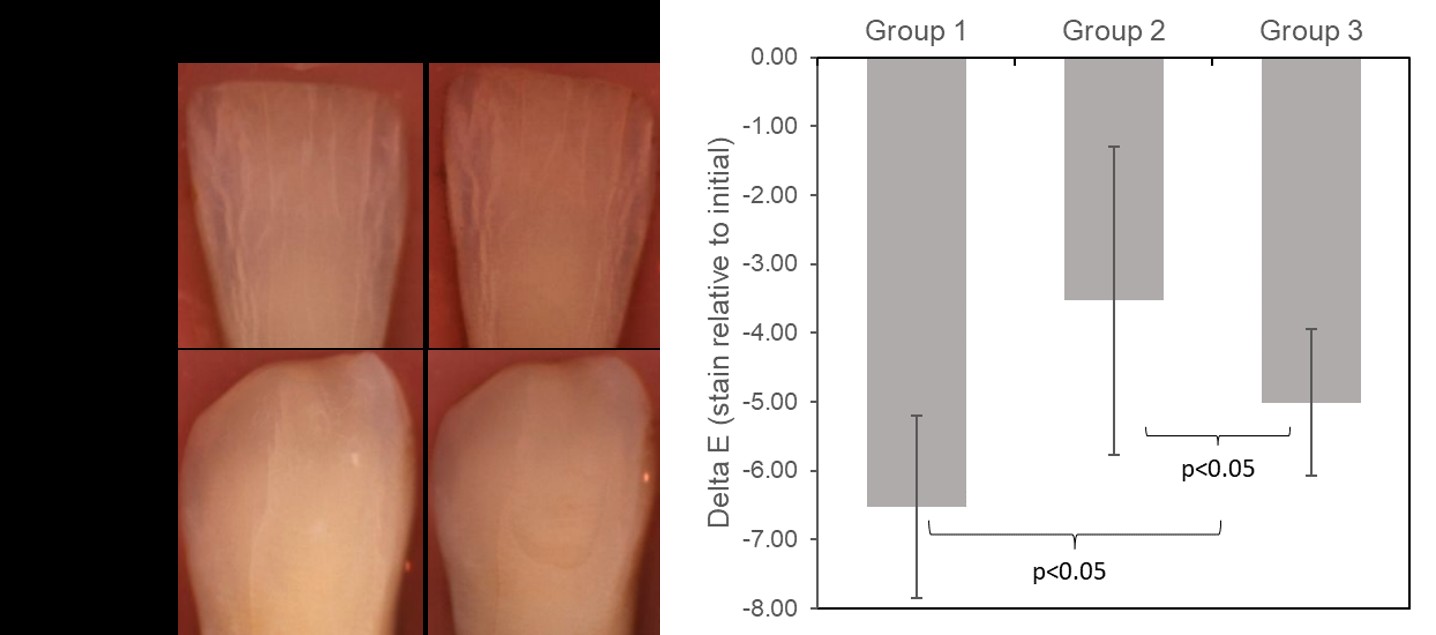
values were all negative. The overall color change for the bleach control group was larger than the irradiated groups, indicating that groups 1 samples experienced a higher degree of staining. Additionally, group 3 showed a larger color change than group 2.
Figure 4: A) Example images of mounted incisors before (left) and after (right) 24 hours of staining in black tea. The teeth irradiated before bleaching gel demonstrated a reduced overall uptake of the stain. B) Inverted delta E values for the three groups after 24 hours in a tea stain. Negative values indicate a shift in brightness and color toward the darker and redder/yellower. The bleach control group showed a higher degree of staining compared to both laser irradiated groups. Group 2, which was exposed only to two applications of bleach at 10 minutes each, demonstrated the lowest decrease in delta E from staining.
Discussion
An enhancement of the whitening effects of a standard 40% whitening bleach gel was obtained by performing a pre-treatment of the enamel surface using sub-ablative 9.3 µm CO2 laser irradiation. Changes to the enamel surface caused by this irradiation include a minor structural effect in the form of superficial crazing [15, 16, 17, 18, 19, 20, 21], and a chemical modification in the form of the removal of water, organic components, and carbonate groups [17, 18, 19, 20, 21, 22].
These changes may temporarily enhance the passage of hydrogen peroxide into the enamel during bleaching, which could explain the increased whitening effect observed using irradiation before bleach gel. Additionally, heat generated from laser irradiation is well in excess of the burning point of organic matter in the enamel [23], and a removal of that potentially stained matter may also help explain the observed whitening benefit of the laser. Furthermore, since this enhancement from irradiation persisted after remineralization, whitening caused solely by desiccation can be ruled out. The effects of desiccation or accumulating heat have been demonstrated to not be an issue using very similar irradiation conditions [15].
Whitening values presented here are similar with power bleaching data reported using other lasers, despite the technique being the opposite workflow of laser irradiation before bleach application [24, 25, 26, 27]. Delta E is a value representing overall color change, but it is hard to interpret its meaning clinically, and it may be easily affected by the particular device used. Traditionally, the effects of whitening were measured using reference shade guides such as the classical Vita shade guide [2]. We found through previous testing (not shown in this study) that the spectroscopy device used here corresponds to a Vita shade guide change of roughly 50% more than the delta E value calculated. For the whitening data, this would indicate that the irradiated groups produced an overall improvement of over 6 shades, while the bleach alone improved whitening by a little over 4 shades. This could be further investigated clinically with direct measurements in a blinded study, along with spectroscopy measurements.
The enhanced acid resistance from 9.3 µm CO2 laser irradiation was expected, but to the authors knowledge, the data presented here is the first to show that this acid resistance is maintained after the use of a whitening bleach gel. This is significant, as it suggests that the effect of laser- enhanced whitening may provide a longer-lasting benefit, since a non-irradiated surface will erode away sooner, rendering the non-bleached underlying enamel or yellower dentin more visible. Since most stain-producing foods are acidic in nature, this acid resistance would be expected to also provide a lasting benefit in preventing stains from penetrating into the enamel subsurface. Indeed, the data shown in this study provides evidence in vitro that this may be the case.
Conclusion
Irradiation with a 9.3 µm CO2 laser before application of a standard whitening gel provides an enhanced whitening by up to 50% in half the application time, and with no significant damage or adverse effects. An additional benefit of this treatment is the generation of an acid-resistant layer which prevents erosive losses and reduces the potential for penetration of stains from acidic foods. Further clinical studies need to be performed to validate the in-vitro study results and evaluate any benefits for reduced tooth sensitivity and irritation.
References
-
Carey CM (2014) Tooth Whitening: What We Now Know. J Evid Based Dent Pract 14: 70-76.
-
Matis BA, Cochran MA, Eckert G (2009) Review of the Effectiveness of Various Tooth Whitening Systems. Oper Dent 34(2): 230-235.
-
Fiorillo L, Laino L, De Stefano R, D’Amico C, Bocchieri S, et al. (2019) Dental whitening gels: Strengths and weaknesses of an increasingly used method. Gels 5(3).
-
D’Arce MBF, Lima DANL, Aguiar FHB, Bertoldo CE dos S, Ambrosano GMB, et al. (2013) Effectiveness of dental bleaching in depth after using different bleaching agents. J Clin Exp Dent 5(2).
-
Goldberg M, Grootveld M, Lynch E (2009) Undesirable and adverse effects of tooth-whitening products: a review. Clin Oral Investig 14(1): 1-10.
-
Farag I, Abouelfotouh I, Mohamed O, Fahmy I, Khairy AE, et al. (2018) A comparative study of different bleaching techniques, regarding the color change, stability and postoperative hypersensitivity: a randomized controlled clinical trial. Stomatol Dis Sci (5): 5.
-
Polonsky M (2018) Shining the Light on Power Bleaching : A Review. In: Oral Health Group 1-10.
-
De Moor RJG, Verheyen J, Diachuk A, Verheyen P, Verheyen P (2015) Insight in the chemistry of laser- activated dental bleaching. Sci World J 2015: 650492.
-
De Moor RJG, Verheyen J, Verheyen P, Diachuk A, De Coster PJ, et al. (2015) Laser teeth bleaching: Evaluation of eventual side effects on enamel and the pulp and the efficiency in vitro and in vivo. Sci World J 2015: 835405.
-
Sulieman M, Addy M, Rees JS (2005) Surface and intra- pulpal temperature rises during tooth bleaching: An in vitro study. Br Dent J 199(1): 37-40.
-
Esteves Oliveira M, Pasaporti C, Heussen N, Eduardo CP, Lampert F, et al. (2011) Rehardening of acid-softened enamel and prevention of enamel softening through CO2 laser irradiation. J Dent 39(6): 414-421.
-
Esteves Oliveira M, Wollgarten S, Liebegall S, Jansen P, Bilandzic M, et al. (2017) A New Laser-Processing Strategy for Improving Enamel Erosion Resistance. J Dent Res 96(10): 1168-1175.
-
Silva CV, Mantilla TF, Engel Y, Tavares JP, Freitas PM, et al. (2020) The effect of CO2 9.3 μm short-pulsed laser irradiation in enamel erosion reduction with and without fluoride applications—a randomized, controlled in vitro study. Lasers Med Sci 35(5): 1213-1222.
-
Rechmann P, Le CQ, Kinsel R, Kerbage C, Rechmann BMT (2020) In vitro CO2 9.3-μm short-pulsed laser caries prevention—effects of a newly developed laser irradiation pattern. Lasers Med Sci 35(4): 979-989.
-
Badreddine AH, Couitt S, Donovan J, Cantor Balan R, Kerbage C, et al. (2021) Demineralization Inhibition by High-Speed Scanning of 9.3 µm CO 2 Single Laser Pulses Over Enamel. Lasers Surg Med 53(5): 703-712.
-
Rechmann P, Rechmann BMT, Groves WH, Rapozo Hilo ML, Kinsel R, et al. (2016) Caries inhibition with a CO2 9.3 μm laser: an in vitro study. Lasers Surg Med 48(5): 546-554.
-
Featherstone JDB, Fried D (2001) Fundamental interactions of lasers with dental hard tissues. Med Laser Appl 16(3): 181-194.
-
Rechmann P, Charland DA, Rechmann BMT, Le CQ, Featherstone JDB (2013) In-vivo occlusal caries prevention by pulsed CO2 -laser and fluoride varnish treatment--a clinical pilot study. Lasers Surg Med 45(5): 302-310.
-
Karadas M, Seven N (2014) The effect of different drinks on tooth color after home bleaching. Eur J Dent 8(2): 249-253.
-
Turkun L, Turkun M (2004) Effect of bleaching and repolishing procedures on coffee and tea stain removal from three anterior composite veneering materials. J Esthet Restor Dent 16(5): 301-302.
-
McNally KM, Gillings BRD, Dawes JM (1999) Dye-assisted diode laser ablation of carious enamel and dentine. Aust Dent J 44(3): 169-175.
-
Corrêa Afonso AM, Bachmann L, De Almeida CG, Corona SAM, Borsatto MC (2012) FTIR and SEM analysis of CO2 laser irradiated human enamel. Arch Oral Biol 57(9): 1153-1158.
-
Zuerlein M, Fried D, Featherstone JDB (1999) Modeling the modification depth of carbon dioxide laser treated enamel. Lasers Surg Med 25(4) 335-347.
-
Fornaini C, Lagori G, Merigo E, Guidotti R, Serraj A, et al. (2011) Analysis of shade, temperature and hydrogen peroxide concentration during dental bleaching: In vitro study with the KTP and diode lasers. Lasers Med Sci 28(1): 1-6.
-
Lagori G, Vescovi P, Merigo E, Meleti M, Fornaini C (2014) The bleaching efficiency of KTP and diode 810 nm lasers on teeth stained with different substances: An in vitro study. Laser Ther 23(1): 21-30.
-
Fekrazad R, Alimazandarani S, Kalhori KAM, Assadian H, Mirmohammadi SM (2017) Comparison of laser and power bleaching techniques in tooth color change. J Clin Exp Dent 9(4): e511-e515.
-
Shahabi S, Assadian H, Nahavandi AM, Nokhbatolfoghahaei (2018) Comparison of tooth color change after bleaching with conventional and different light-activated methods. J Lasers Med Sci 9(1): 27-31.
- Diagnosis and Management of Mental Nerve Paresthesia Secondary to Apical Periodontitis of Mandibular Second Premolar: A CBCT Based Case Report
- A Randomized, Double Blinded Clinical Trial to Compare the Effect of Oral Premedication (Diclofenac Potassium or Dexamethasone) on Post-Operative Pain Following Pulpectomy
- Modified Lip Repositioning Technique for the Management of Excessive Gingival Display
- Integral Role of Non-Dental Providers and Fluoride Dissemination
- Root Canal Treatment Rate in Deciduous Teeth Among 6-Year- Olds in the Era of Discontinuing Water Fluoridation - Historical Cohort Study
- The Impact of the Notch1 on the Migratory Capacity and the Expression of E-Cadherin and CyclinD1 in Ameloblastoma Cells