Stereomicroscope Analysis of the Cleaning Capacity of an Abrasive-Expansive System in Oval Canals: A Laboratory Study
Background: The aim of this study was to evaluate, by means of a stereomicroscope, the influence on the removal of smear layer obtained by the XP-endo Finisher and Tornado Disinfection Kit systems during the final agitation of the irrigating solution, in conservative preparations. Materials and methods: Ten extracted human lower premolars were chemically-mechanically prepared with the ProDesign Logic 2 NiTi rotary system (25/.05) and 2.5% NaOCl. The same specimens were submitted to three different protocols of agitation of the irrigating solution: Conventional irrigation, XP-endo Finisher and Tornado Disinfection Kit. For this, a closed system was made, the roots were cleaved and three holes were made in each third of the root canal and filled with dentinal debris to take initial photos under a stereomicroscope. The amount of debris was rated using a 4-point scoring system. The data obtained were submitted to the Wilcoxon, Kruskal-Wallis and Dunn tests (p<0.05). Results: In the analysis of the effectiveness of each protocol, it was possible to observe a statistically significant difference in the removal of debris in all thirds for all protocols (p<0.05). Regarding the difference in score found before and after application of the protocol, a significant difference was observed between the apical and the middle third in the Tornado Disinfection Kit group (p<0.05) and there was no difference between the same third for different groups (p>0.05). Conclusion: Based on the methodology and the results presented, it was concluded that both evaluated protocols improved the cleaning of root canal recesses in all thirds, with no difference between them.
Introduction
The biomechanical preparation of the root canal system involves cleaning and decontamination through mechanical instrumentation concomitant with irrigating solutions [1], capable to control the root canal infection [2]. But despite this, reconciling root canal system cleaning with dentin preservation has been difficult to achieve [3].
Therefore, the new proposal to avoid excessive pericervical dentin wear is the use of instruments with smaller diameters [4], such as the ProDesign Logic 2 System (Bassi Endo, Belo Horizonte, MG, Brazil), which is an instrument manufactured in NiTi thermally treated and which has a maximum cervical diameter of 1.0 mm, becoming cylindrical instead of conical when it reaches this diameter [5].
These systems, despite advances in endodontic treatment techniques, still do not completely eliminate dentinal debris [6] due to the complexity of the root canal system [7], making endodontic irrigation even more important for proper cleaning and decontamination [8].
The conventional technique, usually performed with syringes and needles, has failed to remove the smear layer and clean the most apical portions of the root canal system [9]. And, to increase irrigation efficiency, other techniques for agitating irrigating solutions have been investigated.
XP-endo Finisher 25./00 (FKG, La Chaux-de-Fonds, Switzerland) is made from a unique NiTi alloy (MaxWire) and has a straight shape in the martensitic phase, but assumes a “spoon shape” when reaches body temperature, in the austenite phase [10]. According to the manufacturer, when it reaches this shape, the instrument is able to remove the debris present in the anatomical complexities of the root canal (FKG 2015).
Another system is the Tornado Disinfection Kit (MedicNRG, Kibbutz Afikim, Israel). It consists of two rotary instruments made of stainless steel (Gentlefile Red and Gentlefile Brush) [11], which promote an abrasion process on the dentin walls. They are driven by an automated and non-customizable handpiece, running at 6,500 rpm [12] and performing a quick and short entry and exit movement. Knowing the importance of maintaining dentin for a good rehabilitation prognosis, it became important to investigate whether the use of abrasive-expansive systems for agitating the irrigating solution could complement root canal debridement, promoting better cleaning rates in recesses through an analysis in stereomicroscope.
The aim of the present study was to verify whether the use of the XP-endo Finisher and Tornado Disinfection Kit systems were able to improve the removal of dentin debris in recesses after conservative preparation of the root canal of single-rooted maxillary premolars, performing a stereomicroscope analysis. The null hypothesis is that there will be no difference in the removal of dentinal debris promoted by the different agitation systems of the irrigating solution.
Material and Methods
Sample Calculation
For sample calculation, G * Power v3.1 for Mac (Heinrich Heine, Universität Düsseldorf, Dusseldorf, Germany) was used and the ANOVA test of repeated measures was selected. Data from a previous study that evaluated agitation of the irrigating solution (13) were used. An alpha type error of
0.05, a beta power of 0.80 and an N2/N1 ratio of 1 were also stipulated. A total of 8 specimens were indicated as the ideal size needed to notice significant differences. A sample of 10 specimens was used, considering a 20% risk of sample loss.
Selection of Specimens
Before starting this study, it was reviewed and approved by the Research Ethics Committee (nº 42281921.3.0000.5417). Ten maxillary premolars extracted with a single root canal, intact crown and complete apex were initially selected based on radiographs taken in the buccolingual and mesiodistal directions (Microimagem, Indaiatuba, São Paulo) and stored in a 0.1% thymol solution.
Chemical-Mechanical Preparation
Access to the pulp chamber was made with diamond burs at high speed, under refrigeration, and an exploration of the canal was performed with a K-file n°10 and n°15 (Dentsply Maillefer, Baillagueis, Switzerland) to recognize its internal anatomy.
After this initial recognition, the incisal surface of the crowns were ground using a double-sided diamond disc (KG Sorensen, Cotia, Brazil) until, when inserting the K-file n°10 in the root canal, its tip was visualized in the apical foramen under stereomicroscope magnification x30 (Carl Zeiss Vision GmbH, Hallbergmoos, Germany), standardizing the sample length at 16mm. This measurement was considered the actual length of the tooth and the working length (WL) was determined by reducing this measurement by 1 mm.
The root canal was filled with 2.5% sodium hypochlorite (NaOCl) solution and a ProDesign Logic 2 25/.05 instrument (BassiEndo) was introduced in a rotary motion (400rpm and 2N). During instrumentation, after three movement cycles (insertion + removal), the instrument was removed from the canal, cleaned with gauze soaked in alcohol, and the process was repeated until instrumentation was completed. At the end, the canal was washed with 10 ml of distilled water, totaling a final volume of 25 ml of irrigating solution. Each instrument was used on 3 teeth and then discarded. The preparation of all root canals was performed by a single operator.
Stereomicroscope Analysis
After chemical-mechanical preparation, the root canals were dried with an absorbent paper point and a 25/.05 calibrated gutta-percha cone (BassiEndo) was inserted into the root canal up to the WL. Two longitudinal grooves were made on the mesial and distal walls using double-sided diamond discs (KG Sorensen) operated on a pneumatic motor without reaching the root canal space.
Then the roots were enveloped in heavy body silicone (Optosil Comfort Putty; Heraeus Kulzer GmbH, Ha-nau, Alemanha) up to the level of the cemento-enamel junction (CEJ) and placed in a muffle. After the silicone had set, the specimens were divided into two parts with a spatula nº 36 (SSWhite Duflex, Rio de Janeiro, Brazil). The half that best presented conditions to evaluate the internal surface in a stereomicroscope was selected.
With a diamond bur 2137F (KG Sorensen) high rotation, a hole of approximately 0.5 mm in diameter was created inside the canal in each of the thirds. The specimens were subsequently washed in running water for 1 minute to remove debris. Dentinal debris was created from the grinding of extracted teeth and was mixed with distilled water and added into the root canal space, filling the created holes.
Then, the specimens were analyzed under a stereomicroscope. After locating the holes, an image for each third was obtained at 50x magnification.
The halves of each specimen were reassembled and stabilized with resin (Topdam; FGM, Joinville, Brazil). The specimens were re-inserted into the heavy body silicone mold and into the muffle to increase stability and prevent leakage of solutions used in the final irrigation protocols
Distribution of Groups
The same 10 samples were used to apply the 3 chemical- mechanical preparation protocols: Conventional Irrigation (CI) – The root canal was irrigated with 40 mL of 2.5% NaOCl using a 5 mL disposable syringe (Ultradent Products Inc.) and a 30-gauge NaviTip needle (Ultradent Products Inc.).
Tornado Disinfection Kit (TDK) – The root canal was irrigated with 5 mL of 2.5% NaOCl and the irrigating solution was initially stirred with GentleFile Red (MedicNRG), gently inserting it into the canal, with a rotational movement with 6,500 rpm with its own contra-angle until resistance was obtained and then it was activated using a gentle in- and-out movement with light apical pressure and lasting 5 seconds. The canal was irrigated and filled again with a 2.5% NaOCl solution and the procedure was repeated until the instrument reached the WL. Then, the canal was again filled with the irrigating solution and the GentleFile Brush (MedicNRG) instrument was introduced and activated for 30 seconds. Each instrument was used on one tooth and then discarded.
XP-endo Finisher (XPF) - The root canal was irrigated with 5 mL of 2.5% NaOCl and the solution stirred with XP- endo Finisher (FKG Dentaire). The instrument was placed on a contra-angle (VDW Silver; VDW, Munich, Bavaria, Germany) and inserted into the canal without rotation. After that, rotation was started (800 rpm and 1 Ncm), and the instrument activated for 1 min using slow and smooth movements of 7 to 8 mm in the long axis direction from the tooth to the WL. Each instrument was used on one tooth and then discarded.
All groups were irrigated at the end with 5mL of distilled water to remove NaOCl. At the end, a total of 45 mL of irrigating solution was obtained. After the protocols of agitation of the irrigating solution with NaOCl, the specimens were submitted to analysis in a stereomicroscope (Carl Zeiss Vision GmbH), following the same parameters used previously.
Rating Criteria
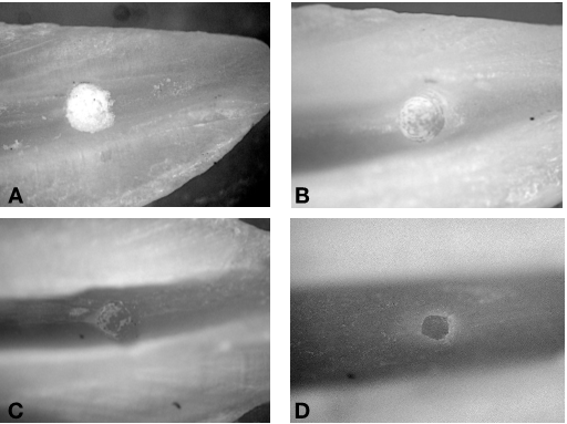
The images were saved and coded according to the group (G1, G2 or G3), the tooth (1-10) and the level (C, M, A) to which it corresponds. All images were loaded into Microsoft Office PowerPoint application (Microsoft Corporation, Redmond, WA). Two independent examiners, calibrated and blinded to the study, analyzed and classified the images using a 4-score scoring system adapted from GAMBARINI and LASZKIEWICZ (2002) (14) as follows: (A) hole covered by debris in 100 % of the examined area; (B) hole with debris covering more than 50% of the area; (C) hole with debris covering less than 50% of the area; (D) orifice without debris (Figure 1).
Statistical Analysis
The reliability of three examiners for classifying the degree of cleanliness of the holes in the dentin ranged from good to excellent (Kappa coefficients ranged from 0.59 to 0.82). The Wilcoxon test was used to compare cleaning ability before and after using the cleaning protocol. Kruskall-Wallis and Dunn tests were used to compare cleaning ability between cleaning protocols. The significance level was set at a = 5% (GraphPad Prism, San Diego, United States).
Results
Table 1 shows the median and percentile values of the root canal score before and after the irrigation protocols. It is possible to verify a statistical difference in cleaning before and after irrigation in all groups and for all root canals (p<0.05).
| Group | Period | A | M | C | ||||||
|---|---|---|---|---|---|---|---|---|---|---|
| 25% | Median | 75% | 25% | Median | 75% | 25% | Median | 75% | ||
| Conventional irrigation | Before | 4 | 4a | 4 | 4 | 4a | 4 | 4 | 4a | 4 |
| After | 2 | 2b | 3 | 2 | 2b | 3 | 2 | 2b | 2,75 | |
| XP-Endo Finisher | Before | 4 | 4a | 4 | 4 | 4a | 4 | 4 | 4a | 4 |
| After | 2 | 2b | 3 | 1 | 2b | 2 | 1 | 2b | 2 | |
| Tornado Disinfection Kit | Before | 4 | 4a | 4 | 4 | 4a | 4 | 4 | 4a | 4 |
| After | 2 | 3b | 4 | 1 | 1,5b | 2 | 1,25 | 2b | 2,75 |
Table 1: Median and percentile of the score of dentinal debris before and after irrigation protocols in the apical (A), middle (M
Table 2 shows the median and percentile values of the root canal score difference before and after the irrigation protocols. When comparing the thirds in the same group, a statistically significant difference was verified between the apical and middle thirds in the Tornado Disinfection Kit group (p<0.05).
| Group | A | M | C | ||||||
|---|---|---|---|---|---|---|---|---|---|
| 25% | Median | 75% | 25% | Median | 75% | 25% | Median | 75% | |
| Conventional irrigation | 1 | 2aA | 2 | 1 | 2aA | 2 | 1,5 | 2aA | 2 |
| XP-Endo Finisher | 1 | 2aA | 2 | 2 | 2aA | 3 | 2 | 2aA | 3 |
| Tornado Disinfection Kit | 0 | 1a* | 2 | 2 | 2,5a* | 3 | 1,25 | 2a | 2,75 |
Table 2: Median and percentile of Difference in dentinal debris scores before and after irrigation protocols in the apical (A), m
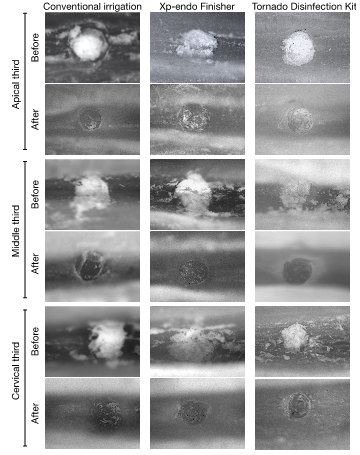
In Figure 2, it is possible to see representative photos of the ability to remove dentinal debris from the holes created in all thirds of the root canal, after applying the irrigation protocols.
Discussion
This study aimed to verify whether the use of expansive systems, XP-endo Finisher and Tornado Disinfection Kit, would be able to improve the removal of debris after agitation of irrigating solutions, when conservative preparations of root canals are performed. Based on the results, the null hypothesis was denied, as all protocols demonstrated improvement in cleanliness in all thirds.
In the evaluation of the ability to remove debris before and after the application of the irrigation protocols, a statistically significant difference was observed between the thirds in all groups (p<0.05, Table 1), demonstrating that all the protocols used are capable of improving cleaning in areas of root canal recesses and confirming the importance of root canal irrigation as an aid in the cleaning process [15].
The alternating use of sodium hypochlorite (NaOCl) and EDTA has been recommended to remove the organic and inorganic portion, respectively [16]. However, in the present research, only sodium hypochlorite solution was used, since, in a pilot study, the properties of EDTA led to the removal of debris in a marked way, preventing the evaluation of the real effect of protocols of agitation of the irrigation solutions.
Comparing the thirds before and after irrigation for the same group, the Tornado Disinfection Kit showed greater removal of debris in the middle third compared to the apical third. In the apical third, the root canal space is smaller than that found in the cervical and middle thirds [16]; therefore, cleaning ability may be decreased.
The specimen preparation procedures were adapted from the method used by Kato, et al. [13] and the use of the same teeth for all experimental groups ensured sample homogeneity, reducing bias and consequently inaccurate or invalid results [17]. In addition, to replicate the clinical conditions, a closed apical system was created to simulate the clinical conditions, since the presence of periapical tissues and possible air trapping in the apical region can hinder the penetration of solutions in this region [18].
Comparing the thirds before and after irrigation for the same group, the Tornado Disinfection Kit showed greater removal of debris in the middle third compared to the apical third. In the apical third, the root canal space is smaller than that found in the cervical and middle thirds [19]; therefore, cleaning ability may be decreased.
Irrigation methods that use syringes and needles have been shown to be incapable of reaching areas of difficult access [20]. Thus, the activation of irrigation solutions by several methods has been proposed to enhance their action and penetration [1, 21]. Furthermore, there is no consensus on which solution should be activated to remove smear layer [16]. Some researchers agitated EDTA [22], NaOCl only [23], or both [24].
Previous studies have demonstrated better efficacy of the XP-endo Finisher and Tornado Disinfection Kit [9, 14, 25] in removing dentinal debris compared to conventional irrigation after 2.5% NaOCl and 17% EDTA agitation. However, in the present study, no difference was observed regarding the cleaning capacity of the root canals between the irrigation protocols used for any of the thirds (p<0.5, Table 2). This can be explained by the applied methodology and mode of action of these instruments.
The evaluation of this study was in holes created in the dentin that function as recesses and that were not included in the biomechanical preparation. Both instruments, Tornado Disinfection Kit and XP-endo Finisher, work by an expansive action method and if the instrument used for the activation of the solution touches the walls of the root canal, more dentinal debris can form during this process [26], accumulating in untouched areas.
The cleaning capacity was examined based on a numerical evaluation scheme for dentinal debris, through a stereomicroscope evaluation of the coronal, middle and apical parts of the canals. However, it should be taken into account that stereomicroscope evaluations have some limitations, as this method only allows the evaluation of limited areas of the canal wall. To overcome this potential limitation, the analysis was limited to the holes created, which were photographed and subsequently analyzed for each third of the canal. Another limitation of stereomicroscope evaluations is the attribution of scores to classify the degree of cleanliness, which can be a very subjective parameter, depending on the different interpretations of each examiner [19, 15] of debris and smear layer. A proper calibration process was performed before image classification [27, 28]. Kappa values from 0.59 to 0.8 were obtained, demonstrating agreement between examiners, which is important for the reliability of the results obtained [29].
Although none of the applied protocols were able to leave root canals with smaller apical sizes free of dentinal debris, the results reveal that they improved the cleaning of recesses in all thirds. This demonstrates the need and importance of irrigating with a greater volume of irrigating solution during endodontic treatment, at the same time that more techniques should be investigated in order to achieve canals completely free of debris.
This study has limitations related to its laboratory nature and the fact that it was restricted to only one anatomical type of root canal. Results were obtained for oval canals with relatively straight roots and further studies are needed to include other types of teeth, including those with curved roots and isthmus areas. However, in the absence of randomized clinical trials, in vitro laboratory studies are important. Given the above, there is a need for caution and common sense in extrapolating the results of the present study to the clinic.
Conclusion
Based on the results and proposed methodology, it can be concluded that both evaluated protocols improved the cleaning of root canal recesses in all thirds, with no difference between them.
Funding
This study was funded by Programa Institucional de Bolsas de Iniciação Científica- Conselho Nacional de Desenvolvimento Científico e Tecnológico (PIBIC/CNPq- 161640/2021-4).
References
-
Haapasalo M, Shen Y, Qian W, Gao Y (2010) Irrigation in endodontics. Dent Clin North Am 54(2): 291-312.
-
Siqueira Junior JF, Rôças IDN, Marceliano Alves MF, Perez AR, Ricucci D (2018) Unprepared root canal surface areas: causes, clinical implications, and therapeutic strategies. Braz Oral Res 32(suppl 1): e65.
-
Wang Z, Shen Y, Haapasalo M (2018) Root Canal Wall Dentin Structure in Uninstrumented but Cleaned Human Premolars: A Scanning Electron Microscopic Study. J Endod 44(5): 842-848.
-
Plotino G, Özyürek T, Grande NM, Gundogar M (2019) Influence of size and taper of basic root canal preparation on root canal cleanliness: a scanning electron microscopy study. Int Endod J 52(3): 343-351.
-
De Deus G, Marins J, Silva EJ, Souza E, Belladonna FG, et al. (2015) Accumulated hard tissue debris produced during reciprocating and rotary nickel-titanium canal preparation. J Endod 41(5): 676-681.
-
Siqueira JF Jr, Alves FR, Versiani MA, Rocas IN, Almeida BM, et al. (2013) Correlative bacteriologic and micro- computed tomographic analysis of mandibular molar mesial canals prepared by self-adjusting file, reciproc, and twisted file systems. J Endod 39(8): 1044-1050.
-
Siqueira JF Jr, Alves FR, Almeida BM, Oliveira JCM, Rocas IN (2010) Ability of chemomechanical preparation with either rotary instruments or self-adjusting file to disinfect oval-shaped root canals. J Endod 36(11): 1860- 1865.
-
Paqué F, Al Jadaa A, Kfir A (2012) Hard-tissue debris accumulation created by conventional rotary versus self-adjusting file instrumentation in mesial root canal systems of mandibular molars. Int Endod J 45(5): 413- 418.
-
Leoni GB, Versiani MA, Silva Sousa YT, Bruniera JFB, Pecora JD, et al. (2017) Ex vivo evaluation of four final irrigation protocols on the removal of hard-tissue debris from the mesial root canal system of mandibular first molars. Int Endod J 50(4): 398-406.
-
Trope M, Debelian G (2015) XP-3D FinisherTM file — the next step in restorative endodontics. Endod Pract US 8: 22-24.
-
Moreinos D, Dakar A, Stone NJ, Moshonov J (2016) Evaluation of Time to Fracture and Vertical Forces Applied by a Novel Gentlefile System for Root Canal Preparation in Simulated Root Canals. J Endod 42(3): 505-508.
-
Neelakantan P, Khan K, Li KY, Shetty H, Xi W (2018) Effectiveness of supplementary irrigant agitation with the Finisher GF Brush on the debridement of oval root canals instrumented with the Gentlefile or nickel titanium rotary instruments. Int Endod J 51(7): 800-807.
-
Kato AS, Cunha RS, da Silveira Bueno CE, Pelegrine RA, Fontana CE, et al. (2016) Investigation of the Efficacy of Passive Ultrasonic Irrigation Versus Irrigation with Reciprocating Activation: An Environmental Scanning Electron Microscopic Study. J Endod 42(4): 659-663.
-
Gambarini G, Laszkiewicz J (2002) A scanning electron microscopic study of debris and smear layer remaining following use of GT rotary instruments. Int Endod J 35(5): 422-427.
-
Duque JA, Duarte MA, Canali LC, Zancan RF, Vivan RR, et al. (2017) Comparative Effectiveness of New Mechanical Irrigant Agitating Devices for Debris Removal from the Canal and Isthmus of Mesial Roots of Mandibular Molars. J Endod 43(2): 326-331.
-
Perez F, Rouqueyrol Pourcel N (2005) Effect of a low- concentration EDTA solution on root canal walls: a scanning eléctron microscopic study. Oral Surg Oral Med Oral Pathol Oral Radiol Endod; 99(3): 383-387.
-
Lima CO, Barbosa AFA, Ferreira CM, Augusto CM, Sassone LM, et al. (2020) The impact of minimally invasive root canal preparation strategies on the ability to shape root canals of mandibular molars. Int Endod J 53(12): 1680- 1688.
-
Tay FR, Gu LS, Schoeffel GJ, Wimmer C, Susin L, et al. (2010) Effect of vapor lock on root canal debridement by using a side-vented needle for positive-pressure irrigant delivery. J Endod 36(4): 745-750.
-
Baumgartner JC, Mader CL (1987) A scanning electron microscopic evaluation of four root canal irrigation regimens. J Endod 13(4): 147-157.
-
Rosenfeld EF, James GA, Burch BS (1978) Vital pulp tissue response to sodium hypochlorite. J Endod 4(5): 140-146.
-
Mozo S, Llena C, Forner L (2012) Review of ultrasonic irrigation in endodontics: increasing action of irrigating solutions. Med Oral Patol Oral Cir Bucal 17(3): 512-516.
-
Chopra S, Murray PE, Namerow KN (2008) A scanning electron microscopic evaluation of the effectiveness of the F-file versus ultrasonic activation of a K-file to remove smear layer. J Endod 34(10): 1243-1245.
-
Mancini M, Cerroni L, Iorio L, Armellin E, Conte G, et al. (2013) Smear layer removal and canal cleanliness using different irrigation systems (EndoActivator, EndoVac, and passive ultrasonic irrigation): field emission scanning eléctron microscopic evaluation in an in vitro study. J Endod 39(11): 1456-1460.
-
Blank-Gonçalves LM, Nabeshima CK, Martins GH, Machado MEL (2011) Qualitative analysis of the removal of the smear layer in the apical third of curved roots: conventional irrigation versus activation systems. J Endod 37(9): 1268-1271.
-
Nangia D, Nawal RR, Yadav S, Talwar S (2020) Influence of Final Apical Width on Smear Layer Removal Efficacy of Xp Endo Finisher and Endodontic Needle: An Ex Vivo Study. Eur Endod J 5(1): 18-22.
-
Goel S, Tewari S (2009) Smear layer removal with passive ultrasonic irrigation and the NaviTip FX: a scanning electron microscopic study. Oral Surg Oral Med Oral Pathol Oral Radiol Endod 108(3): 465-470.
-
Kuah HG, Lui JN, Tseng OS, Chen NN (2009) The effect of EDTA with and without ultrasonics on removal of the smear layer. J Endod 35(3): 393-396.
-
Mancini M, Armellin E, Casaglia A, Cerroni L, Cianconi L (2009) A comparative study of smear layer removal and erosion in apical intraradicular dentine with three irrigating solutions: a scanning electron microscopy evaluation. J Endod 35(6): 900-903.
-
Machado R, Garcia LDFR, da Silva Neto UX, Filho AMC, Silva RG, et al. (2018) Evaluation of 17% EDTA and 10% citric acid in smear layer removal and tubular dentin sealer penetration. Microsc Res Tech 81(3): 275-282.
- Diagnosis and Management of Mental Nerve Paresthesia Secondary to Apical Periodontitis of Mandibular Second Premolar: A CBCT Based Case Report
- A Randomized, Double Blinded Clinical Trial to Compare the Effect of Oral Premedication (Diclofenac Potassium or Dexamethasone) on Post-Operative Pain Following Pulpectomy
- Modified Lip Repositioning Technique for the Management of Excessive Gingival Display
- Integral Role of Non-Dental Providers and Fluoride Dissemination
- Root Canal Treatment Rate in Deciduous Teeth Among 6-Year- Olds in the Era of Discontinuing Water Fluoridation - Historical Cohort Study
- The Impact of the Notch1 on the Migratory Capacity and the Expression of E-Cadherin and CyclinD1 in Ameloblastoma Cells