Rare Occurrence of ‘Tuberculum Sextum’ (Cusp 6 or Entoconulid) in the Permanent Mandibular First Molar-A Case Report
Morphology of the human tooth with variation in cusp numbers and patterns are of great interest to the anthropologist, morphologist, and the clinical dentist. Very rarely an extra cusp known as Cusp 6 can be seen in the permanent or primary mandibular molars particularly the first molars. Cusp 6 is a supernumerary cusp seen at the distal margin of the crown, lingual to the distobuccal cusp. Cusp 6 is also known by other names like “Tuberculum Sextum” or “Entoconulid” which are anthropologic terms as evident from the existing literature. The present article reports the occurrence of such a rare cusp in the permanent mandibular first molar of an 8-year-old Indian boy.
Introduction
Variations in cusp number, size and shape in human teeth are the topic of interest to both the anthropologists and anatomists as they signify the importance of biological dental evolution [1, 2, 3, 4, 5]. The occlusal surface morphology of the permanent mandibular molars varies from square to a mesiodistally elongated oval shape. The first molars have four major cusps, arranged symmetrically around the major axes of the tooth. The four primary mandibular cusps are arranged in buccal and lingual rows, as in the maxillary molars. The two major buccal cusps are the protoconid (mesiobuccal cusp) and the hypoconid (distobuccal cusp). The two major lingual cusps are the metaconid (mesiolingual) and the entoconid (distolingual). Mandibular molars often possess a smaller fifth cusp, and more rarely a sixth cusp. The frequently occurring fifth cusp is the hypoconulid (more distal than the hypoconid) [6]. In some teeth, the hypoconulid is positioned more near the buccal-lingual midline. And it is more commonly seen buccally toward the hypoconid. When six cusps are present, the additional cusp is usually the entoconulid, which is a small cusp sandwiched between the entoconid and the hypoconulid. The pattern created by the grooves separating the cusps is classically called as the Y-5 pattern, because the groove separating the lingual and buccal cusp rows bifurcates into two grooves which run around the hypoconulid in a Y-shaped pattern [6]. The purpose of this article is to present a rare occurrence of ‘Tuberculum Sextum’ which is also called as ‘Entoconulid’ or ‘Cusp 6’ in an Indian male patient.
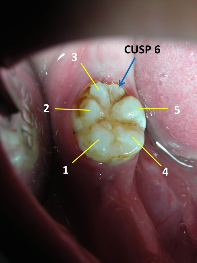
An 8-year-old body along with their parents visited a private dental clinic complaining of some missing teeth and delay in eruption of the permanent teeth in the lower right back region. Physical examination revealed a moderately built and well-nourished boy with good normal behavior. There were no signs and symptoms of any systemic, syndromic or metabolic disorders. Intraoral examination showed mixed dentition stage with erupted first permanent molars in both upper and lower arches (including both right and left quadrant). History elicited previous removal of some primary teeth in the right mandibular region after that there were no eruption of permanent teeth. Examination of oral cavity showed presence of only right first molar and mesial to it there was an edentulous space due to absence of some permanent teeth which were still not erupted. On careful examination, an additional supernumerary cusp was noticed on lingual side distal to the distolingual cusp. Total six cusps were noticed giving the molar tooth a ‘flower’ appearance (Figure 1). Finally based on literature search this extra cusp was diagnosed as “Entoconulid” or “Cusp 6” or “Tuberculum Sextum.” Following radiographic examination, patient was assured about the presence and eruption of permanent premolars.
Discussion
Normal Occlusal Anatomy of Permanent Mandibular First Molar
Permanent mandibular first molars usually have 5 cusps, named mesiobuccal, centrobuccal, distobuccal, mesiolingual, and distolingual [6, 7]. However, there may be four or three cusps. In four cusped molars, the distobuccal cusp appears to be missing, while the distolingual cusp is additionally missing in three-cusped forms [8]. The cusp 6 can only be distinguished when there are five other cusps are present. There is no way of knowing whether a single distal cusp is actually cusp 5 or 6. This procedure requires that there should be two distal cusps to define cusp 6.
Grading of Tuberculum Sextum/Cusp 6/ Entoconulid trait
Turner in 1970 [9] developed the standard reference plaque to denote different expression of this trait [Arizona State University Dental Anthropology System (ASUDAS)] which is shown in detail in Table 1.
| Scoring | Description of Cusp 6 expression |
|---|---|
| 0 | Cusp 6 is absent |
| 1 | Cusp 6 is much smaller than cusps 5 |
| 2 | Cusp 6 is smaller than cusp 5 |
| 3 | Cusp 6 is equal in size to cusp 5 |
| 4 | Cusp 6 is larger than cusp 5 |
| 5 | Cusp 6 is much larger than cusp 5 |
Table 1: Arizona State University Dental Anthropology System (ASUDAS)’s reference plaque on different expression of Cusp 6 trait
Table 1: Arizona State University Dental Anthropology System (ASUDAS)’s reference plaque on different expression of Cusp 6 trait [9]. Townsend, et al. [10] investigated the expression and genetic basis of the sixth cusp or entoconulid in mandibular molars among geographically isolated group of aboriginals from Northern Territory of Australia. They examined the dental casts of 399 patients and suggested four grades of trait expression, ranging from trace to small, medium, and large cusps of entoconulid [10]. The frequency of occurrence of entoconulid was about 80% for primary mandibular second molars compared to the frequencies of occurrence in permanent dentition ranging from around 50% on mandibular second molars to 70% on first molars and 80% on mandibular third molars. The degree of expression increased distally along the molar teeth, with only 3% of primary second molars showing large cusps compared with 25% of permanent mandibular third molars. Highest fluctuating asymmetry was observed in permanent mandibular second molar and lowest in primary second molars. No strong evidence for sexual dimorphism in occurrence or degree of expression was found [10].
Author of the current paper has performed few investigations on different dental morphological traits including both metric and non-metric tooth traits constituting both crown and roots of Indian pediatric patients [1, 2, 3, 4, 5, 11, 12, 13, 14, 15, 16, 17, 18, 19]. Nagaveni, et al. [13], published four cases of cusp 7 or Metaconulid in permanent lower first molars of Indian patients which is also rarely reported in the literature. Following this, in 2009, the same author reported on occurrence of “Paramolar Tubercle” in the primary dentition [12]. Again in 2012, Nagaveni [14] evaluated the occurrence of supernumerary roots in the permanent mandibular molars of Indian children.
One quasi-continuous threshold model performed in a study [10] showed that genetic contribution to occurrence of entoconulid variability was strongest in mandibular first molars. Significant associations were noted between entoconulid expression on mandibular molars and metaconule expression on maxillary molars, indicating that similar developmental mechanisms may influence these traits. The entoconulid and the metaconule both provide additional bulk on the distal occlusal surface of molar teeth, an area subjected to early wear during mastication in aboriginals [10].
In 2011, Kharaisat, et al. [20] studied the entoconulid, metaconulid, post-metaconulid and pre-entoconulid expression on permanent mandibular first molar in the living Jordanian population and inter-trait interactions in permanent mandibular first molars. Authors examined 360 school children consisting of 176 males and 184 females with age ranging from 15.5 to 16 years. Dental casts were prepared from these children and they were studied for the above-mentioned dental traits. They found 15.83% of cusp 7 in mandibular first molars and about 21.6% of cusp 6 in the same tooth. Authors concluded from their investigation stating that both cusp 6 and 7 on mandibular first molars were expressed in a relatively higher rate among the Jordanian Arabs compared to other studied Western Eurasians, indicating a significant gene flow from Sub-Saharan Africans and Mongolians to the Middle East. They also suggested that both cusp 6 and 7 dental morphological traits are genetically and phenotypically independent [20].
The occurrence of cusp 6 has no much clinical importance other than in forensics and in the dental anthropology related importance. Only few clinical considerations can be given importance which is related to the presence of deep pit and fissures and grooves around this cusp and its attachment with other cusps [7]. The prophylactic sealing of these retentive pit and fissure can be carried out using pit and fissure sealants. Apart from this prophylactic measure, regular topical fluoride application using any topical agents can be performed in these teeth having cusp 6 to prevent occurrence of dental caries [1]. If the cusp 6 is big and sharp it may cause chronic irritation to the soft tissue or tongue leading to chronic ulcers. In such circumstances grinding of the sharp edges of this cusp 6 can be done using dental finishing burs to make it smooth. There is no correlation with occurrence of cusp 6 and presence of extra or supernumerary third roots like Radix entomolaris or paramolaris in this clinical entity. However, during root canal treatment in a tooth having cusp 6, it is essential to keep an eye in the search of extra canals or orifices to avoid non-attending extra canals [14].
Conclusion
Rare dental morphological traits including both metric and non-metric traits are rarely encountered during clinical practice. Although occurrence of “Tuberculum Sextum” or “Entoconulid” is not associated with clinical problems, it is of highest interest to the anthropologists or morphologists as they provide more research evidence about biological dental evolution in human teeth of present Homo Sapiens compared to pre-historic people.
References
-
Nagaveni NB, Umashankara KV, Radhika NB, Praveen Reddy B, Manjunath S (2010) Maxillary paramolar: report of a case and literature review. Arch Orofac Sci 5(1): 24-28.
-
Nagaveni NB, Umashankar KV (2023) Report of a rare Odonto-Stomatologic Anomaly – Maxillary Paramolar. Series Clin Med Case Rep Rev 1(6): 1-3.
-
Nagaveni NB, Umashankara KV (2012) Radix entomolaris and paramolaris in children: A review of the literature. J Indian Soc Pedod Prev Dent 30(2): 94.
-
Nagaveni NB, Umashankar KV (2009) Radix entomolaris in permanent mandibular first molars: Case reports and literature review. Gen Dent 57(3): e25.
-
Nagaveni NB, Umashankara KV (2014) Maxillary molar with dens evaginatus and multiple cusps: Report of a rare case and literature review. Int J Oral Health Sci 3(2): 92.
-
Nelson SJ Wheeler’s dental anatomy, Physiology and occlusion, 2nd (Edn.), South Asia.
-
Nagaveni NB, Umashankara KV, Radhika NB, Mohan M (2015) Molarization of the mandibular second premolar in an Indian patient: Report of a rare case. Annals Bioanthropol 3(1): 33-45.
-
Nagaveni NB (2023) Premolarization of the permanent of the permanent maxillary second molars – Report of a rare case. J Dent Oral Health 10: 1-8.
-
Turner CG II, Nichol CR, Scott GR (2018) Scoring procedures for key morphological traits of the permanent dentition: the Arizona State University Dental Anthropology System. In: Kelley M, Larsen CS (Eds.), Advances Dental Anthropol. Wiley-Liss, New York, USA, pp: 13-31.
-
Townsend G, Yamada H, Smith P (1990) Expression of the entoconulid (sixth cusp) on mandibular molar teeth of an Australian aboriginal population. Am J Phys Anthropol 82(3): 267-274.
-
Nagaveni NB (2023) Protoconidal Cingulum/Protostylid in permanent mandibular first molar – Report of a rare dental morphological trait. Clin Pathol 7(1): 1-5.
-
Nagaveni NB, Umashankara KV (2009) “Paramolar Tubercle” in the primary dentition: Case reports and literature review. Int J Dent Anthropol 14(1): 12-18.
-
Nagaveni NB (2008) Occurrence of cusp 7 (Metaconulid) in permanent lower first molars – Report of 4 cases and review of literature. Int J Dent Anthropol 13: 22-27.
-
Nagaveni NB (2012) A retrospective analysis of accessory roots in mandibular molars of Indian pediatric patients. Int J Dent Anthropol 20: 38-46.
-
Nagaveni NB, Umashankara KV (2014) A clinical and radiographic retrospective analysis of talon cusps in ethnic Indian children. J Cranio Max Dis 3(2): 79.
-
Gupta S, Nagaveni NB, Chandranee NJ (2012) Three- rooted mandibular first primary molar: Report of three cases. Contemp Clin Dent 3(1): S134-136.
-
Nagaveni NB, Sreedevi B, Praveen BS, Reddy P, Vidyullatha BG (2010) Survey of mesiodens and its characteristics in 2500 children of Davangere city, India. Eur J Paediatr Dent 11(4): 185-188.
-
Nagaveni NB (2023) Huge Double Tuberculum Dentale (Lingual Tubercle) in maxillary central incisor – Report of a rarest dental morphological trait. Glob J Res Dent Sci 3(5): 13-15.
-
Nagaveni NB, Radhika NB (2012) Prevalence of taurodontism in primary mandibular first molars of ethnic Indian children. Gen Dent 60(5): e335-340.
-
Khraisat A, Alsoleihat F, Sawair FA, Shaweesh AI (2011) Entoconulid (cusp 6), metaconulid (cusp 7), post-metaconulid and pre-entoconulid expression on permanent mandibular first molar in the living Jordanian population and inter-trait interactions. Odontostomatol Trop 34(136): 11-19.
- Diagnosis and Management of Mental Nerve Paresthesia Secondary to Apical Periodontitis of Mandibular Second Premolar: A CBCT Based Case Report
- A Randomized, Double Blinded Clinical Trial to Compare the Effect of Oral Premedication (Diclofenac Potassium or Dexamethasone) on Post-Operative Pain Following Pulpectomy
- Modified Lip Repositioning Technique for the Management of Excessive Gingival Display
- Integral Role of Non-Dental Providers and Fluoride Dissemination
- Root Canal Treatment Rate in Deciduous Teeth Among 6-Year- Olds in the Era of Discontinuing Water Fluoridation - Historical Cohort Study
- The Impact of the Notch1 on the Migratory Capacity and the Expression of E-Cadherin and CyclinD1 in Ameloblastoma Cells