Sharp Rise in Fungal Infections on Platanus Orientalis-Needs Serious Introspections
Canker stain symptoms in alarming proportions were observed on beneath of the leaf surfaces in 21 differentially located Platanus orientalis kashmeriana populations similar to what has been reported earlier in United States and Europe. Genetic interpretations of these isolates, using ITS region of rDNA as unique DNA barcoding site for molecular identification showed the evolution of new fungal species, Alternaria kashmeriana sp.nov. From closely related species under geographically distinct conditions encountering 0.5 amount of genetic change in ITS region of rDNA. The pathogen is having a dramatic lethal effect on the natural populations of Platanus orientalis kashmeriana in Northern India and solid containment measures should be imposed to restrict its spread throughout the natural range of this ecologically, economically and historically precious host.
Introduction
Platanus orientalis L., the oriental plane perennial woody tree with important riparian species is widely spread in Crete and other southern islands [1, 2]. Its natural spread ranges from the southern Balkans, Crete, mountains of Turkey, Syria, Iran and Iraq, eastwardly to Kashmir valley, where it is familiarly known as the ‘chinar’. Chinar, Platanus orientalis kashmeriana, is the lone species of family Platanaceae found in India and growing throughout the valley of Kashmir. The family, Platanaceae consists of only a single living genus Platanus and is a native of the eastern Mediterranean portion from where it spread eastwards. It is a large deciduous tree, native to temperate regions. Pharmacologically, the buds are used in folk medicine as antiseptic and antimicrobial medication of the urinary system [1]. Plane has distinct advantages such as rapid growth rate, easy propagation, lush foliage and also strong air purification ability.
Canker stain of plane tree, caused by the fungus Ceratocystis platani is a lethal disease that attacks Platanus sp. was earlier reported from the United States and Europe [3, 4, 5, 6, 7]. The disease results in staining of the xylem, distraction of osmotic movement, cankers, and finally death of the tree [8]. The first report of the disease was observed in Greece in the 2003 on oriental plane in the southwestern Peloponnese, where C. platani has caused considerable mortality of the oriental plane in natural stands such as streams, rivers as well as in ornamental plantings in London plane [9, 10].
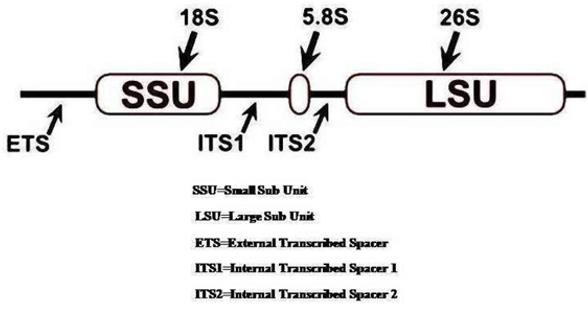
During the mycological explorations to the northern India (Kashmir), similar canker stain type symptoms were observed on beneath of the leaf surfaces of Platanus orientalis kashmeriana to what has been reported earlier in United States and Europe. In this study, the genetic variation was assessed in nuclear rDNA in order to recruit the pathogens classification purely relied on the molecular mycotaxonomic characterization based on fungal ITS DNA sequencing and its phylogenetic interpretations. The current revolutions in molecular biology has provided advanced techniques in mycotaxonomy. Molecular methods exploit the naturally occurring variations in the DNA. In eukaryotes, the ribosomal DNA (rDNA) found in the nuclear genome in general consists of tandem repeats of three RNA genes which undergoes transcription as a single unit and code for the 18S, 5.8S, and 28S subunits of the RNA (Figure 1) [11]. The internal transcribed spacer (ITS) region of the nuclear ribosomal DNA is the unique DNA barcoding site for molecular identification of fungi. Analysis of DNA sequences from rRNA genes and the ITS region have been used in studies of phylogenetic relationships and evolution of a wide range of organisms. The gold standard for fungal ITS sequence based classification rely on the concept of phylogenetic recognition of species. This systematic approach is commonly used with conserved large or small subunit of rDNA across taxonomically diverse fungi [12, 13]. The inclusion of taxonomically sundry ITS sequences into a single alignment is hectic because of considerable amounts of sequence variation, normally only the highly conserved 5.8S region can be considered for phylogenetic interpretations. Specific ITS primers (ITS1 and ITS4) were used in PCR to amplify a portion of the 18S rRNA gene, the ITS1 and ITS2 regions. Variation within the 18S rDNA and ITS1 region was interpreted viz sequence analysis and by digesting the amplified rDNA to give allele specific restriction fragment patterns. Molecular characterization of ITS region of rDNA of causal organism revealed the emergence of a new fungal species, Alternaria kashmeriana sp.nov_._, which may have evolved from closely related taxa encountering the ‘0.5’ of genetic change encountered by different bootstrapping values (random sampling with replacement) under geographically different environment.
Materials and Methods
Tissue Sampling
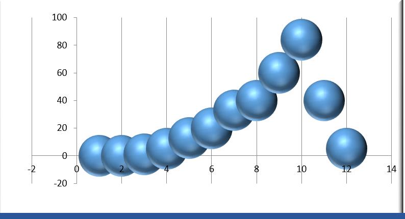
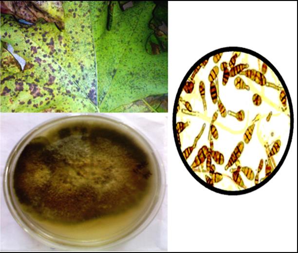
Periodical mycological visits were carried out to the selected locations. The fungal strains isolated from infected samples of Platanus orientalis kashmeriana collected from the different selected sites viz. Anantnag (04 isolates), Baramulla (03 isolates), Kupwara (05 isolates) and Srinagar (07 isolates). The Symptoms appeared on lower leaf surfaces as light dark brown irregular canker spots (like rust), later becoming deep grayish brown on the upper surface. Mostly the infected trees were prematurely defoliated. It was also observed that the extent of infection changes parabolically with the fluctuating environmental conditions in different seasons in the prevailing area shown in Figure 2.
(dictyoconidia) and are produced sympodially (Figure 3). Obclavate, obpyriform conidia sometimes ovoid or ellipsoidal with a short conical beak. On the 16th day fungal cultures were examined for CFU using hemocytometer and the spore suspension of 1.103 concentration were inoculated in potato dextrose broth medium.
DNA Isolation
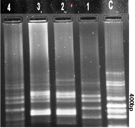
According to Moller et al 50 mg of dry mycelium mat was transferred in a sterile eppendorf tube. It was Crush with sterile scissor and made fine powder. 600μl of 60°C pre-warmed CTAB extraction buffer was added. The resulted mycelial powder was incubated in a water bath at 50°C for 3-4 hours with vortexing at the interval of 10 minutes. The resulting samples were centrifuged at 10000 rpm for 20 minutes at room temperature. The supernatant transferred to sterile eppendorf tube. 600μl of Chloroform and isoamyl alcohol (24:1) were added and subjected to vortexing again for 5 minutes. 0.5μl 3 M of sodium acetate (pH 5.2) was added and mix gently and stored on ice for 15 minutes. The solution was again centrifuged at 16000 rpm at 4°C for 10 minutes. 600μl of chilled isopropanol was added to eppendorf tube and stored at -50°C for 10 minutes. The supernatant decanted and 600μl 70% ethanol was added and solution was kept at room temperature. The overnight pellet was dried. 25μl of distilled water was added and kept at room temperatures for 2 hours followed by electrophoresis and ITS gene was amplified by selecting specific primer and optimizing PCR conditions. The primers (ITS1 and ITS4) showed highest amplification efficiency (Table 1). PCR reactions were performed using Kapabiosystems kit in 96-well plates. The reaction master mix constitute of 9.6 µl of 10% trehalose, 7.0 µl H2O, 2.5 µl 10X PCR buffer ‘B’, 0.8 µl MgCl2, 2.0 µl 2.5mM DNTP, 1.0 µl 10mM forward and reverse primers each and 0.1 µl taq polymerase (5 units/µl). This master mix is then distributed in each PCR tube 24 µl of mastermix in each PCR tube with 1 µl DNA template having concentration of 30-100ng/µl. The PCR reaction thermal cycles includes an initial step of 3 min at 95⁰C and 35 cycles of 30 sec at 94⁰C, 40 sec at 52°C, and 1 min at 72°C, with a final extension at 72°C for 10 min. Amplicons were visualized on 1.5% agarose gel shown in Figure 4.
nano drop spectrophotometry. The DNA samples were stored at -20°C.
PCR Amplification and Digestion of rDNA (ITS Gene Amplification)
Results
Internal Transcribed Spacer (ITS) Sequencing
These PCR amplicons were processed for cleanup to remove unincorporated nucleotide and residual primers using Exo-SAP enzyme. These amplicons were forwarded for cycle sequencing reaction using BigDye® Terminator v.3.1 Cycle Sequencing Kit (Applied Biosystems, Inc.) with 16th fold dilution. For sequencing of ITS1 & ITS4 primers were used for gene amplification were successfully used here. The thermal cycler conditions were an initial 5 step of 2 min at 96°C and 35 cycles of 30 sec at 96°C, 15 sec at 55°C and 4 min at 60°C. The Cycle sequencing is followed by sequencing cleanup by ethanol precipitation method followed by dissolving template in HiDiformamide. These samples were bi directionally sequenced in ABI 3130 Genetic analyzer.
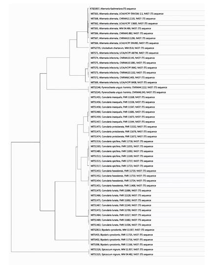
likelihood analysis carried out by MOLE BLAST (http://blast.ncbi.nlm.nih.gov/moleblast/moleblast.cgi) and statistical values of relatedness given by bootstrapping (random sampling with replacement) using another bioinformatic program phylogeny.lirmm.fr. The bottom line segment with the numeric number '0.5' reflects the length of branch which represents an amount of genetic change. The branch length units (nucleotide substitutions/site) are the number of genetic changes or substitutions divided by the sequence length (Figure 5). A branch length fluctuates proportionally to the nucleotide differences, and the numbers indicated on respective branches reflects the percentages of frequencies with which a given branch appeared in different 100 bootstrap replications.
Nucleotide Sequence Accession Numbers
The Gene Bank accession number for the sequence of the ITS region product using primers ITS1 and ITS4 in PCR amplification from Alternaria kashmeriana sp.nov. strain 150b (NCBI unique submission Id BankIt1858984) were assigned NCBI GenBank accession number KT825857.
Phylogenetic Analysis
The Phylogenetic tree has documented enough information about the inferred evolutionary relationships among the different allied Alternaria species. The ITS sequence was BLAST using http://www.ncbi.nlm.nih.gov data base to render the species similarities. The maximum
BLAST Score (E-Value Summary)
The E-value or Expect value is a metric tool that has shown the significant alignment in a particular homology ITS sequence match. In the specific BLAST search results are designated an E-value which reflects how significant the BLAST hit is. Statistically, the E- value is calculated as:
E = Kmne-λS Where, K and λ are constants which purely depend on the used scoring matrix, while as m is the query length and n is the database length and S is the alignment score. The calculated E-values of different hits are given in Figures 6a & 6b.
Discussion
Direct analysis/interpretations of DNA polymorphisms is a useful method to ascertain genetic variation in Alternaria species. Sequence analysis from ITS regions has made some progress towards isolation and better understanding the taxonomy of Alternaria kashmeriana sp.nov but further molecular analysis using more sensitive techniques to assess genetic variation have been implemented to determine the extent to which different taxa are present within this complex. In the present study ITS region of rDNA gene complex was used to evaluate genetic diversity/variation of geographically distributed Alternaria isolates. Sequence specific primers were designed at conserved genic regions of rDNA gene complex to amplify ITS regions. Phylogenetic analysis emphasized the evolutionary distance between different Alternaria species and related taxa. Alternaria kashmeriana sp.nov and Alternaria alternata are more closely related to each other than to Alternaria infectoria MITS71 isolates. It is sometimes difficult to differentiate the Alternaria kashmeriana sp.nov and from the rest known Alternaria species on the phenotypic bases but reflects the marked contradictions when compared on genetic grounds. Further the bootstrapping allowed to assign the measures of accuracy to sample estimates of Alternaria kashmeriana sp.nov with the rest of Alternaria species. The variance of 0.12655 of Alternaria kashmeriana sp.nov. from the closely related Alternaria isolates has been recorded with boot values of 0.2531, 0.07668, and 0.21472 (marked as SEQ00004, SEQ00002, SEQ00003 respectively (Figure 6b). Therefore it is concluded that the causal organism of canker stain in India is Alternaria kashmeriana sp.nov which has been evolved with genetic variance/mutation of 0.5 in ITS region of rDNA from closely related species under geographically distinct region (The great Himalayan mountains) [14, 15, 16, 17, 18].
Conclusion
The invasion or evolution and diversification of new strains or species under geographically distinct regions with different and extremely harsh environments is a lethal threat to disease prone regions which are comparatively less explored. The present study revealed the isolation of a new fungal pathogen, its identification and classification supported by both classical as well as molecular techniques. To our knowledge, this is the first report of canker stain disease of Platanus orientalis kashmeriana so far, caused by new fungal species. The disease is currently limited to the northern India where it was first observed. However, it may become epidemic due to its wide host range.
Acknowledgement
We thank Chief Conservator, Jammu & Kashmir for access to forest surveys, Central laboratory Instrumentation Centre (CLIC), Dr. Harisingh Gour Central University, Sagar, India and Director, Paul Hebert Centre, Aurangabad for DNA Barcoding and genetic analysis. This
work was financially supported by the University Grants Commission (UGC), India, sponsored Central University fellowship.
References
-
1 Mitrokotsa D, Mitaku S, Demetzos C, Harvala C, Mentis A, et al. (1993) Bioactive compounds from the buds of Platanus orientalis and isolation of a new kaempferol glycoside. Planta Med 59(6): 517-520. 2 Schnitzler A, Hale BW, Alsum E (2005) Biodiversity of floodplain forests in Europe and eastern North America: a comparative study of the Rhine and Mississippi Valleys. Biodivers Conserv 14: 97-117. 3 Walter JM, Rex EG, Schreiber R (1952) The rate of progress and destructiveness of canker stain of plane- trees. Phytopathology 42(5): 236-239. 4 Baker CJ, Harrington TC, Krauss U, Alfenas AC (2003) Genetic variability and host specialization in the Latin American clade of _Ceratocystis_ _fimbriata_. Phytopathology 93(10): 1274-1284. 5 Engelbrecht CJ, Harrington TC (2005) Intersterility, morphology and taxonomy of _Ceratocystis fimbriata_ on sweet potato, cacao and sycamore. Mycologia 97(1): 57-69. 6 Engelbrecht CJ, Harrington TC, Steimel J, Capretti P (2004) Genetic variation in eastern North American and putatively introduced populations of _Ceratocystis_ _fimbriata_ f. platani. Mol Ecol 13(10): 2995-3005. 7 Panconesi A (1999) Canker stain of plane tree: a serious danger to urban plantings in Europe. J Plant Pathol 81(1): 3-15. 8 CAB International (2001) Ceratocystis fimbriata. In: Baker CJ, Harrington TC (Eds.), Crop Protection Compendium. CAB International, Wallingford, UK. 9 Tsopelas P, Angelopoulos A (2004) First report of canker stain disease on plane trees, caused by Ceratocystis fimbriata f. sp. platani in Greece. Plant Pathol 53(4): 531. 10 Tsopelas P, Harrington TC, Angelopoulos A, Soulioti N (2006) Canker stain disease of oriental plane in Greece. In: Tjamos UE, Paplomatas E (Eds.), Proc. 12th Congr. Mediterr. Phytopathol. Rhodes Island, Greece, pp: 55-57. 11 Belkhiri A, Klassen GR (1996) Diverged 5S rRNA sequences adjacent to 5S rRNA genes in the rDNA of Pythium pachycaule. Curr Genet 29(3): 287-292. 12 O’Brien HE, Parrent JL, Jackson JA, Moncalvo JM, Vilgalys R (2005) Fungal community analysis by large-scale sequencing of environmental samples. Applied and Environmental Microbiology 71(9): 5544-5550. 13 Arnold AE, Henk DA, Eells RL, Lutzoni F, Vilgalys R (2007) Diversity and phylogenetic affinities of foliar fungal endophytes in loblolly pine inferred by culturing and environmental PCR. Mycologia 99(2): 185-206. 14 Altschul SF, Madden TL, Schaffer AA, Zhang J, Zhang Z, et al. (1997) Gapped BLAST and PSI-BLAST. A new generation of protein database search programs. Nucleic Acids Research 25(17): 3389-3402. 15 Abarenkov K, Nilsson RH, Larsson KH, Alexander IJ, Eberhardt U, et al. (2010) The UNITE database for molecular identification of fungi–recent updates and future perspectives. New Phytol 186(2): 281-285. 16 Dar RA, Rai AN, Surywanshi J (2014) Comparative kinetics and effect of different media on the growth of three foliicolous fungi Alternaria alternata, Tricothecium roseum and Fusarium oxysporium. Int J Bot 4: 1-10. 17 Ocasio Morales RG, Tsopelas P, Harrington TC (2007) Origin of Ceratocystis platani on native Platanus orientalis in Greece and its impact on natural forests. Plant Dis 91(7): 901-904. 18 Tedersoo L, Nilsson RH, Abarenkov K, Jairus T, Sadam A ,et al. (2010) 454 Pyrosequencing and Sanger sequencing of tropical mycorrhiza fungi provide similar results but reveal substantial methodological biases. New Phytologis 188(1): 291-301.
- Diversity of Candida sp and Antifungal Susceptibility Patterns in Digestive Candidiasis among People Living with HIV in CHU of Libreville, Gabon
- Vulvovaginal candidiasis: Retrospective study (2019- 2021) at the Centre Hospitalier National de Pikine, Suburban Dakar, Senegal
- Identification of Environmental Fungal Species in Clinical Services of University Hospital of Angre, Abidjan (Cote d’Ivoire)
- New Location of some Gasteroid Basidiomycetes in Western Kazakhstan
- Evaluation of Various Extracellular Enzymes of Ectomycorrhizal Mushrooms
- Morphology and Phylogeny of Lactarius Wallichianae sp. nov and Xerula magnispora sp. nov. from India