Ocular Sarcoidosis as a Presenting Symptom of the Systemic Disease
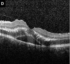
A 32-year-old man with no systemic complaints presents with flashers, floaters and blurry vision in the left eye for one week. Fundus examination reveals right eye (Figure 1A) intraretinal hemorrhages (black arrowhead) and perivascular sheathing of retinal veins (black arrow). The left eye (Figure 1B) shows a macular star (white arrowhead) and an elevated inferior granuloma (white arrow) with distal hemorrhages. Testing revealed an elevated angiotensin-converting enzyme (ACE) level, chest X-ray showed bilateral hilar lymphadenopathy (Figure 1C) and OCT imaging of the left macular star revealed hard exudates and subretinal fluid (Figure 1D). Biopsy via bronchoscopy showed granulomatous disease secondary to sarcoidosis
Introduction
A 32-year-old man with no systemic complaints presents with flashers, floaters and blurry vision in the left eye for one week. Fundus examination reveals right eye (Figure 1A) intraretinal hemorrhages (black arrowhead) and perivascular sheathing of retinal veins (black arrow). The left eye (Figure 1B) shows a macular star (white arrowhead) and an elevated inferior granuloma (white arrow) with distal hemorrhages. Testing revealed an elevated angiotensin-converting enzyme (ACE) level, chest X-ray showed bilateral hilar lymphadenopathy (Figure 1C) and OCT imaging of the left macular star revealed hard exudates and subretinal fluid (Figure 1D). Biopsy via bronchoscopy showed granulomatous disease secondary to sarcoidosis.
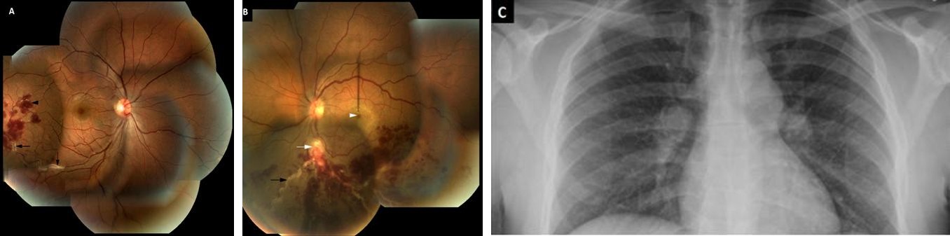
Image Article
- Screening of Hospital Staff During World Glaucoma Week in a Tertiary Eye Care Centre
- Angioid Streaks with Macular Neovascularization: Clinical Insights from Two Cases
- Giant Kissing Naevus: An Oculoplastic Challenge
- Why Freedom of Vision Should Not Cost the Freedom of Feeling - LASIK in the Climate of Change
- Asymmetric Optic Nerve with Small Disc and Large Cup: A Rare and Challenging Case of Unilateral Optic Nerve Hypoplasia
- Large Angle Exotropia in a Child: A Case Report