Asymmetrical Fundus Autofluorescence Findings in Parkinson’s Disease
Objective: Our aim was to show that retinal asymmetric degeneration was also a finding in Parkinson's disease (PD) Methods: The files of 20 PD patients diagnosed at other neurology clinics were examined. Fundus autofluorescence (FAF) images of these patients were observed by two ophthalmologists in a masked fashion. Results: In 15 patients hypo and hyper fluorescent lesions were on the nasal side of the retina. 1 L- handed patient had changes in the temporal retina. Conclusion: Our study suggests that imaging of the retina early in the disease by FAF may reveal findings consistent with the asymmetric nature of PD.
Introduction
Motor asymmetry is one of the criteria for the diagnosis of Parkinson’s disease (PD) [1]. A right-sided symptom on set is usually associated with a more favorable outcome in terms of cognitive impairment while a left-sided symptom on set appears to be associated with a better outcome in terms of motor progression. PD symptoms emerge more often on the dominant hand-side [2]. The mammalian retina contains dopaminergic neurons with in the inner retinal layer. Visual alterations are associated with PD and seem to be caused by dysfunction of the in traretinal dopaminergic circuits [3]. Inter ocular symmetry on spectral domain optical coherence tomography (SD-OCT) of the retina and possibly nerve fiber layer has also been documented [3]. As the neurosensory retina is impacted in PD, it is plausible that the earliest changes are also asymmetrical and retina screening would thus be useful and be a good marker of disease presence and progression.
Methods
The files of 20 PD patients diagnosed at other neurology clinics were examined. Fundus auto fluorescence (FAF) images of these patients were observed by two ophthalmologists in a masked fashion. Hyper or hypo fluorescent lesions which suggested neurodegeneration were taken into consideration. The mean age of the patients was 69. 16 patients were right- handed while 4 were left- handed.
Results
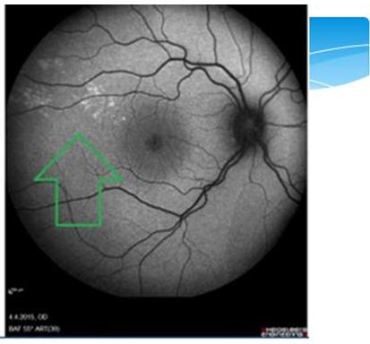
In 15 patients hypo and hyper fluorescent lesions were on then as al side of the retina. (Figures 1 & 2).
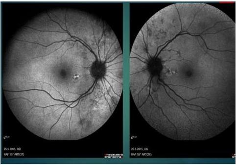
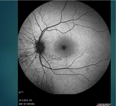
Figure2: Nasal neurodegeneration on FAF. In 14 R- handed patients who had early PD, degenerative lesions were on then as al side. 1 L- handed patient had changes in the temporal retina (Figure 3). None of the R- handed patients had neurodegeneration on the temporal side. Diffuse degeneration on FAF was detected in the middle –late stages of the disease. 4patients had this kind of distribution. The early asymmetric appearance was in parallel with the asymmetric findings in motor functions. The possibility to detect hemi-retinal neurodegeneration in patients with unilateral motor findings was statistically significant (P: 0,00).
Discussion
Since FAF detects lipofuscin in the retina, the images were consistent with retinal damage. Lipofuscin gives damage to the tissues by mechanically obstructing the flow into and out of the cells and slowing down the elimination of waste materials. Photo receptor degeneration unmasks the auto fluorescent signal of the underlying RPE and thus creates hyper-auto fluorescent images. In contrast, hypo-auto fluorescence arises from decreased lipofuscin or blockage by material anterior to the RPE and photo receptors [4]. In the brain there is an important component of hemispheric lateralization over the course of PD. The motor asymmetry is associated with a severe contra lateral nigrostriatal degeneration [5]. Some studies suggest in creased “left hemi sphere susceptibility,” in that the left nigrostriatal pathway is more affected than the right [6]. Some suggest this may be an effect of handedness, but handedness does not seem to account for this observation entirely. The etiology of this left hemisphere‐predominant atrophy across the spectrum of neurodegenerative disease remain sun clear, although the rear several hypo theses involving genetics, lateralized vulnerability, and disease‐specific factors [6]. Similar to the brain, the retina is also affected asymmetrically. FAF detected neurodegenerative changes seem to affect one side of the retina early in the disease. In our study, the nasal retina was more affected than the temporal part. The predilection of neurodegeneration for one side of the retina and the asymmetric appearance on FAF was not reported before, to the best of our knowledge. Studies with larger series may give important information about the early detection of PD by ophthalmological examination.
Conclusion
Our study suggests that imaging of the retina by FAF may reveal findings consistent with the asymmetric nature of PD.
References
-
Baumann CR, Held U, Valko PO, Wienecke M, Waldvogel D (2014) Body side and predominant motor features at the onset of Parkinson's disease are linked to motor and nonmotor progression. Mov Disord 29(2): 207-213.
-
Tomer R, Levin BE, Weiner WJ (1993) Side of onset of motor symptoms influencies cognition in Parkinson's disease. Ann Neurol 34(4): 579-584.
-
Lee JY, Ahn J, Kim TW, Jeon BS (2014) Optical coherence tomography in Parkinson's disease: is the retina a biomarker. J Parkinsons Dis 4(2): 197-204.
-
Kayabasi U, Sergott RC, Rispoli M (2014) Retinal Examination for the Diagnosis of Alzheimer’s Disease. Int J Ophthalmic Pathol 3(4).
-
Daniel OC, Katherine EM, Manus D, Shiv R, Scott A Wylie, et al. (2016) Cortical asymmetry in Parkinson's disease: early susceptibility of the left hemi sphere. Brain Behav 6(12): e00573.
-
Christoph S, Klaus S, Katherina JM, Eveline D, Irene V, et al. (2012) Left hemispheric predominance of nigrostriatal dysfunction in Parkinson’s disease. Brain 135 (11): 3348-3354.
- Screening of Hospital Staff During World Glaucoma Week in a Tertiary Eye Care Centre
- Angioid Streaks with Macular Neovascularization: Clinical Insights from Two Cases
- Giant Kissing Naevus: An Oculoplastic Challenge
- Why Freedom of Vision Should Not Cost the Freedom of Feeling - LASIK in the Climate of Change
- Asymmetric Optic Nerve with Small Disc and Large Cup: A Rare and Challenging Case of Unilateral Optic Nerve Hypoplasia
- Large Angle Exotropia in a Child: A Case Report