Misfolded Tau Protein in the Retina
Introduction: Tau protein plays a crucial role in many neurodegenerative diseases including Alzheimer's disease (AD). Tau inclusions and amyloid beta (AB) depositions have been described in the post-mortem retina exams of AD patients. Cryo- electron microscopy (Cryo- EM) was recently used to detect the detailed structure of Tau filaments. Methods and Result: We examined the retinas of PET-proven live AD patients by spectral domain optical scanning tomography (SD- OCT) and fundus auto fluoresce in (FAF). The hyper or hypo- fluorescent lesions in the retina were scanned by OCT and images that completely corresponded with the histopathological and Cryo- EM shapes of Tau filaments were observed. Conclusion: Retinal Tau is a very promising target to detect early changes in AD and retinal imaging may be an exciting and trustable technique to predict and monitor the disease.
Introduction
Tau protein plays a crucial role in many neurodegenerative diseases including Alzheimer’s disease (AD). Tau dysfunction includes abnormal tau phosphorylation, protein aggregation, neurofibrillary tangle formation and neurotoxicity [1]. The retina is integrated with the central nervous system (CNS) and has been considered a window to the brain. Similar neurodegenerative processes affect the retina causing impaired contrast sensitivity, reduced visual acuity and abnormal motion perception. Approximately 50% of AD patients present with visual deficits that go along with RGC loss, thinning of the retinal nerve fiber layer, abnormal electroretinogram response and reduced blood flow [2]. Tau inclusions and amyloid beta (AB) deposition have been described in the post-mortem retina exams of AD patients. Phosphorylated and misfolded Tau accumulation has also been observed in RGC soma, dendrites and in traretinalaxons in animal models. These pathological changes may cause retinal neurondys function and sub sequent death and suggest a prominent role for abnormal tau in visual deficits [2].
Cryo- Electron Microscopy
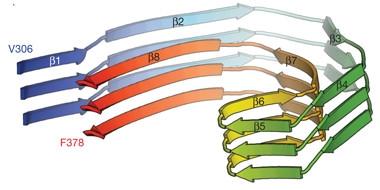
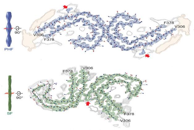
Cryo- electron microscopy (Cryo- EM) was recently used to detect the detailed structure of Tau filaments [3]. There searchers discovered C- shaped paired helical filaments (PHF) composed of Tau- subunits in the brains of live AD patients and they presented the C-shape in striking detail (Figure 1). Tau aggregation consisted of a mixture of PHFs and straight filaments (SF), the former making up about 90 precent of the total [3]. A C-shaped core defined the common proto filament of PHFs and SFs although the two Cs could have been arranged differently (Figure 2).
Retinal Imaging of Tau
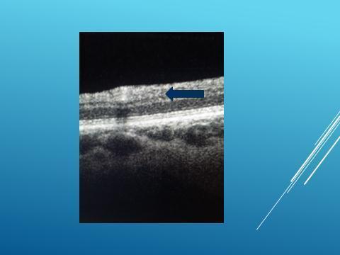
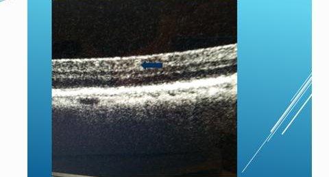
We examined the retinas of PET-provenlive AD patients by spectral domain optical scanning tomography (SD- OCT) and fundus auto fluoresce in (FAF). The hyper or hypo- fluorescent lesions in the retina were scanned by OCT and images that completely corresponded with the histopathological and Cryo EM shapes of Tau filaments were observed. PHFs that were identical with the Cryo- EM images in terms of shape and diameter were detected (Figures 3 & 4).
Discussion and Conclusion
Since the eye is the most easily access able part of CNS, retinal exam is quick, non-invasive and no radiation is involved. The results of a very important study revealed that in a group of patients with mild cognitive impairment and full-blown AD dementia, although all patients had extensive amyloid plaques, their brain amount of Tau was highly individual [4]. This finding could explain why the disease progressed at such a varying rate from one patient to the other stressing the importance of Tau in AD. Retinal Tau is a very promising target to detect early changes in AD and retinal imaging may be an exciting and trustable technique to predict and monitor the disease.
References
-
Marius Chiasseu, Luis Alarcon-Martinez, Nicolas Belforte, Quintero H, Dotigny F, et al. (2017) Tau accumulation in the retina promotes early neuronal dysfunction and precedes brain pathology in a mouse model of Alzheimer’s disease. Mol Neurodegener 12(1): 58.
-
Javaid FZ, Brenton J, Guo L, Cordeiro MF (2016) Visual and ocular manifestations of Alzheimer's disease and their use as biomarkers for diagnosis and progression. Front Neurol 7: 55.
-
Fitzpatrick AWP, Falcon B, He S, Shaoda He, Alexey G Murzin, et al. (2017) Cryo-EM structures of tau filaments from Alzheimer's disease. Nature 13(547): 185-190.
-
Chiotis K, Saint-Aubert L, Rodriguez-Vieitez E, Leuzy A, Almkvist O, et al. (2017) Longitudinal changes of tau PET imaging in relation to hypometabolism in prodromal and Alzheimer’s disease dementia. Molecular Psychiatry.
- Screening of Hospital Staff During World Glaucoma Week in a Tertiary Eye Care Centre
- Angioid Streaks with Macular Neovascularization: Clinical Insights from Two Cases
- Giant Kissing Naevus: An Oculoplastic Challenge
- Why Freedom of Vision Should Not Cost the Freedom of Feeling - LASIK in the Climate of Change
- Asymmetric Optic Nerve with Small Disc and Large Cup: A Rare and Challenging Case of Unilateral Optic Nerve Hypoplasia
- Large Angle Exotropia in a Child: A Case Report