Effect of Colour Vision in Different Amount of Deviation of Exotropia
Purpose: The aim of the present study is to compare the effect of colour vision in different amount of deviations in Exotropia. Methods: A pilot, cross sectional, observational study was performed at tertiary eye care centers. Subjects with Ocular deviation between 10 to 40 prism diopters, Corrected distance Visual Acuity should be greater than 6/18 and Age should be between 10 to 40 years of age were included in the study. Colour vision was assessed with Fransworth D 15 colour vision test. Results: 30 subjects were included in the study. Out of that, 16 subjects were in the age group of 11-20 years, 12 subjects were in the age group of 21-30 years and 2 subjects were in the age group of 31-40 years. 60% subjects were Female and 40% subjects were Male. The mean colour vision was considered in each amount of deviation. It shows that colour vision will be deteriorated more as ocular deviation increases in cases of Exotropia. Conclusions: In ocular deviation of Exotropia, as amount of deviation increases, Colour vision decreases gradually. Mild to Moderate Tritanopia occurs in increased amount of deviation.
Introduction
In case of ocular deviation images of an object are falling on parafoveal region and due to inequality of the number of the cone cells in the macular region there may be chances of the Colour vision deterioration with increasing ocular deviation. In case of Esodeviation the images of an object is placed at the nasal retinal side and due to its intermittent stages is very low compare to Exodeviation [1]. Thus there may be strong reason with increasing ocular deviation Colour vision deterioration is also being deteriorated.
Methodology
A pilot, cross sectional, observational study was performed at tertiary eye care centers [2, 3, 4, 5, 6, 7]. Subjects with Ocular deviation between 10 to 40 prism diopters, Corrected distance Visual Acuity should be greater than 6/18 and Age should be between 10 to 40 years of age were included in the study. Individuals with any other systemic disease(specially which can affect study),Individuals with any other Ocular Pathology, with any active ocular infection, any ocular anomalies like Corneal Scar etc ,ocular deviation if less than 10 degree and Significant amount of amblyopic patient were excluded from the study. Full refractive correction along with detailed fundus evaluation was performed in each and every patient. Colour vision was assessed with Fransworth D 15 colour vision test in different amounts of deviation in Exotropia. Data was analyzed using SPSS software version 20.
Results
30 subjects were included in the study. Graph 1 shows distribution of subjects in various age groups.16 subjects were in the age group of 11-20 years, 12 subjects were in the age group of 21-30 years and 2 subjects were in the age group of 31-40 years. Graph 2 shows gender wise distribution of the subjects. 60% subjects were Female and 40% subjects were Male. Graph 3 shows comparison of Colour vision for different amount of deviation for Exotropia. 1 is indicated for normal colour vision, 2 are indicated for mild tritanopia, 3 are indicated for moderate tritanopia and 4 are indicated for severe tritanopia.
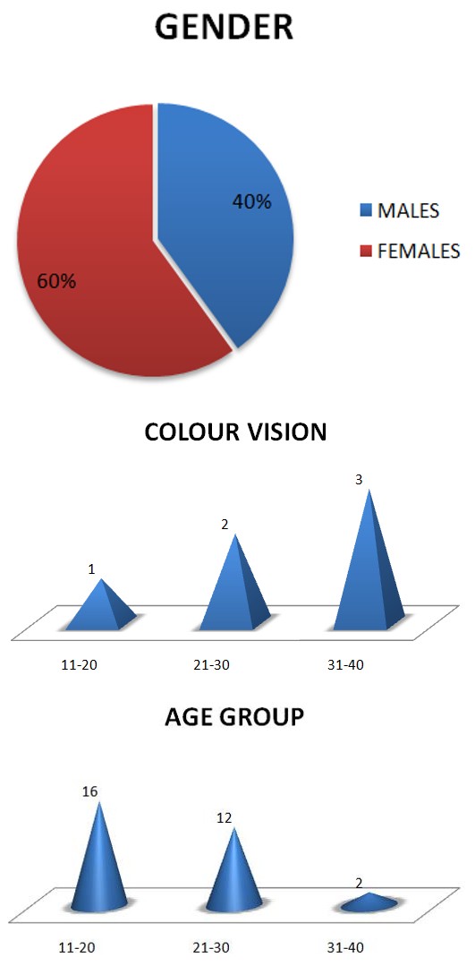
Graph 1: Shows Age Wise Distribution of the Subject.
Graph 2: shows gender wise distribution of the subjects.
Graph 3: Shows comparison of Colour vision for different amount of deviation for Exotropia.
Discussion
In this study, as per statistical analysis it shows that there is normal colour vision in the ocular deviation of 11-20 prism diopters, mild tritanopia in 21-30 prism diopters and moderate tritanopia in 31-40 prism diopters. According to the present study, it has been showed that in cases of ocular deviation, images of an object fall on the Para foveal region and deteriorates the colour vision. In case of Exodeviation there have less chances to become Amblyopia. Due to most of cases intermittent timing is high compare to Eso deviation. In case of Exodeviation images of an object is fall on the temporal parafoveal region and according to the deformity of the anatomical arrangement of the photoreceptor cells in the macula that’s why in the parafoveal region number of cone cells is less compare to foveal region. Just for this reason in case of Exodeviation images is shifted towards parafoveal region and due to less number of cone cells and according to the statistics it has been proved that with increasing Exodeviation color vision is been deteriorated
Conclusion
In ocular deviation of Exotropia, as amount of deviation increases, colour vision decreases gradually. Mild to Moderate Tritanopia occurs in increased amount of deviation.
References
-
Kenneth W Wright, Peter H Spiegel, Lisa Thompson (2006) Handbook of Pediatric Strabismus and Amblyopia. 1st (Edn.).
-
Hui Zhu, Jia-Jia Yu, Rong-Bin Yu, Hui Ding, Jing Bai, et al. (2015) Association between Childhood Strabismus and Refractive Error in Chinese Preschool Children. Journal of Plos One 10(6): e0130914.
-
Zhale Rajavi, Sabbaghi H, Baghini AS, Yaseri M, Sheibani K, et al. (2015) Prevalence of Colour Vision Deficiency and its Correlation with Amblyopia and Refractive Errors among Primary School Children. J Ophthalmic Vis Res 10(2): 130-138.
-
Anika K Tandon, Federico G Velez, Stacy L Pineles (2014) Binocular Inhibition in Strabismic Patients is Associated with Diminished Quality of Life. Journal of American Association for Pediatric Ophthalmology and Strabismus 18(5): 423-426.
-
XC Ye, Pegado V, Wasserman WW (2014) Strabismus genetics across a spectrum of eye misalignment disorders. Journal of clinical genetics 86(2): 103-111.
-
Koçak Altintas AG, Satana B, Koçak I, Duman S (2000) Visual Acuity and Colour Vision deficiency in Amblyopia. Eur J Ophthalmol 10(1): 77-81.
-
Freeman AW, Nguyen VA, Jolly N (1996) Components of Visual Acuity Loss in Strabismus. Vision Res 36(5): 765-774.
- Screening of Hospital Staff During World Glaucoma Week in a Tertiary Eye Care Centre
- Angioid Streaks with Macular Neovascularization: Clinical Insights from Two Cases
- Giant Kissing Naevus: An Oculoplastic Challenge
- Why Freedom of Vision Should Not Cost the Freedom of Feeling - LASIK in the Climate of Change
- Asymmetric Optic Nerve with Small Disc and Large Cup: A Rare and Challenging Case of Unilateral Optic Nerve Hypoplasia
- Large Angle Exotropia in a Child: A Case Report