The Retino-Hypothalamic Ultrastructure aspects of Traumaticoptic Neuropathy’s Pathogenesis and Treatment
Purpose: The purpose was to study the retino-hypothalamic ultrastructural changes in traumatic optic neuropathy’s pathogenesis and treatment. Methods: There was reproduced the experiment traumatic to the optic nerve crush by surgical clips for 90 mature rabbits. There were next groups of animals: intact (I), experimental (II) and two groups with two types of treatment (III and IV). There were 30 individuals in each group (there were 120 animals).The group III took infusions of Methylprednisolone in dose 30 mg/kg for three days. The group IV took infusions Methylprednisolone in dose 15 mg/kg for 3days in combination with phosphine-elecric stimulation (PES) from the third till the 13thday. The power of electrical impulsive supply was 800 mРin affected side and 300 mРin opposite. The morphological analysis included electron microscopy of the semi-thin and ultrathin sections and the morphometry of the retina and suprachiasmatic nucleus of the hypothalamus of all groups. It was performed one month after the injury whom the animals were removed from the experiment. It was conducted an analysis of the content of cortisol and adreno-corticotrope hormone (ACTH) in the blood serum of experimental animals of all groups in the dynamics up to one month after an injury. Results: There was reproduced that traumatic damage to the orbital part of the optic nerve. It caused colicvative necrosis of ganglion cells and swelling of the layer of nerve fibers of the ipsilateral retina. It is established also that traumatic damage to the orbital part of the optic nerve causes structural changes in the suprachiasmatic nucleus of the hypothalamus. Combined treatment with phosphine electro stimulation characterized by reduction of retina thickness, reduction of cytokaryometric indices and regeneration processes in bipolar and ganglionic neurons of retina. There was found changing architectonics and increasing the number of neurosecretory granules of the suprachiasmatic nucleus of the hypothalamus under combine treatment. The content of ACTH in the peripheral blood decreases and the content of cortisol increases in the III group. In the IV group, the content of hormones is more consistent with the group without treatment. Conclusion: Thus, the complex treatment of TON with the use of phosphine electrostimulation can be an alternative to traditional treatment, since it allows reducing the dose of infusion of corticosteroids and provides the necessary neuroprotective effect.
Introduction
The possibility of the suprachiasmatic hypothalamus nucles activation using by light was shown by Bremer [1]. The peptides of supraoptic and paraventricular nucleus activate of the synthesis of nerve tissue growth factor [2, 3]. The mutation of this factor promotes growth differentiation of vegetative system’s elements and change its properties neurosecretory [4]. It is known else that suprachiasmatic nucleus regulates light depended corticosteroids’ secretion at night and day time [5, 6].
The neural secretion of hypothalamic nucleus activates due to phospin electric stimulation (PES). There was detected dose independence of PES influence for cerebral vessels’ to use and for neural secrete’s synthesis by magnocellular are neurons of the supraoptic hypothalamic nucleus. Course application of stimulation with current of 100 μA and 300 μA causes redistribution of the contents of different structural-functional types of neurons in the supraoptical nucleus of the hypothalamus [7, 8].
These mechanisms formed the basis of our assumption about the possibility of using retino- hypothalamic stimulation in traumatic optical neuropathy (TON) to ensure the development of endogenous cortecosteroids, which would reduce the dose of proven use of Methylprednisolone, which in the dose of 30 mg/ kg is a standard TON treatment [5]. This would have the ability to reduce the toxic effects of megadoses of corticosteroids, while preserving their neuroprotective effect. The purpose was to study the importance of retino- hypothalamic ultrastructural changes in traumatic optic neuropathy’s pathogenesis and treatment.
Materials and Methods
There experimental traumatic optic nerve crush by surgical clips was reproduced in 90 sexually mature rabbits of the chinchilla breed, according to the conclusion of the Bioethics Commission approved by the Ivano-Frankivsk National Medical University in 2016 [9].
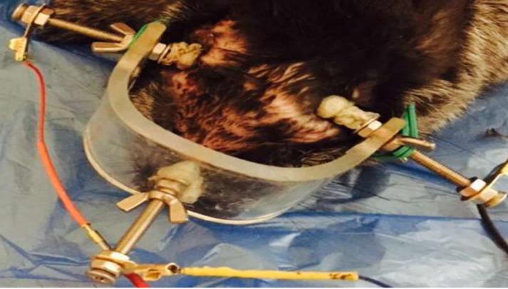
Animal groups were as follows: intact (I), experimental (II) and 2 groups with different treatments (I and II) for 30 individuals in each (120 animals). Group III received (Methylprednisol) at a dose of 30 mg/kg for three days. Group IV received methylprednisolone 15 mg/kg with a combination of PES from the third till the 13thday (Figure 1) with a current of 800 mA on the side of the lesion and 300 mA on the opposite side.
The morphological analysis (electron microscopy of the semithin and ultrathin sections and morphometry) of the retina and suprachiasmatic (synonyms of supraoptic) nucleus of the hypothalamusin all groups after removing from the experiment (using guillotine)was done in one month after the trauma (in the electron microscopy laboratory of the Anatomy Department of Ivano- Frankivsk National Medical University). The maintenance of animals (up to 1 month after injury) and their remove from the experiment were performed in accordance with the "Requirements of Bioethics of the Helsinki Declaration on the Ethical Regulation of Medical Research". An analysis of the content of cortisol and adeno-corticotropic hormone (ACTH) in blood serum of experimental animals of all groups in the dynamics up to one month after injury was carried out at 9.00.
Result
In the retina of the side with crushed optic nerve II group of animals, there is find a significant thickening of the retina in comparison with the control group (I) from 178,5±11,47 μm to 246,85±23,69 μm. There is an increase
| Retinal | Groups of animals | |||||||
|---|---|---|---|---|---|---|---|---|
| layers | І | ІІ | ||||||
| PSL | 32,43±4,85 | 55,41±13,64 | ||||||
| ↑*70,8% | ||||||||
| ONL | 33,14±4,08 | 51,33±8,52 | ||||||
| ↑*54,9% | ||||||||
| INL | 27,52±7,92 | 38,57±4,16 | ||||||
| ↑*40,1% | ||||||||
| GCL | 20,02±3,61 | 30,61±11,64 | ||||||
| ↑*53% | ||||||||
| LNF | 16,36±4,72 | 22,08±4,35 | ||||||
| ↑*35% |
Table 1: Retinal morphometric dates for TON (M±m, μm).
Note: * significant difference with I group, P<0,05.
Also in animals of group ІІ it was noted swelling in ONL, INL and GCL (Figure 2б, 2B). There were observed microcystal degeneration of ganglionic neurons and pericellular edema in the GCL. There was seen the significant decrease in the number of multipolar neurons in the GCL compared with the intact group of animals (Figure 2a). The area of these neurons increases compared with intact from 58,81±9,01 μm2 to 71,68±8,87 μm2 (P<0,01), whereas the core area does not significantly change and is 37,06±36,20 μm2 (intact group - 36,20±6,63 μm2), which leads to a decrease in nuclean- cytoplasmic index (NCI) from 1,14±0,36 to 1,14±0,36 (P<0,03). There are also thickening and enligtnment of NFL.
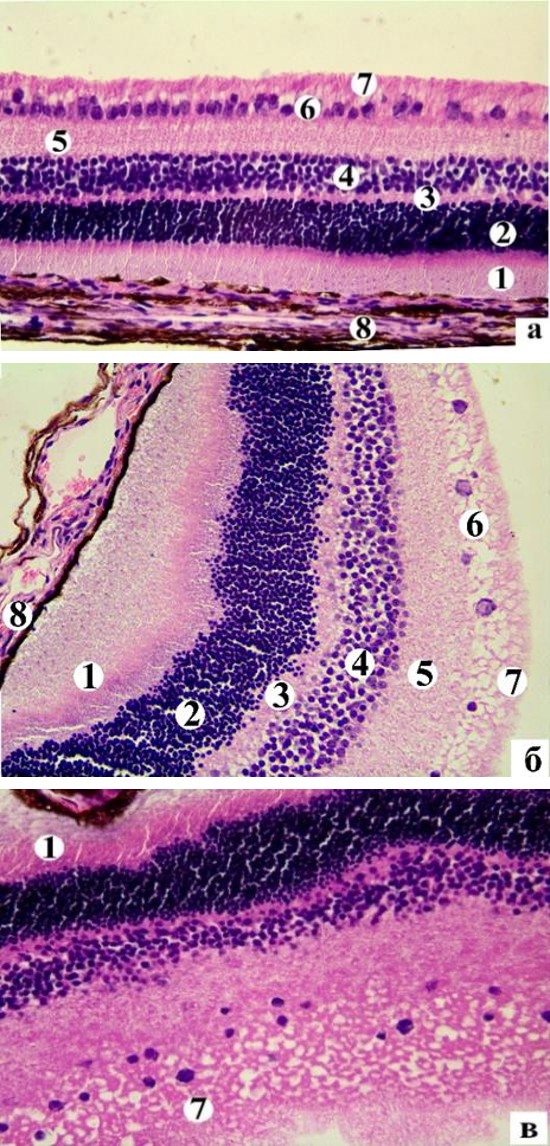
in the thickness of the photosensory layer (PSL) by 70,8%, outer nuclear layer (ONL) by 54,9%, internal nuclear layer (INl) by 40,1%, ganglion cell layer (GCL) by 53%, and layer of nerve fibers (NFL), by 35% (Table 1).
There were number of neural cells (NC) with the phenomena of peripheral chromatolysis and hypochromic nuclei increases, "shadow cells" appear (Figure 3) in the hypothalamus suprachiasmatic nucleus (SN) in the experimental group (II) at the 30th day after injury the right optic nerve. There were found some NC with small vacuoles on the pericarp of the pericarion. There are karyopicnosis and somewhere carriolysisin majority NC. The area of pericarion SN in comparison with the intact group increases to 276, 59±38, 02 μm2 (P<0,05), whereas the area of the core field of nuclei does not significantly change 71,93±15.67 μm2 (P>0,05). NCI decreases to 0, 35±0,07 (P<0,01). This does not indicate NC’s functional activation. It means faster the swelling of the nucleus and cells and their destructive changes.
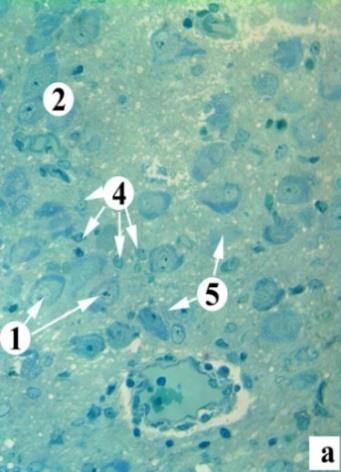
Notes: 1 - normochromic neurons with central chromatolysis, 2 - hyperchromic neurons, 3 – chiasmic fibers, 4 - glyocytes, 5 - shadow cells.
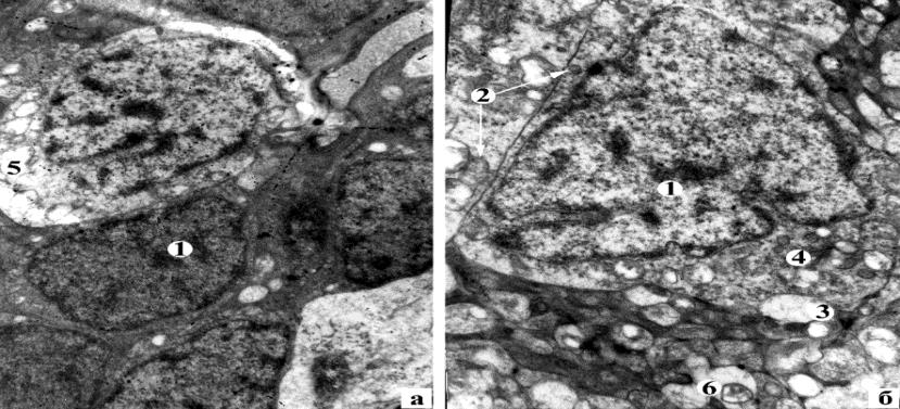
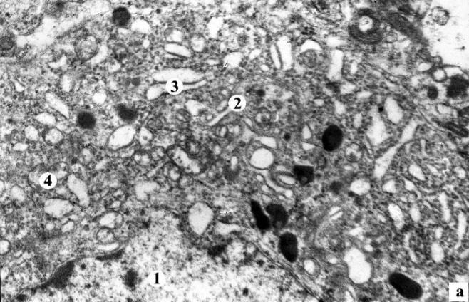
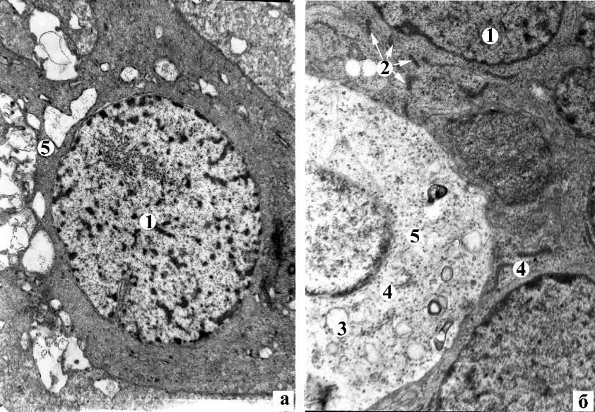
At the ultrastructural level, in the retina, there were observed neurons with dystrophic and destructive changes in the INL (Figure 4a). There were noted karyopicnose, invaginations of the nuclear shell, enlightenment and vacuolation of the cytoplasm. There were single neurons in the state of colicvative necrosis. In GCL, the great path of neurons has the cytoplasm with small vacuoles (Figure 4b). The nuclei acquire a triangular and irregular shape due to significant invasions of the nuclear shell, indicating reactive changes in the cell. The granular endoplasmic net tanks are expanded, some of them destroyed, others shortened, contain single attached ribosomes. A part of the mitochondria in the cell has a normal ultrastructural organization, other partially disorganized crystals and the rest - with a broken inner membrane. There were seen capillaries with significant edema of endothelial cells, which blocked the lumen of the vessel, were encountered in the NFL. Around these capillaries was found perivascular edema. In the part of nerve fibers of NFL there was observed axonal degeneration, which morphologically manifested itself: electron-transparent axoplasm, decrease in the number of neurofilaments on the background of the complete absence of microtubules; disorganization and destruction of mitochondrial crists.
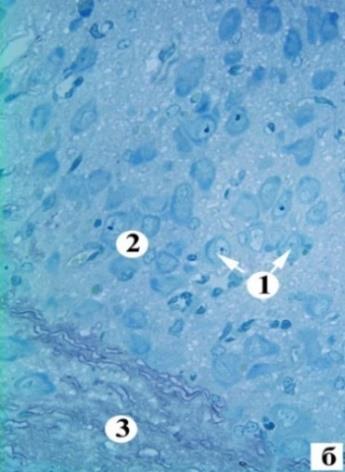
Figure 4: The bipolar cell’s (а) and ganglion (б) neuron dystrophy and destructive changes for animal of II group at the 30th day after experiment Electronmicroscopy. Magn: а) x4800, б) x6400 Notes: 1 – neuron’snuclei, 2 – mitochondria, 3 – vacuole, 4 – granular endoplasmicnet, 5 – colicvativenecrosisofneuron, 6 – enlightenment of the axoplasm in NFL.
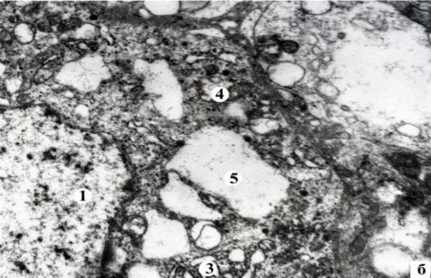
The cytoplasm, an expansion of the granular endoplasmic mesh tanks, illumination of the mitochondrial matrix and the destruction of crystals with the subsequent formation of vacuoles were found at the ultrastructural level in the majority of light NC of SN’s hypotalamus. An increase in the number of primary and secondary lysosomes, and the destruction of the Golgi complex are observed (Figure 5). Nucleis have low electron-optical density with minor invasions of the nuclear shell. There are isolated light NC with the phenomena of hydropic dystrophy. The volume density of NG in lightneurons is significantly reduced to 2,24±0,19% (P<0,02) compared to the intact group of animals (Table 2). The small vacuoles, lysosomes, an increase in the number and expansion of granular endoplasmic mesh tanks, multivascular corpses, and single NG are found in the neuroplasm of dark NCs. Their nuclei are electron- cubic with one or two nucleolis. The volumetric density of dark NG is significantly reduced to 1, 14±0,04% (P<0,02) compared with intact animals.
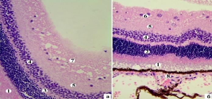
Notes: 1 – PSL, 2 – ONL, 3 – ORL, 4 – INL, 5 – IRL, 6 – GCL, 7 – NFL, 8 – pigment layer.
They are respectively 60,03±11,7 μm2 (P<0,01), 36,33±8,69 μm2 (P<0,01), 1,70±0,79 (P<0,05). Such quantitative indices of ganglionic neurons of the IV group of rabbits do not significantly different from intact animals (in all cases, P<0,05). There was a recovery of SN’s hypotalamus structure in rabbits III and IV groups, who received treatment during a month. There are central chromatolysis neurons. Most of the neurons had diffusely located tigroid granules (a sign of regeneration) in animals of the IV group. Such NCs were single in animals of the group III.
The area of pericarions in animals of groups III and IV significantly decreased to 232,14 ± 56,81 μm2 (P<0,04) and 224,25±58.26 μm2 (P<0,05) compared with the pathology(II) and were not significantly differed from the intact (I) group of animals (P<0,05).It indicates as edema. The nuclei in animals of groups III and IV did not significantly change and amounted to 63,46±14,38 μm2 and 76,51±16,54 μm2 (in all cases, P<0,05) compared to pathology and intact animals. The NCI significantly increased to 0, 56±0,23 (P<0,03) compared to the pathology and did not differ significantly from intact animals (P>0,05)in the animals in the IV group. There is not significantly different from the animals in the III and II group. The NCI of the group III is 0,39±0,09 (P>0,05). It is statistically significantly lower than intact animals (P<0,03).
In animals, after treatment at the ultrastructural level, traces of regenerative processes in the retina (Figure 7). There is an enlightenment of the neuroplasia of the individual pericarions, the disorganization of the mitochondria in the neurons of the INL. In the GCL of III group most of the neurons are in a state of vacuolic dystrophy, in some cases phenomena of apoptosis are observed. There are compensatory regeneration processes in the ganglionic neurons of the IV group. They characterized by: hypertrophy of the granular endoplasmatic mesh tanks and an increase in the surface of the attached ribosomes; restoration of fine-grained neuroplasm of moderate electron density; the appearance in the pericarion of new electron-density mitochondria with densely packed crystals. Nucleuses of round-shaped ganglionic neurons with diffusely located granules of euchromatin.
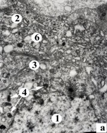
Figure 7: Ganglionic cell of III (а) and IV (б) groups of animals Electromicroscopy. Magn.: а) x4800, б)x6400 Notes: 1 – neuron’snuclei, 2 – new mitochondria, 3 – vacuole, 4 – endoplasmaticnet, 5 – neuron with vacuole dystrophy indirect sign of more intensive activation of neurosecretory processes under the action of combined treatment using PES, in comparison with monotherapy with corticosteroids.
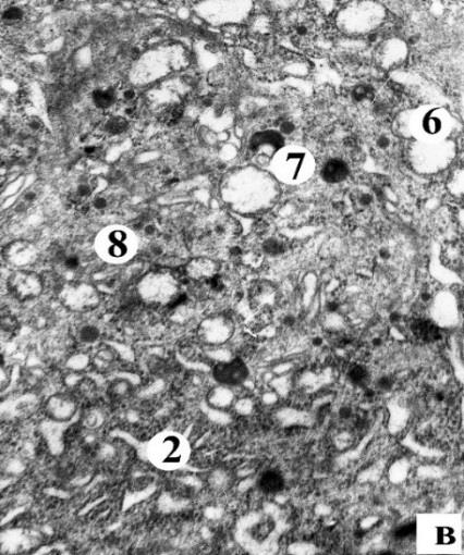
Notes: 1 - nucleus of the neuron, 2 - GEN, 3 - Golgi complex, 4 - lysosomes, 5 - mitochondria, 6 - vacuoles, 7 - autophagosomes, 8 – NG There were also observed recovery processes in dark NC. The confirmation of this is an increase in volumetric density of NG in dark NC, there were compared with animals without treatment, in animals of the group III to 1, 40±0,13% (P<0,01), in animals of the group IV to 1,71±0,17 (P<0,003), But these data was remained statistically significantly lower than in intact rabbits (in all cases P>0,05) (Table 2). It should be noted that the volume density of NG in the animal of the group IV was significantly higher than in IIІ (P<0,01), which also confirms the more pronounced activation of neurosecretory processes under the influence of combined treatment with the use of PES, than with monotherapy with corticosteroids. The "pycnomorphic" neurons are observed along with the dark NCs with regeneration processes. They are at the final stages of their life cycle [10, 11].
- Group of
- The NC’s area,
- The NC nucleus area, μm2
- NCI
- The volumetric density of NG in
- The volumetric density of
- NG in hyperchromic NC, %
- I (n=30)
- 244,12±35,50
- 78,58±14,30
- 0,47±0,07
- 6,95±0,36
- 3,56±0,12
- II (n=30) 276,59±38,02*
- 71,93±15,67
- 0,35±0,07*
- 2,24±0,19*
- 1,14±0,04*
- III (n=30) 232,14±56,81▫ 63,46±14,38▫ 0,39±0,09*
- 3,11±0,18▫*
- 1,40±0,13*
- IV (n=30) 224,25±58,26▫
- 76,51±16,54 0,56±0,23▫
- 4,27±0,29▫*
- 1,71±0,17*▪ animals μm2 normochromic NC, %
Table 2: Morphometric dates of suprachiasmic nuclei for TON (M±m, μm).
The above morphological changes in the structure of the hypothalamus there were occurred on the background of changes in the concentration of hormones in the blood. The content of cortisol decreases from 92, 31±3, 26 μg/dl to 11,79±0,12μg/dl (P<0,05) in the II group in comparison with I. The content of ACTH decreases from 11, 64±0, 43 pg/ml to 6,91±0,09 pg/ml (P<0,05) in the II group in comparison with I. The cortisol’s level increases to 290,12±6,72 μg/dl and the ACTH values decreases to 0,32±0,13 pg/ml (P<0,05) in the group III compared with I group of animals. The content of cortisol was reduced to 6, 93±0.14 μg/dl in the group IV compared to the I group and the III group (P<0,05). The ACTH’ scontent in the group IV (6,13±0,12 pg/ml)was higher than in the group III (P<0,05).
Discussion
Thus, in the retina of the eye after traumatic orbital part optic nerve crush, are observed dystrophic and destructive changes. Mainly they were found in the neurons of the INL, GCL and in nerve fibers of the NFL. There are reactive axonal reaction in ganglionic neurons. Bipolar neurons have vacuolic dystrophy due to disturbance of the blood supply to the retina.
In that time, it was seen reactive edema and destructive changes in the suprachioscular nucleus of the hypothalamus. This leads to a decrease in the production of corticosteroids. Among the dark NCs there are cells of all groups with pronounced destructive changes that do not contain NG, but only lysosomes. A number of authors relate these hypothalamic neurons to pycnomorphic cells that are in the final stages of their life cycle [12, 13]. A characteristic feature of them is high osmophilia and total shrinkage of cells in general. Other researchers, like us, are isolated in the population of dark neurons "chromatophilic" and "pycnomorphic" [10, 11]. The chromatophilic neurons are characterized by a high level of RNA in the nucleus and nucleolis, and the absence of irreversible destructive changes, from which the authors conclude that these cells are more functionally active. The pycnomorphic neurons are in the final stages of cellular destruction. We, like other researchers, tend to attribute these neurons to functionally active, which are characterized by a high level of RNA in the nucleus and nucleoli, and the absence of irreversible destructive changes [10, 11]. These authors conclude that these cells are more functionally active than light, but according to our research, the volumetric density of NG in light cells is significantly higher than that of dark ones.
The treatment led to a partial restoration of the morphological parameters of the ipsilateral retina. The neuroprotective therapy contributes to the development of regenerative processes of the nucleus (more pronounced in combination with PES than with corticosteroid monotherapy). There becomes changing of retinal and hypothalamus architectonics. The number of neurosecretory granules increases. This can be morphological sign of the neurohumoral processes activation. The most optimal treatment is with the use of phosphine electro stimulation, which according to our research leads to compensatory regeneration processes, also described in the works of other researchers, which are characterized by: reduction of the thickness of the retina, restoration of cytokaryometric indices and regenerative processes in bipolar and ganglionic neurons [3].
The peripheral blood ACTH’ scontent decrease with cortisol’s content increase in the group III apparently become due to the use of corticosteroid mega doses. This is likely to result in a disturbance of feedback between hormones. This may be a sign of the depletion of the suprachiasmic nucleus of the hypothalamus. This is confirmed by the presence of pycnomorphic neurons that are not capable to product of hormones. The content of hormones is more closely related to the group without treatment in the group II. Therefore, obviously, it is more physiological, because it corresponds to endogenous processes of the organism. A lot of authors point out such a rearrangement of the hypothalamic NC under different pathological conditions [1, 7, 12].
Conclusion
Thus, retino-hypothalamic neurohumoral dysfunction is an important mechanism of pathogenesis of damage and possible negative consequences of treatment in traumatic optical neuropathy. Contralateral phosphine electric stimulation leads to activation of the neurosecretory processes of the suprachiosmic nucleus of the hypothalamus and the normalization of the content of cortisol and ACTH, which ensures the development of restorative processes in more physiological conditions, compared with monotherapy of corticosteroid mega doses, reducing their toxic effects and retaining retinal neuroprotective properties.
References
-
G Said, D Baudoin, K Toyooka (2008) Sensory loss, pains, motor deficit and axonal regeneration in length-dependent diabetic polyneuropathy. J Neurol 255(11): 1693-1702.
-
Toshinai K, Nakazato M (2009) Neuroendocrine regulatory peptide-1 and -2: novel bioactive peptides processed from VGF. Cell Mol Life Sci 66(11-12): 1939-1945.
-
Koji Toshinai, Hideki Yamaguchi, Kageyama H, Matsuo T, Koshinaka K, et al. (2010) Neuroendocrine regulatory peptide-2 regulates feeding behavior via the orexin system in the hypothalamus. Am J Physiol Endocrinol Metab 299(3): 394-401.
-
Ferri GL (1996) A Neurotrophin-Inducible Gene Expressed. Trends Endocrinol Metab 7: 233-239.
-
Li Y, Irwin N, Yin Y, Lanser M, Benowitz LI (2003) Axon regeneration in goldfish and rat retinal ganglion cells: differential responsiveness to carbohydrates and cAMP. J Neurosci 23(21): 7830-7838.
-
Silke Kiessling, Patricia J Sollars, Gary E Pickard (2014) Light Stimulates the Mouse Adrenal through a Retinohypothalamic Pathway Independent of an Effect on the Clock in the Suprachiasmatic Nucleus. PLOS ONE 9(3): 1-11.
-
Alexandrova VA, Lebedev VP, Rychkova SV (1996) Stimulation of the endorphin structures of the brain is a new non-drug method of treatment. Journal of Neurology and Psychiatry. Ss Korsakov 9(26): 101- 103.
-
Sergeev PV (1987) Receptors of physiologically active substances. M Meditsina.
-
Moiseenko NM (2015) Morphofunctional aspects of restorative processes in the optic nerve after traumatic injury under the influence of high doses of corticosteroids. J ophthalmol (Ukraine) 6: 37-41.
-
Zhurakivska Oуа (2014) The age features of morphological changes of the pituitary neurohypophysis system in the later stages of streptozotocin diabetes. Journal World of Medicine and Biology 4: 123-127.
-
Kalymullyna LB (2002) K voprosu o «temnыx» y «svetlыx» kletkax. Morfolohyya 122(4): 75-80.
-
Beetle of the CE (2008) Ultrastructure of neurons of suprachiazmatic nuclei of the hypothalamus in conditions of light deprivation [Electronic resource]. Bulletin of scientific research No 1(50): 78-80.
-
Zhurakivska OYa (2013) Morphofunctional changes of arcuate nucleus of the hypothalamus at early stages of streptozotocininduced diabetes. Morfolohyya 143(1): 16-22.
-
Levytskyi VA, Shovkova NI (2009) Histoultrastructure of facial nerve in normal and under experimental neuropathy. Vis-nyk morfologii 1: 38-43.
-
Baver SB, Pickard GE, Sollars PJ, Pickard GE (2008) Two types of melanopsin retinal ganglion cell differentially innervate the hypothalamic suprachiasmatic nucleus and the olivary pretectal nucleus. Eur J Neurosci 27(7): 1763-1770.
-
Bremer F (1976) Photic responses of the basal preoptic area in the cat. Brain Res 115(1): 145-149.
- Screening of Hospital Staff During World Glaucoma Week in a Tertiary Eye Care Centre
- Angioid Streaks with Macular Neovascularization: Clinical Insights from Two Cases
- Giant Kissing Naevus: An Oculoplastic Challenge
- Why Freedom of Vision Should Not Cost the Freedom of Feeling - LASIK in the Climate of Change
- Asymmetric Optic Nerve with Small Disc and Large Cup: A Rare and Challenging Case of Unilateral Optic Nerve Hypoplasia
- Large Angle Exotropia in a Child: A Case Report