Tuberculosis Interstitial Keratitis
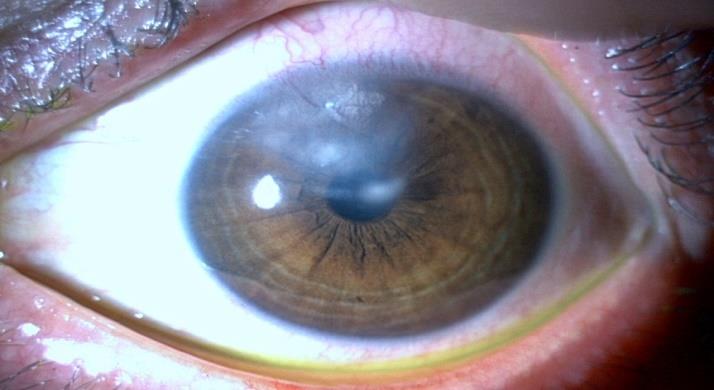
A 42-year-old male with no clinical relevant medical history presented with nonspecific ocular pain in the left eye over a period of 5 days. Although the visual acuity of the left eye was 20/20 (Snellen scale) without correction, the slit-lamp examination showed upper conjunctival injection, superior-sectorial stromal white cell infiltration with deep neovascularization and circumferential haziness (Figure 1). The fundoscopic examination revealed no pathological alterations.
Introduction
A 42-year-old male with no clinical relevant medical history presented with nonspecific ocular pain in the left eye over a period of 5 days. Although the visual acuity of the left eye was 20/20 (Snellen scale) without correction, the slit-lamp examination showed upper conjunctival injection, superior-sectorial stromal white cell infiltration with deep neovascularization and circumferential haziness (Figure 1). The fundoscopic examination revealed no pathological alterations.
Rafael SG and Inês A. Tuberculosis Interstitial Keratitis. J Ophthalmol 2019, 4(1): 000174.
Image Article
These findings were suggestive of interstitial keratitis; empiric topical dexamethasone and artificial tears were initiated and laboratory tests were requested. IGRA was positive which suggests that Mycobacterium tuberculosis infection was the likely etiological cause. After nearly a month of follow-up the cell infiltration and neovascularization were reduced.
Interstitial keratitis by M. tuberculosis infection is a rare condition that affects few numbers of individuals including those with active pulmonary infection. Normally it develops several years after systemic infection and primary ocular tuberculosis is absolutely exceptional, with scarce information described in literature [1, 2, 3].
References
-
Thompson MJ, Albert DM (2005) Ocular tuberculosis. Arch Ophthalmol (Chicago, Ill 1960) 123(6): 844-849.
-
Gupta V, Shoughy SS, Mahajan S, Khairallah M, Rosenbaum JT, et al. (2015) Clinics of ocular tuberculosis. Ocul Immunol Inflamm 23(1): 14-24.
-
Kamal S, Kumar R, Kumar S, Goel R (2014) Bilateral Copyright© Rafael SG and Inês A.
- Screening of Hospital Staff During World Glaucoma Week in a Tertiary Eye Care Centre
- Angioid Streaks with Macular Neovascularization: Clinical Insights from Two Cases
- Giant Kissing Naevus: An Oculoplastic Challenge
- Why Freedom of Vision Should Not Cost the Freedom of Feeling - LASIK in the Climate of Change
- Asymmetric Optic Nerve with Small Disc and Large Cup: A Rare and Challenging Case of Unilateral Optic Nerve Hypoplasia
- Large Angle Exotropia in a Child: A Case Report