Felis Punctatis Oculis: Cat Claw-Induced Occult Scleral Perforation
This is a case report of a 59-year-old woman who experienced minimal trauma, in this case from a house cat which resulted in an open globe. This case shows how an open globe injury can result after minimal trauma and a method for non-invasive treatment. During this case ophthalmic imaging was used to assist in the diagnosis of occult open globe injury. There were many causes of open globe injury found during out search of the literature, but scleral perforation from animal claw was not yet described.
Case Study
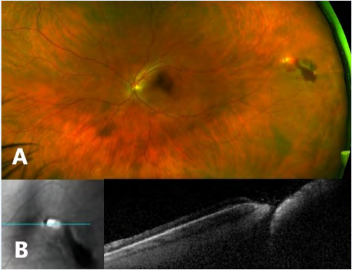
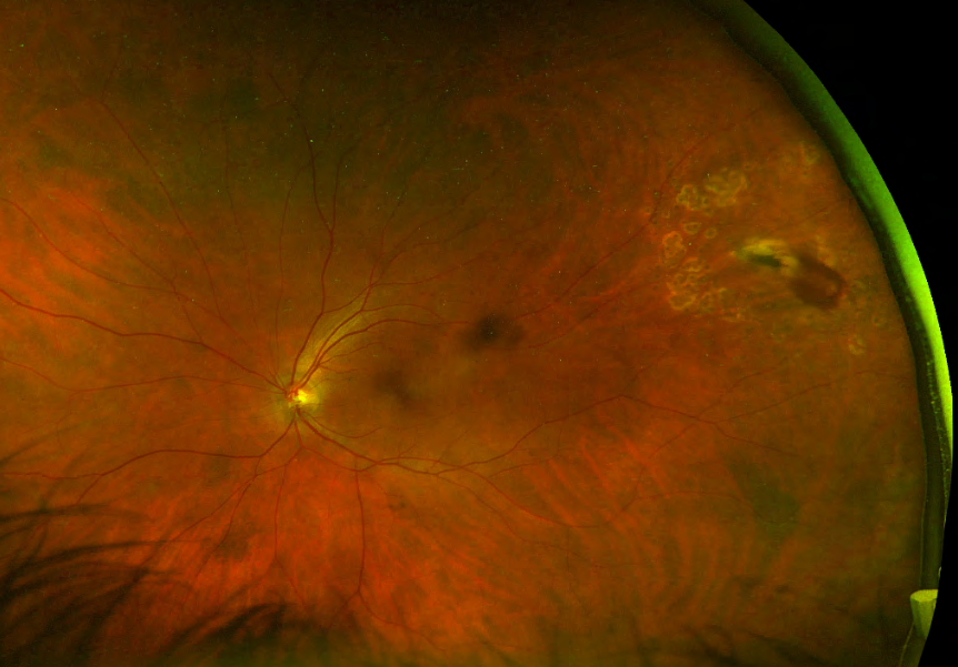
A 59-year-old female presented with left eye pain, conjunctival injection, and floaters five days after her cat pounced on her face. On examination of the left eye, the visual acuity was 20/20, intraocular pressure was 16 mmHg, and no afferent pupillary defect was detected. A small, temporal sub-conjunctival hemorrhage was observed with no anterior chamber cell or flare. Fundus exam and peripheral spectral-domain optical coherence tomography (SD-OCT) are described on (Figure 1: A & B). The patient underwent laser retinopexy and was treated with oral clindamycin and topical moxifloxacin. At her two-month follow up visit, no signs of infection had been noted and vision remained stable (Figure 2).
Open globe injuries commonly occur secondary to trauma. The causative nature of the injury differs between males and females. The most common cause is work related injury in males, and female injury is more likely to be caused by falls [1]. Immediate evaluation of a globe injury is crucial for accurate prognosis. Poor prognosis was found to be associated with the following exam findings: poor visual acuity, prior penetrating keratoplasty, vitreous hemorrhage, retinal detachment, or lens dislocation [1]. Open globe injuries involving intraocular foreign bodies, the most common cause of which was hammering, were associated with additional poor prognostic factors: age greater than 50 and wound length great than 4 mm [2]. Radiographic signs on CT, such as loss of eye contour or volume loss, are good predictors of open globe [3]. However, radiology is inferior to surgical evaluation and not diagnostic alone [3]. The use of intravitreal antibiotics at the time of repair may be helpful in preventing endophthalmitis and, therefore, improve visual outcomes [2]. In one study from an ophthalmic trauma referral centre of 660 eyes, only 8.3% of eyes that presented with open globe injury had to undergo enucleation or evisceration [4]. The most common reason for enucleation was pain, with a rate 0.3% for sympathetic ophthalmia [4].
Open globes, although common, rarely need enucleation and visual outcomes can be improved by prompt surgical evaluation and antibiotics.
References
-
Fujikawa A, Mohamed YH, Kinoshita H, Matsumoto M, Uematsu M, et al. (2018) Visual outcomes and prognostic factors in open-globe injuries. BMC Ophthalmol 18(1): 138.
-
Liu Y, Wang S, Li Y, Gong Q, Su G, et al. (2019) Intraocular Foreign Bodies: Clinical Characteristics and Prognostic Factors Influencing Visual Outcome and Globe Survival in 373 Eyes. J Ophthalmol 2019: 1-7.
-
Arey ML, Mootha VV, Whittemore AR, Chason DP, Blomquist PH (2007) Computed tomography in the diagnosis of occult open-globe injuries. Ophthalmology 114(8): 1448-1452.
-
Savar A, Andreoli MT, Kloek CE, Andreoli CM (2009) Enucleation for open globe injury. Am J Ophthalmol 147(4): 595- 600.
- Screening of Hospital Staff During World Glaucoma Week in a Tertiary Eye Care Centre
- Angioid Streaks with Macular Neovascularization: Clinical Insights from Two Cases
- Giant Kissing Naevus: An Oculoplastic Challenge
- Why Freedom of Vision Should Not Cost the Freedom of Feeling - LASIK in the Climate of Change
- Asymmetric Optic Nerve with Small Disc and Large Cup: A Rare and Challenging Case of Unilateral Optic Nerve Hypoplasia
- Large Angle Exotropia in a Child: A Case Report