Pneumosinus dilatans Presenting with Optic Atrophy in a Gambian Adolescent
Pneumosinus dilatans is an abnormal but benign expansion of one or more of the paranasal sinuses, as a result of hyperpneumatization. Though it is often asymptomatic and diagnosed incidentally on imaging of the cranium. Rarely, it presents with ocular, neurological, cosmetic or rhinological complications. Pneumosinus dilatans has the potential to cause progressive and irreversible vision loss.
Abbreviations
HM: Hand Motion.
Case Report
A 19-year old Gambian boy presented with a history of progressive painless loss of vision in both eyes. Presenting vision was hand motion (HM) in both eyes. There was no history of headache, sinusitis or head trauma. Though he had a history of seizures starting in the neonatal period, there was no history of birth trauma, perinatal asphyxia or severe jaundice. He was a product of a full-term uneventful pregnancy. There was no family history of seizures. The seizures were controlled with oral anticonvulsant therapy, and he had been seizure free for 1 month before presentation. Vision did not improve with refraction.
Clinical examination revealed 45o exotropia, unremarkable anterior segments and bilateral pale, cupped discs with normal intraocular pressures. There was no obvious ocular cause for the optic atrophy. Magnetic resonance imaging of the brain and orbits was therefore requested.
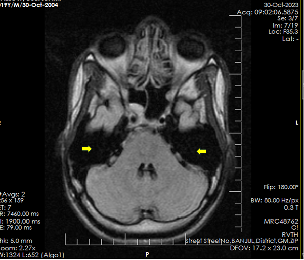
Brain MRI showed marked enlargement and hyperpneumatization of the frontal, and sphenoid sinuses and grossly dilated mastoid air cells bilaterally (Figure 1). In addition, there were abnormal signal changes in the cuneus of the occipital lobe (visual cortex) with loss of white matter bilaterally (demyelination) and localized dilatation of the posterior horns of the lateral ventricles bilaterally, shown in Figures 2a & 2b. There was no intracranial space occupying lesion. A diagnosis of Pneumatosinus dilatans with chronic bilateral occipital lobe infarction was made. Doppler ultrasound studies of the vertebral and carotid arteries were within normal limits. The patient was offered brimonidine eye drops for neuroprotection then referred for ENT and neurosurgical assessment and visual rehabilitation, because of limited expertise with invasive procedures such as optic nerve decompression. In nine months of follow up, the patient’s vision remained unchanged.
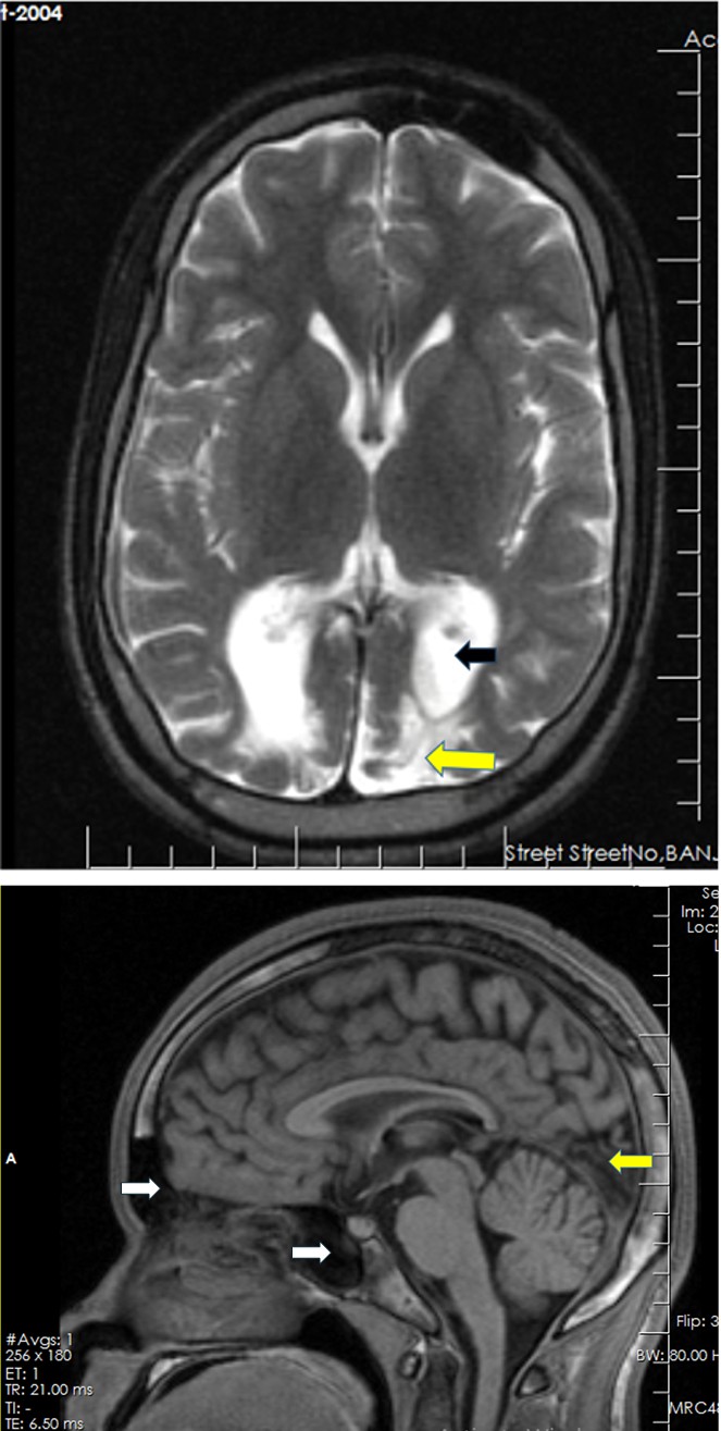
Figure 2a: Focal dilatation of the occipital horns of the lateral ventricles is shown (black arrow) in this T2 weighted axial image associated with occipital cortical atrophy and demyelination (yellow arrow) bilaterally, worse on the left.
Figure 2b: White arrows point to dilated frontal and sphenoid sinuses while yellow arrow points to the chronic occipital infarct affecting the visual cortex. Note: The full extent of the frontal sinus dilatation is not visible in this section).
Discussion
Pneumosinus dilatans is a rare and benign condition characterized by progressive enlargement of the paranasal sinuses. It was first described by Benjamin in 1918 [1, 2]. It most frequently occurs in young men aged between 20 and 40 years but can affect any age or sex [1]. Clinical presentation of pneumosinus dilatants ranges from an asymptomatic incidental diagnosis, to presentation with complications such as nasal obstruction pain, visual disturbance and craniofacial deformity [1, 2, 3, 4, 5, 6, 7]. Pneumosinus dilatans may affect a single sinus, multiple sinuses or all paranasal sinuses. It most commonly affects the frontal (63%), sphenoid (25%) and maxillary sinuses (19%) with the ethmoid sinus being the least commonly involved (18%) [1]. Mastoid air cells may also be involved in the dilatation [3]. Diagnosis is usually confirmed on radiological imaging of the skull either using plain x-rays, computerized tomography or magnetic resonance imaging of the head.
Our patient is a young male in late adolescence, about the age of presentation of most cases described in literature [2, 3, 4, 8]. He was largely asymptomatic except for the visual disturbance and history of seizures.
Visual disturbance in pneumosinus dilatans has been attributed to compression of the optic nerves by a grossly enlarged sphenoid sinus, splaying of the optic chiasm or constriction within the narrowed optic canals (optic canal stenosis) along the walls of the sphenoid sinus [2].
The aetiology of seizures in most cases, including our case, is less obvious. It is possible that unnoticed birth trauma could have predisposed to seizures especially as the onset occurred early in the neonatal period [9]. Cerebral infarcts have been implicated in the aetiology of early-onset neonatal seizures and these may be related to clinical or subclinical hypoxic events. In such a case, the seizures may just be coincidental to the Pneumosinus dilatans as is likely to be the case in our patient. This is further supported by the presence of occipital infarcts in our patient. The presence of bilateral almost symmetrical occipital lobe infarction is in itself a curious finding in this young man because bilateral occipital infarction or stroke is extremely rare and usually affects the elderly in the sixties and above and not neonates or young children [10]. This may suggest an underlying congenital vascular anomaly in the terminal portion of the vertebrobasilar arteries. Although this was not identified on our limited imaging within the limits of our low resource setting, it does not mean that such an anomaly does not exist.
Furthermore, if the basis of the vascular compromise was a connective tissue deficiency of some sort, the same deficiency may be hypothetically related to the development of the hyperpneumatized sinuses.
The evidence of demyelination seen in the occipital cortex, of our patient, is attributed to a previous insult which, we suspect to have been a vascular event. Demyelination can result from any insult to cerebral white matter, whether inflammatory, traumatic or vascular [11]. In this case, we do not believe the demyelination was directly a consequence of the Pneumosinus dilatans, but an associated finding. While demyelinated cortical lesions may be the foci of epilectic seizures, as demonstrated in some patients with MS, the seizures in this adolescent appear to be unrelated to these occipital lesions.
In terms of treatment and visual prognosis in this patient, the visual outcome in our patient was limited by the fact that he presented very late, vision was down to hand motion in both eyes, and he already had marked atrophy, this precludes recovery of good or near normal vision. Had the patient presented earlier, a procedure such as optic nerve decompression could have offered potential relief of the pressure and recovery of some vision. However, the expertise for optic nerve neurotomy and optic canal decompression are currently not available in The Gambia. This patient was therefore managed conservatively with a focus on rehabilitation [11].
In conclusion, Pneumosinus Dilatans, though benign, is a multifaceted anomaly with the potential to cause moderate to severe vision impairment. It is important for ophthalmologists to be aware of this condition, and to facilitate early intervention before irreversible vision loss occurs.
References
-
Seigell S, Singhal S, Gupta N, Verma RR, Gulati A (2022) _Pneumosinus Dilatans_: A Myriad of Symptomology. Indian J Otolaryngol Head Neck Surg 74(Suppl 2): 1305- 1309.
-
Stretch JR, Poole MD (1992) _Pneumosinus dilatans_ as the aetiology of progressive bilateral blindness. British Journal of Plastic Surgery 45(6): 469-473.
-
Kiroglu Y, Karabulut N, Sabir NA, Yagci AB, Gakmak V, et al. (2007) _Pneumosinus dilatans_ and multiplex: report of three rare cases and review of the literature. Dentomaxillofac Radiol 36(5): 298-303.
-
Reicher MA, Bentson JR, Halbach VV, Lufkin R, Hepler RS (1986) _Pneumosinus dilatans_ of the sphenoid sinus. AJNR Am J Neuroradiol 7(5): 865-868.
-
Danassegarane G, Bretonnier M, Tinois J, Proisy M, Riffaud L (2021) _Pneumosinus dilatans_ of the sphenoid and visual loss: when should the optic nerve be decompressed. Childs Nerv Syst 37(8): 2677-2682.
-
Aghdam KA, Aghajani A, Sanjari MS (2021) Bilateral Visual Loss Caused by _Pneumosinus dilatans_: Idiopathic Cases are not Always Reversible. J Curr Ophthalmol 33(2): 197-200.
-
Bouguila J, Rejeb BM, Omezzine M, Mani R, Khochtali H (2015) _Pneumosinus dilatans_: rare cause of slowly changing frontal contours. Aesthet Surg J 35(3): NP47- 53.
-
Aryan S, Thakar S, Jagannatha AT, Channegowda C, Rao AS, et al. (2017) _Pneumosinus dilatans_ of the spheno- ethmoidal complex associated with hypovitaminosis D causing bilateral optic canal stenosis. Child’s Nervous System 33(6): 1005-1008.
-
Lien JM, Towers CV, Quilligan EJ, de Veciana M, Toohey JS, et al. (1995) Term early-onset neonatal seizures: obstetric characteristics, etiologic classifications, and perinatal care. Obstet Gynecol 85(2): 163-169.
-
Satija L, Singh AP, Khanna SK, Baijal V, Verma BS (1999) Rare cause of complete blindness: Bilateral occipital cortical infarction: A Report on three cases. Med J Armed Forces India 55(2): 149-150.
-
Trapp BD, Vignos M, Dudman J, Chang A, Fisher E, et al. (2018) Cortical neuronal densities and cerebral white matter demyelination in multiple sclerosis: a retrospective study. Lancet Neurol 17(10): 870-884.
- Screening of Hospital Staff During World Glaucoma Week in a Tertiary Eye Care Centre
- Angioid Streaks with Macular Neovascularization: Clinical Insights from Two Cases
- Giant Kissing Naevus: An Oculoplastic Challenge
- Why Freedom of Vision Should Not Cost the Freedom of Feeling - LASIK in the Climate of Change
- Asymmetric Optic Nerve with Small Disc and Large Cup: A Rare and Challenging Case of Unilateral Optic Nerve Hypoplasia
- Large Angle Exotropia in a Child: A Case Report