Bladder Melanoma
Metastatic bladder tumors constitute <5% of all bladder tumors, however metastatic malignant melanoma of the urinary bladder is very rare. The determination of the original focus of metastatic melanomas in urinary bladder is convenient in some cases, where the primary focus is already known from patients reported clinical history. In the presenting case, we report a case of a patient with ocular melanoma with bladder metastasis. To our knowledge, this is the first described case in Morocco. The patient underwent a cystoscopy with Trans Urethral Resection of the bladder, immunohistochemical analysis revealed that the tumor cells were positive for Melan-A.
Introduction
Melanoma is an aggressive form of cancer that cans metastasis anywhere in the body, most commonly affected organs are the lungs liver and brain. The discovery of metastasis of malignant melanoma in the urinary bladder is extremely rare in living patients. Metastatic melanomas in urinary bladder used to be mainly autopsy findings [1], and currently, apart from case reports and small case series, no comprehensive cohorts of patients with metastatic melanoma in the urinary bladder have been reported [2]. The determination of the original focus of metastatic melanomas in urinary bladder is convenient in some cases, where the primary focus is already known from patients reported clinical history. In the presenting case, we report a case of a patient with ocular melanoma with bladder metastasis, to our knowledge; this is the first described case in Morocco.
Case Presentation
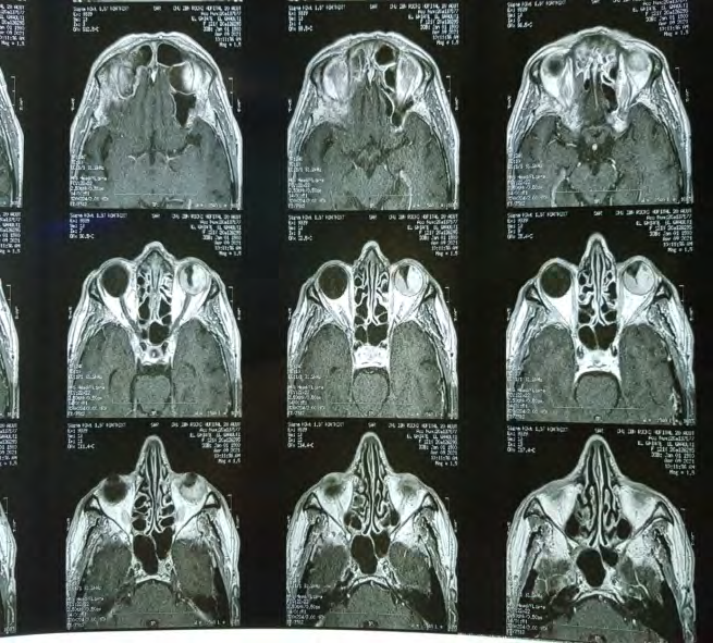
A 65-year-old patient, followed in ophthalmology for a decrease in visual acuity and amputation of the visual field of the left eye for 1 year. A cranio-orbital MRI was performed objectifying the appearance of a choroidal melanoma (Figure 1). The extension workup performed by thoraco-abdomino- pelvic CT revealed a thickening of the right superolateral parietal bladder 14 mm thick extended over 9 mm (Figure 2).
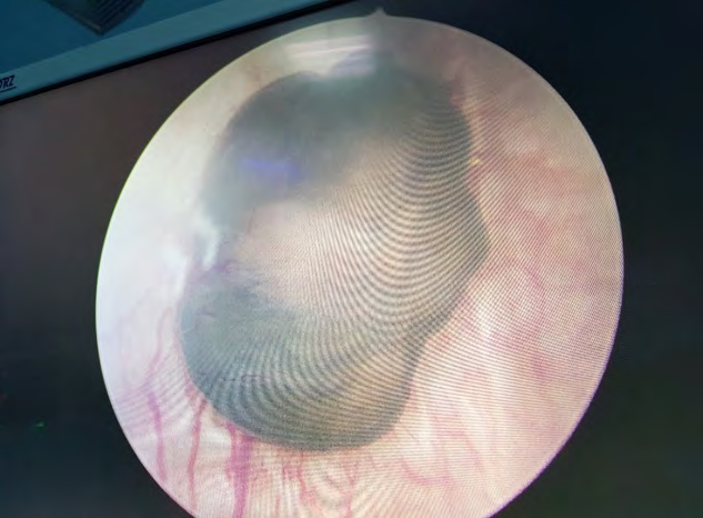
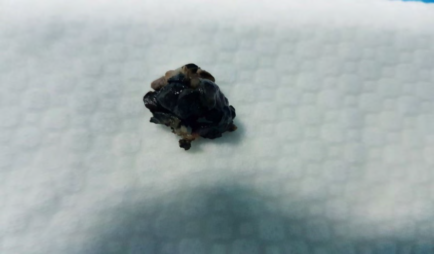
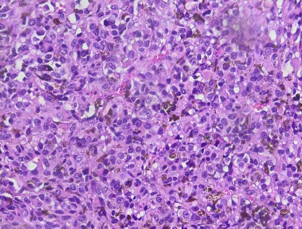
The patient underwent a cystoscopy with Trans urethral resection of the bladder, the cystoscopy objectified the presence of a polypoidal tumor measuring 2 cm, lashed blackish at the bottom level of the bladder (Figure 3), the resection was complete (Figure 4). Histopathological examination showed a malignant tumor proliferation of high cell density arranged in layers. Tumor cells are large in size taking an epithelioid appearance or as round or nevoid cells, often loaded with melanin pigment.
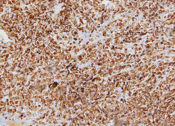
The nuclei are nucleolated anisokaryotic with a mitotic index located at 9 mitoses / mm2. The stroma is poorly observed without intratumoral lymphocytes (Figure 5). Immunohistochemical analysis revealed diffuse positivity for melanin A and heterogeneous positivity for HMB45, PS100 is negative (Figures 6,7). Based on the patient’s ophthalmologic history, we then made the diagnosis of metastatic malignant melanoma of the bladder. Patient referred to ophthalmology for enucleation, and oncology for chemotherapy


Discussion
Metastatic bladder tumors constitute less than 5% of all bladder tumors. Metastatic malignant melanoma of the urinary bladder is very rare. Only 30 cases have been published in the English literature [3]. Similar to cutaneous melanomas, conjunctival melanomas originate from melanocytes that are derived from the neural crest.
Clinically evident metastatic melanoma of the bladder has been rarely described, and it typically presents as painless, macroscopic hematuria. A subsequent diagnosis is usually made on the basis of the cystoscopic findings and histopathological features combined with a clinical history of previous melanomas. Metastasis of malignant melanoma may present as soon as the diagnosis of the primary lesion is made or it may remain dormant for a period of 15 years or more [3].
This is only the fourth reported case in the literature describing a urinary bladder tumor resulting from a primary ocular melanoma [3]. Several treatments have been proposed to deal with metastatic malignant melanoma. TUR, partial cystectomy, radical cystectomy, chemotherapy, and radiation therapy have all been used to treat melanoma of the bladder [3, 4]. Metastatic malignant melanoma has a very poor prognosis, with an overall median survival of 6-7.5 months [3, 5]. Our patient was treated with TUR and enucleation with chemotherapy.
Conclusion
Melanoma of the bladder is typically a secondary recurrence in patients with widespread metastatic melanoma originating from the skin or visceral tissue. In most cases, a detailed patient history, careful examination of the patient’s skin, and evaluation of other visceral primary sites are necessary to determine the primary or metastatic nature of the tumor.
References
-
Meyer JE (1974) Metastatic melanoma of the urinary bladder. Cancer 34(5): 1822-1824.
-
Velcheti V, Govindan R (2007) Metastatic cancer involving bladder: a review. The Canadian Journal of Urology 14(1): 3443-3448.
-
Topal CS, Kir G, Daş T, Sarbay B, Tosun MI (2016) Metastatic malignant melanoma of the urinary bladder: A case report and review of the literature. Indian journal of pathology and microbiology: 59(4): 532-534.
-
Pacella M, Gallo F, Gastaldi C, Ambruosi C, Carmignani G (2006) Primary malignant melanoma of the bladder. Int J Urol 13: 635-637.
-
Crosby T, Fish R, Coles B, Mason MD (2000) Systemic treatments for metastatic cutaneous melanoma. Cochrane Database Syst Rev 2: CD001215.
- Results of 6-Month Follow-Up of Patients After B-Turp and Thulep
- The Effect of Drinking Water with a High Content of Antimony and Arsenic on the Dynamics of their Distribution in the Kidneys and the Renal Excretory Function in Rats
- Effectiveness and Safety of Tansurethral Thulium Laser Enucleation of the Prostate in the Treatment of BPH: Review
- A Systematic Review on Molecular Pathophysiology Involved in Chronic Kidney Disease and the Role of Animal Models in Drug Discovery to Manage in Chronic Kidney Disease - An Update
- Functional Development of Kidneys in Human Ontogenesis
- Testicular Metastasis: Uncommon Prostate Cancer Case Report