Epidemiology of Rotavirus Infection in Dairy Calves in Central Ethiopia
Rotavirus is a major pathogen responsible for diarrheal disease in calves and neonates resulting in loss of productivity and economic losses in dairy farms in developed and developing countries. In Ethiopia, the diarrheal disease caused by Rotavirus is not so far studied adequately. Across-sectional study was conducted from October 2019 to May 2020 to estimate the prevalence, assess associated factors, and isolate Rotavirus in calves below two months of age in the central part of Ethiopia, namely Holeta, Sululta, Bishoftu, and Sebeta. A total of 404 fecal samples, comprising 213 diarrheic and 191 non-diarrheic cases, were collected from calves of less than two months of age by simple random sampling technique. Rotavirus infection was detected using antigen detection Enzyme-linked immunosorbent assay technique. All samples positive for ELISA were propagated in Madin Darby bovine kidney cells. Data was analyzed using STATA version 13. Fisher’s exact test and logistic regression analysis were used to determine the association between prevalence of rotavirus and potential risk factors. An overall rotavirus prevalence of 1.24% (95%CI 0.40 – 2.86%) was found. The prevalence of rotavirus in diarrheic calves (1.41%) was not significantly different from the prevalence of rotavirus in non-diarrheic calves (1.04%) (p>0.05). Similarly, the origin, breed, sex, age, management, and type of farm had no significant association with the prevalence of rotavirus infection of calves (p>0.05). After 3 subsequent passages, progressive cytopathic effect i.e. Cell swelling and obscure cell boundaries, detachment and floatation of cells, cytoplasmic vacuolationas, increased cell granularity and cytoplasmic stranding were observed in all samples. The presence of rotavirus in dairy calves might entail morbidity and mortality in calves. Moreover, rotavirus infection might also lead to zoonotic transmission. Therefore, further detailed molecular epidemiological studies are warranted. Good hygienic and husbandry practices are essential to limit the spread of infection.
Introduction
Rotaviruses are a member of the family Reoviridae in which seven (A to G) species of rotavirus have been identified according to the specificity of VP6 inner shell polypeptide. Furthermore, rotavirus strains are grouped into electro hero types based on the relative migration rates of genome segment in Poly acrylamide gel electrophoresis (PAGE) [1]. Point mutations, re-assortment, and rearrangements within the genome of double-stranded 11 segmented RNA have been identified as the major cause of variation and distribution within rotavirus [2]. Among this species Rotavirus A is the major cause of diarrhea in children and calves [3].
Genetic re-assortment is one of the important mechanisms for generating genetic diversity of rotaviruses and eventually for viral evolution [4, 5]. There is no treatment for BRV, but early and confirmatory diagnosis helps to make appropriate prevention and control measures, which could prevent the great economic losses to farmers and the livestock industry [6].
It suggested that causes of rotavirus could be management/environmental factors such as overfeeding, low temperature, poor hygiene, colostrum deprivation and individual animal susceptibility. Infectious agents, either singly or in combination, may be associated with field outbreaks with considerable economic burden [7].
In animals, rotavirus infection causes an enormous economic loss associated with high mortality and morbidity and treatment costs. Different species of animals like bovine, pigs, horses, dogs, monkeys, cats, rats and chicken are also susceptible to Rotavirus although the severity varies according to the species of the animal [8]. Rotavirus infections have a worldwide distribution and are a common cause of neonatal diarrhea in many mammalian and avian species [9]. Bovine rotavirus is a major cause of calf diarrhea, usually occurring in calves at 1-3 weeks of age [10, 11]. In addition to clinical rotavirus infections, subclinical infections are also common in calves [10, 11]. The agent multiplies in the intestine and spreads with the infected animals faces [12].
Rotavirus is environmentally distributed worldwide and was extensively studied [13, 14] starting from the detection of the virus using an electron microscope (EM) in diarrheic cattle and children in 1968 and 1973 respectively [15]. In different studies BRV infection rates of 20-60% in samples of diarrhea have been reported [16, 17]. Prevalence of rotavirus was estimated ranging from 11.8% to 26.8% in India among diarrheic calves [18, 19, 20, 21]. Also in European countries rotavirus infection was widely examined. In Sweden between 1993 and 2006 estimated prevalence was 24-47% [22], 42% in diarrheal outbreak in the UK [23], and 37 to 47.4% in France [24, 25]. In Asian countries like Bangladesh, prevalence of rotavirus infection in calf feces varied from 0 to 7% [26]. In developing country like Ethiopia the prevalence of bovine rotavirus was 16.7% [27]. Although recently published data’s for the status of calf rota virus in Ethiopia was very rare, Beksisa, et al. [28] reported that 12.5% prevalence of the disease in one selected farm. The vaccine coverage status of the disease in children of Ethiopia showed that it was lower than the targets set by WHOM [29].
The etiology of calf diarrhea is multifactorial and may include infective, environmental, nutritional, and management factors such as calves being born from a heifer [30], being born during the summer [31, 32], suckling [31, 32, 33], low serum IgG concentrations and large herd size [34].
Rota virus infection is associated with huge mortality and morbidity of calve particularly in low income African countries including Ethiopia. The fact that Ethiopia is a leading country in livestock population in Africa and the dairy farms are concentrated in the central parts of the country highlighting the importance of comprehensive epidemiological studies of RV infections in the area is crucial. There is few (no) published data which shows prevalence of rotavirus in cattle so far in Ethiopia. As rotaviruses are zoonotic which transmit from animals to humans the implication of the disease is important both economically and socially. Hence, this study is therefore, articulated in determining the prevalence of RVs and associated factors to bridge the information gaps on epidemiology of the diseases and its isolation from dairy calves in the study area.
Materials and Methods
Description of the Study Area
The current study was conducted in four selected areas of central Ethiopia (Bishoftu, Sebeta, Holeta, and Sululta) from November 2019 to May 2020 (Figure 1). There are 165 small scale farms, 57 medium, and 25 large-scale dairy farms in the selected areas that supply milk and milk products to consumers of the towns and surrounding areas. These dairy farms contain local zebus particularly from extensively and semi-intensively reared herds, hybreeds of Holstein- Friesian and jersey, or exotic breeds of either holestein-friesianor jersey breeds which attain 100% genetic quality.
Bishoftu: The town is found in east Shewa Zone, Oromia Regional State, located about 45 km South-east of the capital city, Addis Ababa. The area is located at 9°N latitude and 40°E longitude at an altitude of 1850 m. above sea level. Bishoftu receives annual rainfall of 866 mm of which 84% is in the long rainy season (June to September). The annual minimum and maximum temperature of Bishoftu is 11and 29°C, respectively. The domestic animals reared in Bishoftu town are 30887cattle, 43138 poultry, 9322 equines, 9294 sheep, and 4753 goats [35]. Sebeta: It is located in the Oromia Special Zone Surrounding Finfinne (Addis Ababa) of the Oromia Region. The district is located 25 km south-west of Addis Ababa at an altitude of 1800-3385 m above sea level and latitude and longitude of 8°55-8.917°N and 38°37-38.617°E respectively. It receives an average annual rainfall of 1073 mm and temperature that ranges from 11.3-280C. It has a total area of 102,758 km2. According to the information obtained from Sebeta Hawas district Administration Office, both livestock rearing and crop production are the main economic activities of the majority of communities. Holeta: The town is located in the central part of the country, 31 km West of Addis Ababa in Oromia Special Zone Surrounding Finfinne Oromia Regional state. The area is bounded between latitude 8o53’ 75” to 9o14’ North and longitude 38o 21’ 40” to 38o 36’ 14” East. The Town has an area of 5550 hectares. Holeta town is found at an altitude of 2449 m above sea level. The annual mean maximum and minimum temperatures are 25.9 and 7.20C, respectively [36]. Sululta: This is a town under the Oromia Special Zone Surrounding Addis Ababa, Oromia region. Sululta is 35.7 km North West of Addis Ababa. It has an altitude and longitude of 9° 11′ 747″ N 38° 45′ 338″ E. The elevation of Sululta is 2500 meters above sea level. The annual average temperature in Sululta is 14.7 °C with an average rain rainfall of 1119 mm per year.
In general, these study areas were purposively selected and identified based on transport accessibility, potential area for commercial dairy farms and on the abundance of dairy farms to get sufficient samples from less than two months aged calves.

Study Animals
Dairy calves less than 2 months of age found in the study areas constitute the study populations. The study animals were randomly selected. This was conducted for both diarrheic and non-diarrheic calves without distinguishing their breed type. Diarrhea was considered if feces are semi-liquid to liquid, with or without other abnormal characteristics such as the presence of blood or mucous. Any calf with feces without these characteristics was considered non-diarrheic or apparently healthy [37].
Study Design
A cross-sectional study was conducted at various dairy farms of Central Ethiopia from November 2019 to May 2020 to study the Epidemiology of Rotavirus infection in dairy calves.
Sample Size Determination and Sampling Technique
As there was no previous report on the epidemiology of bovine rotavirus infection in the study area, the sample size was calculated by considering the expected prevalence of 50% according to [38].
n= 1.962*Pexp (1-Pexp) d2 Where: n = required sample size; Pep = expected prevalence; d =desired absolute precision. 5% desired precision; at 95% confidence level was considered. Accordingly, the total calculated sample size was 384 however, to increase the precision the sample size is extended to 404. This sample size was generated nearly proportionally with insignificant difference at the study areas (Holeta, Sebeta, Sululta, and Bishoftu) using simple random sampling technique.
Fecal Samples Collection
Approximately 30 grams of fecal sample was collected from each calf in a sterile epic doff tube after cleaning the anal area with a paper towel and beats by rectal stimulation with the index finger using disposable sterile plastic gloves [37]. Collected fecal samples were placed into icebox containing ice packs and transported to the Virology Laboratory at National Animal Health Diagnostic and Investigation Center (NAHDIC), Sebeta, and were stored at -20 °C until processing.
Questionnaire Survey
A close ended questionnaire was developed and filled at a farm or household level by face to face interviewing with herd owners or attendants during sampling in order to assess potential risk factors for the disease. On the questionnaire the potential risk factors included were; study areas, farm hygiene, first time of milking, start of suckling, practice of udder washing, habit of feed supplement for calves, awareness about colostrum, uses of colostrum’s, fecal consistency, isolation of dichroic calves and treatments provided for dichroic calves.
Laboratory Techniques
Detection of bovine rotavirus antigen by ELISA: Multi- screen Ag ELISA Calf digestive (BIO K 314/1, Belgium) sandwich ELISA was used to detect BRV Ag in the fecal suspensions. This monoclonal antibody coated plates were used to capture the corresponding Ag in the fecal samples if any. Test procedure was performed according to the manufacturer’s instruction (Kit reference BIO K 314/1, Belgium).
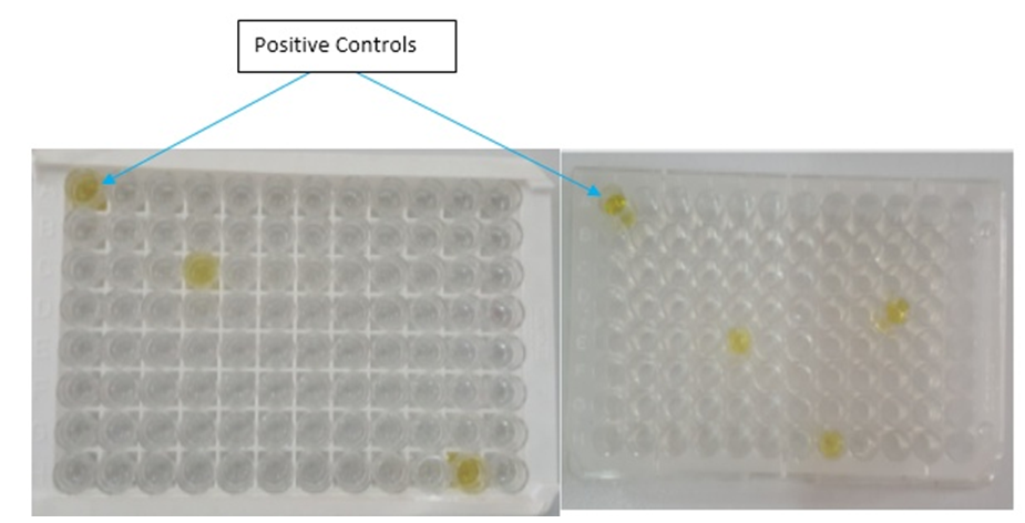
The 96 well plates provided in the kit contained two different capture antibodies. All wells of the plates were coated with rotavirus specific capture antibodies in which well A and B acted as controls (positive and negative control). These control wells ensures the test reliability between specific immunological reactions and non-specific bindings to eliminate false positives. Feces were diluted in the dilution buffer provided in the kit. A volume of 100μl of diluted sample was added to corresponding wells of specific and non-specific antibody-coated rows, respectively. Similarly, both the positive and negative controls were added to their respective well per plate. The plate was incubated at 25°C for 30 minutes and washed 3 times with a washing solution (diluted in the ratio 1:20 with distilled water) provided in the kit. A 100μl ready-made conjugate for rotavirus specific monoclonal antibody tagged with horse radish peroxidase enzyme was added per each well. The plates were covered with a sealant and incubated at 25°C for 30 minutes in the room temperature (22-25°C). Then, 100 μl of the chromogenic (2, 2, 5, 5, tetra methyl Benzedrine) solution was added to each well. The plates were then incubated for 10 minutes at 25°C in the dark to prevent occurrence of oxidation. Finally, the reaction was stopped by adding 50μl stop solution (1M phosphoric acid) per well. The optical density was measured at 450 nm using an ELISA plate reader immediately after stopping the reaction.
The test was validated using the positive control and data sheet provided by the kit. The net optical density of each sample was calculated by subtracting the reading for each sample well from corresponding negative control. Net optical density (OD) = (OD of specific binding -OD of non-specific binding). The end color formed after adding a substrate was blue which is changed to yellow color after intervention with a stop solution. Indeed, a sample was positive when an optical density value was > 0.15 Elisa units (EU) and negative for OD value < 0.15 Elisa units (EU) for bovine rotavirus.
Cell culture preparation: Madine-Darby Bovine Kidney Epithelial cells (MDBK, passage 82) obtained from Athens Veterinary Diagnostic laboratory, University of Georgia, USA, were revived from liquid nitrogen and re-cultured in 25cm2 tissue culture flask. The confluent flask was then sub-cultured to multiple 25cm2 TC flasks and maintained in Dulbecco’s modified Eagle’s medium (DMEM) supplemented with 10% fetal bovine serum, 2% Glutamax and 1% Antibiotic- Antimycotic solution at 37oC in a humidified incubator at 5% CO2.
Sample preparation: 10 % suspension of the fecal material is made using the transport medium with a screw-cupped bottle. Coarse glass beads are added and shaken vigorously. The suspension is poured in to centrifuge tube and centrifuge at 3000 rpm for 10 min at 4°C in a refrigerated centrifuge. The upper two-thirds of the supernatant fluid is collected, filtered using 0.45µm Millipore filters, and stored at – 20 °c until inoculation.
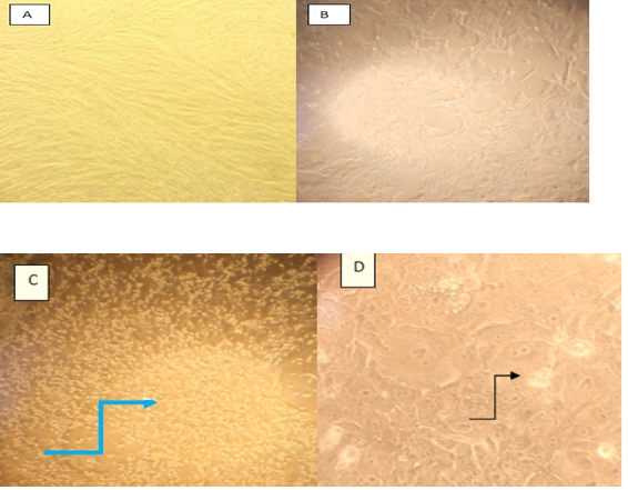
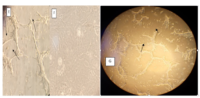
Virus isolation: The fecal suspensions prepared and stored at -20°C were thawed and 0.5ml of the samples inoculated into each sub confluent MDBK culture in 25cm2 flasks. After 60min adsorption at 37°C, maintenance medium is added to each flask including one un-inoculated flask as negative control and incubated at 37°C in a humidified incubator at 5% CO2. Cells were monitored every 24 hrs. post-infection and inspected for cytopathic effects (CPEs) using an inverted microscope. If no CPE was observed on the third passage after 48 hours inoculation, then the sample was considered as negative for rotavirus. On the fifth day, the cultures were freeze-thawed and the resulting lysate was inoculated into fresh cultures until the third passage.
Data Management and Analysis
The collected data were entered and coded in Microsoft Excel Spread sheet. The data was transported to State version 13 software (state Corp College station, USA) for analysis. The prevalence of rotavirus infection was calculated as number of Ag ELISA positive fecal samples divided by total samples examined and multiplied by 100. The association of various risk factors was explored first using Fisher’s exact test. Then the strength of association of potential risk factors (origin, age, sex, breed, farm hygiene, farm management, farm size and fecal consistency) with the outcome variable was analyzed using a logistic regression analysis. Non- collinear variables with a invariable p<0.25 were offered to a multivariable logistic regression model. A p-value <0.05 was considered significant.
Results
Prevalence of Rotavirus
Out of the total 404 fecal samples examined, the prevalence of rotavirus infection was found to be 1.24% (5/404, 95% confidence interval [CI]: 0.40 – 2.86%). Highest animal level seroprevalence was recorded in Bishoftu (2/88, 2.27%) followed by Sululta (2/96, 2.08%) and Sebeta (1/98, 1.02%).
Association Of Risk Factors At Animal Level
The results of the association of different risk factors with the prevalence of rotavirus using Fisher’s exact test. Slightly higher prevalence of rotavirus infection was found in Bishoftu (2.27%) followed by Sululta (2.08%), and Sebeta (1.02%). No positive case was recorded in dairy calves of Holeta. Sera prevalence of rotavirus infection was not significantly associated with the investigated risk factors; breed, sex, age and fecal consistency (p>0.05) as presented in Table 1.
| Variables | Category | No of samples tested | No of Positive (%) | Fisher’s Exact test |
|---|---|---|---|---|
| Origin | 0.33 | |||
| Holeta | 122 | 0(0.00) | ||
| Sululta | 96 | 2(2.08) | ||
| Bishoftu | 88 | 2(2.27) | ||
| Sebeta | 98 | 1(1.02) | ||
| Breed | 1 | |||
| Local | 164 | 2(1.22) | ||
| Cross | 233 | 3(1.29) | ||
| Exotic | 7 | 0(0.00) | ||
| Sex | 0.261 | |||
| Female | 180 | 1(0.56) | ||
| Male | 224 | 4(1.79) | ||
| Age | 1 | |||
| 3days-2Weeks | 104 | 1(0.96) | ||
| 2Weeks-1month | 137 | 2(1.46) | ||
| 1 – 2 months | 163 | 2(1.23) | ||
| Feces consistency | 0.551 | |||
| Diarrheic | 213 | 3(1.41) | ||
| Non diarrheic | 191 | 2(1.04) | ||
| Total | 404 | 5(1.24) | ||
Table 1: Animal level prevalence of rotavirus infection in dairy calves.
Association of Risk Factors at Farm Level
The overall farm level sero prevalence of rotavirus infection was 2.02% (5/247; 95%CI: 0.84, 4.80). Based on origin the infection was high in Bishoftu (2/42, 4.76%) followed by Sululta (2/68, 2.94%) and Sebeta (1/71, 1.41%) as compared to Holeta district (0/66, 0%). Larger farms were slightly higher in Rotavirus infection 8% (2/25) when compared to medium 0% and small farm sizes 1.82% (3/165) with no statistical significance (p>0.05). Similarly, farm hygiene and farm managements were also have no statistical difference among their categories as illustrated in Table 2.
| Variables | Category | No. of farms samples taken | No. of positive (%) | Fisher Exact test |
|---|---|---|---|---|
| Origin | 0.341 | |||
| Holeta | 66 | 0(0) | ||
| Sululta | 68 | 2(2.94) | ||
| Bishoftu | 42 | 2(4.76) | ||
| Sebeta | 71 | 1(1.41) | ||
| Total | 247 | 5(2.02) | ||
| Farm size | 0.077 | |||
| Small | 165 | 3(1.82) | ||
| Medium | 57 | 0(0) | ||
| Large | 25 | 2(8) | ||
| Total | 247 | 5(2.02) | ||
| Farm hygiene | 0.222 | |||
| Inadequate | 92 | 4 (4.35) | ||
| Good | 121 | 1(0.83) | ||
| Very good | 34 | 0(0) | ||
| Total | 247 | 5(2.02) | ||
| Farm management | 1 | |||
| Extensive | 120 | 3(2.5) | ||
| Semi-intensive | 37 | 0(0) | ||
| Intensive | 90 | 2(2.22) | ||
| Total | 247 | 5(2.02) |
Table 2: Distribution of rotavirus infection at farm level.
Association of Potential Risk Factors with Rotavirus Infection at Animal Level
During the statistical analysis, for all the risk factors, the first level of each independent variable (the category of a risk factor with lowest prevalence) was used as a reference category. Animal level seroprevalence of Rotavirus was not statistically significant (P >0.05) between the various risk factors for the disease. Univariable logistic regression analysis showed that the risk of infection in Sululta (OR = 0.48, 95% CI: 0.04, 5.43) and Bishoftu (OR = 0.44, 95% CI: 0.04, 4.97) was not significantly higher than in Sebeta. Rotavirus infection in males 1.79% (4/213) was slightly greater than in females 0.56% (1/191). Invariable logistic regression analysis also showed no significant association (P>0.05) between sera-positivity and Breed, Age and fecal consistency.
| Risk factors | No. of samples tested | No. of Positive (%) | Crude OR (95%) | p-Value |
|---|---|---|---|---|
| Origin | ||||
| Holeta | 122 | 0(0.00) | 1 | |
| Sullta | 96 | 2(2.08) | 0.48(0.04, 5.43) | 0.557 |
| Bishoftu | 88 | 2(2.27) | 0.44(0.04, 4.97) | 0.51 |
| Sebeta | 98 | 1(1.02) | * | 0.45 |
| Breed | ||||
| Local | 164 | 2(1.22) | * | |
| Cross | 233 | 3(1.29) | 0.95(0.16, 5.73) | 0.952 |
| Exotic | 7 | 0(0.00) | 1 | |
| Sex | ||||
| Female | 180 | 1(0.56) | * | |
| Male | 224 | 4(1.79) | 0.31(0.03, 2.77) | 0.293 |
| Age | ||||
| 3days-2Weeks | 104 | 1(0.96) | * | |
| 2Weeks-1month | 137 | 2(1.46) | 0.65(0.06 7.33) | 0.732 |
| 1month-2month | 163 | 2(1.23) | 0.78(0.70 8.73) | 0.841 |
| Feces consistency | ||||
| Diarrheic | 213 | 3(1.41) | 0.74(0.12, 4.48) | 0.744 |
| Non diarrheic | 191 | 2(1.04) | * |
Table 3: Relationship of rotavirus infection and potential risk factors in dairy calves at animal level.
Association of risk factors with rotavirus infection at farm level
At farm level analysis, the variable entered into the multivariate model was farm size and farm hygiene. Origin Variable No. of farms No. of Positive Crude OR (CI: 95%) p-value Adjusted OR (CI: 95%) P-value Origin Holeta 66 0(0) 1 - - Sululta 68 2(2.08) 0.47(0.04, 5.32) 0.543 - - Bishoftu 42 2 0.29(0.03, 3.25) 0.313 - - Sebeta 71 1 * Farm size Small 165 3(1.82) 4.69(0.74, 29.62) 0.1 5.27(0.79, 35.27) 0.087 Medium 57 0 1 1 Large 25 2(8) * * Farm hygiene and farm management were excluded due to univariate analysis p >0.25. Based on farm level all independent variables were statistically insignificant both with univariate and multivariate analysis as shown in Table 4.
| Inadequate | 92 | 4 | 0.18(0.02, 1.67 | 0.132 | 0.21(0.02, 1.99) | 0.175 |
|---|---|---|---|---|---|---|
| Good | 121 | 1 | * | * | ||
| Very good | 34 | 0 | 1 | 1 | ||
| Farm management | ||||||
| Extensive | 120 | 3 | 0.89(0.14, 5.42) | 0.896 | - | - |
| Semi-intensive | 37 | 0 | 1 | - | - | |
| Intensive | 90 | 2 | 1 | * | - | - |
Table 4: Farm level logistic regression analysis of rotavirus infection and potential risk factors for the study animals.
Questionnaire Survey
Of the total 134 interviewed volunteer respondents from the four study areas who were participated on dairy farming environments, 47.76% (64/134) had contracted diahrric calves in the farm previously. Based on fecal consistency, diahrric calves were slightly higher at Sebeta 66.7% (10/15) followed by Sululta 52.3% (23/44), Holeta 42.4 (25/59) and Bishoftu 37.5% (6/16). Regarding farm hygien, 38% were inadequate, 46.3% good and 15.7% very good hygienic condition. On the basis of colostrum knowledge 92.5% knows what a colostrums is and 7.5% don’t know. Regarding udder washing, 74.6% of the respondents had a practice of udder washing prior to milking and suckling (Table 5).
| Risk factors | Categories | Frequency | Percent (%) |
|---|---|---|---|
| Farm hygien | Very good | 21 | 15.7 |
| good | 62 | 46.3 | |
| Start of milking | Inadequate | 51 | 38 |
| Immediately after parturition | 9 | 6.7 | |
| After 3days | 18 | 13.4 | |
| Start for suckling | After a week | 99 | 73.9 |
| Left for the calf | 8 | 6 | |
| Within 1hr of parturition | 129 | 96.3 | |
| After 3 hrs | 5 | 3.7 | |
| Udder washing | After 6 -24hrs | 0 | 0 |
| Yes prior to milking & suckling | 100 | 74.6 | |
| Supplement feeding within 2 months age | Not washed | 34 | 25.4 |
| Yes | 25 | 18.7 | |
| Knowledge of colostrum | No | 109 | 81.3 |
| Know | 124 | 92.5 | |
| If you know, its use for calf? | Don’t know | 10 | 7.5 |
| For nutritive | 17 | 5.6 | |
| Fecal consistency | As a vaccine | 63 | 50.8 |
| For growth | 30 | 24.2 | |
| Don’t know | 14 | 11.3 | |
| Observed diarrhea | 64 | 47.8 | |
| Dichroic state, based on study areas | Not Observed | 70 | 52.2 |
| Holeta | 25/59 | 42.4 | |
| Sex mostly affected by diarrhea | Sululta | 23/44 | 52.3 |
| Bishoftu | 10-Jun | 37.5 | |
| Sebeta | 15-Oct | 66.7 | |
| Male | 13/64 | 20.3 | |
| Age | Female | 18/64 | 28.1 |
| No difference | 33/64 | 51.6 | |
| <1week | 15/64 | 23.4 | |
| Do you isolate diarrheic calves | 1-3 weeks | 35/64 | 54.7 |
| 1-2month | 19/64 | 29.7 | |
| Yes | 21/64 | 32.8 | |
| Measurement taken | No | 43/64 | 67.2 |
| Antibiotic treatment | 30/64 | 46.9 | |
| Traditional treatment | May-64 | 7.8 | |
| Not treated | Mar-64 | 4.7 | |
| Milk provision reduced | 26/64 | 40.6 |
Table 5: Results of questionnaire survey on Rotavirus infection in the study areas.
Isolation of Rotavirus In Cell Culture
In the present study, viral growth in cell culture was assessed by examining inoculated cells for CPE. All Rotavirus positive samples for Ag ELISA were inoculated on MDBK cells were isolated for the virus with various cell morphological changes. This was evident by development of visible cytopathic effect of different degree on MDBK cell lines such as cell swelling and obscure cell boundaries observed on 48hrs of culture, detachment and floating of cells after 72hrs, cytoplasmic vacuolation and increased cell granularity on the fourth day and cytoplasmic stranding observed on the fifth day due to the effect of the virus (Figure 3).
Discussion
Newly borne calves represent worldwide an important resource in livestock production for milk, meat or breeding i.e. replacement stock [39]. The livestock industry faces many series of disease problems like calf diarrhea, which usually affect it dramatically. Neonatal calf diarrhea is a prime disease affecting newborn calves leading to morbidity and mortality in, causing economic losses due to the costs of treatment, diagnostics, weight loss or death in infected animals, and poor growth performance [40]. Indeed, the long-term effects of diarrhea on the health and efficiency of calves cured of clinical courses of the disease may cause more economic loss [41]. A crucial period for these calves is the first few days following birth. In developing countries like Ethiopia, domestic animals are the major income source for poor families. These families suffered badly due to the neonatal calf mortality. Among numerous viral, bacterial, and parasitic causative agents, bovine rotavirus is the foremost cause of neonatal calf diarrhea in domestic animals. The cause of neonatal calf mortality is specifically related to group A bovine rotaviruses [42]. Fecal contamination plays an important role in the transmission of rotavirus infection and the infection is widespread globally in cattle populations. Because of the disease impact both economically and socially through its public health importance, studying the disease distribution is essential the effective control measures [8].
In general, the overall prevalence in the current study was found to be 1.24% in calves. This result is lower when compared to the reports of Pérez E [43] in Costa Rica (7%), [44] in Turkey (8.5%), [45] in Turkey (8.92%), and [46] in India (5.5%). Such a higher prevalence of rotavirus have also been reported from many countries including Ethiopia (16.7%) [26], Algeria (14.63%) [36], Vietnam (15%) [47], Southern Italy (16.8%) [48], India (27.02%) [18] and Switzerland (58.7%) [16]. this difference could be attributed to the number of samples investigated, livestock management style, geographical variations, and due to differences in the test method applied. The prevalence of rotavirus infection also varies depending on the country and region under study [17, 39]. However, the current study was in accord with the reports of [49] in Oldenburg (1.96%).
In the present study, 1.24% (5/404) calves were positive for Rotavirus fecal excretion as detected using Ag-ELISA. The prevalence was likely higher in Bishotu (2.27%) and Sululta 2.08%). The difference in prevalence across the study areas, although not significant, might be due to farm management practices exercised in different areas, hygienic status [50, 51], environmental conditions, and geographical locations (climate condition).
In this study, rotavirus positivity has no significant association with age of calves and fecal consistency unlike the reports of Dhanaraj V, et al. [52] who reported higher prevalence of Rotavirus infection in calves during the first week of life and when liquid feces was present. However, in this study, among positive samples 57.4% were from clinically diarrheic calves and the remaining 42.6% were from subclinical calves which are inconsistent with reports of McNulty S, et al. [53] where diarrhea or excretion of abnormal feces was associated with rotavirus infection in 58% of infected calves, while in the remaining 42% infection was subclinical. This shows the excretion of the virus is not limited only to clinically diahrriec cases and investigating subclinical cases is also crucial to identify calves shedding rotavirus in the feces.
The current result showed that the prevalence of rotavirus in male calves (1.79%) was higher than female calves (0.56%). Dash K, et al. [54] and Sharma R [55] reported much higher prevalence of rotavirus in male bovine calves (20.37%) and (42.85%) as compared to female calves (12.76%) and (28.2%), respectively. The possible justification for this could be due to higher anti-rotavirus IgG concentrations for female calves compared to male calves [56]. It could be due to the management practices due to the fact that in most of the dairy farms female calves are better looked after than male calves. Previously, Ammar M [37] and Dash K, et al. [54] also reported higher susceptibility of male bovine calves in comparison to female calves against rotavirus infection. In line with this, Derso T, et al. [29] noticed that male calves were more susceptible to diarrhea as compared to female calves.
Age-wise susceptibility revealed that 0.96%, 1.46% and 1.23% prevalence of rotavirus infection for calves of less than 2 weeks, 2weeks-1month and 1-2 months, respectively. This result disagrees with the reports of Dhanaraj V, et al. [52] who reported the prevalence of infection with Rotavirus is greater during the first week of life and when liquid feces are present. The possible reason could be in the first week there is maternally derived antibody through colostrums consumption which prevents the occurrence of the disease [57] and when this antibody wanes the calves loses natural resistance against the infection within 2weeks to 1 month of age period. After the age of 1 month, shedding rotavirus by calves had decreased. This might be due to an increased natural resistance against infection [37]. Unlike the present study Bellinzoni R, et al. [58] reported a peak prevalence (53%) of rotavirus infection in calves between 10-19 days. This study disclosed that there was no significant difference in the detection rate of Rotavirus infection among calves of different age groups which is in line with reports of Garcia A, et al. [59] who reported Rotavirus infection detected in age groups of; 1-7, 8-14, 15-21 and 22-30 days were 46.9%, 45.6%, 33.8% and 48.3% respectively with no significant differences in the detection rate of rotavirus among calves on various age-groups.
Based on the hygienic status of dairy farms, there was variation in rotavirus positivity in that in farms with very good hygiene no case of rotavirus was detected while a relatively higher prevalence was recorded in farms with inadequate hygiene (2.05%) than good hygiene (1.18%). This indicates the shedding of the virus is more common in dairy farms where farm hygiene is inferior. This could be attributed to fecal contamination that plays an important role in intensively and crowdedly gathered calves under inadquate hygienic conditions [49, 50]. Also poor water quality and sharing living space with other domestic animals in developing countries could explain the greater risk of rotavirus transmission [60].
Based on farm size a slightly higher sero-prevalence in large size category 8% (2/25) when compared with medium size 0% and small size farm 1.82% (3/165) were observed. This result was in line with the report of Erdoan H, et al. [61] who reported when the number of calves in the herd increased, the risk of Rota virus infection also increased. This could be estimated due to calves born in crowded herd or grouped in large number were more likely to be positive for Rota virus. Large herd size and younger weaning age predisposed to rotavirus infection [62].
The Cytopatic effects (CPEs) produced in this study were in agreement with previous reports [63, 64, 65]. In general, diarrhea of neonatal calf causes major economic loss directly through mortality and indirectly from poor growth and therapy costs after clinical disease. It has been estimated that neonatal calf diarrhea accounts for approximately 75% of the mortality of dairy calves fewer than three weeks of age [66]. The average annual loss of calves from rotavirus and other neonatal disease between 1970 and 1976 was estimated at $ 95,500,000/year [67]. More over the possible long term effect of neonatal diarrhea on the health and performance of calves that survive clinical episodes could constitute an even greater loss [68, 69]. In terms of public health significance calf rotavirus had its own implication. Close contact between man and domestic animals may promote exposure to rotaviruses. All farm workers handling livestock, especially young animals, get contaminated continuously with livestock faces. Viruses in excreta deposited in fields could pass via run-off water into fresh waters such as rivers or lakes. Aerosolized virus could be produced through disturbance of excreta, during cleaning of premises, for example [70]. The limitations of this study could be failure to identify and characterize the isolated viruses using molecular techniques and failure not to include the public health importance of rotavirus infection on children which might be essential to make a comparison.
Conclusion
The current study revealed that the prevalence of bovine Rotavirus in the study areas was very low. All the investigated potential risk factors were not found to be predictors of rotavirus infection. Rotaviruses were isolated from both diarrheic and non-diarrheic calves. Of the 5 Ag-ELISA positive samples cultured 3 samples showed cytopathic effect (CPE) for the virus. Although the prevalence of rotavirus is very low, it might gradually increase on the conductive situation like inadequate hygienic practices, thereby posing risk for animals as well as contact to human beings. Hence, based on the current finding public education and awareness creation among the communities should be warranted, Proper hygienic practices and good husbandry management should be exercised and further large scale epidemiological and molecular studies in both veterinary and public health deplanes should be recommended.
Acknowledgement
The study was supported by National animal health diagnostic and investigation center, Ethiopia. All contributions and supports are gratefully acknowledged.
Funding: The cost of this research work was covered by national animal health diagnostic and investigation center, Ethiopia from its regular budget.
References
-
Kapikian A, Chanock R (2001) Rotaviruses. Fields Virology, 4th (Edn.), Lippincott.
-
Balvinder KM, Minakshi P, Anju M, Baldev RG, Gaya P (2008) A novel genomic constellation (G10P [3]) of group A rotavirus detected from buffalo calves in northern India. In Virus Research 138(1-21): 36-42.
-
Fagiolo A, Cristina R, Ogla L, Antonio B (2005) In: Antonio B (Eds.), Buffalo.
-
Martella V, Banyai K, Matthijnssens J, Buonavoglia C, Ciarlet M (2010) Zoonotic aspects of rotaviruses. Vet Microbiol 140 (3-4): 246-355.
-
Zhou X, Wang Y, Ghosh S, Tang W, Pang B, et al. (2015) Genomic characterization of G3P [6], G4P [6] and G4P [8] human rotaviruses from Wuhan, China: Evidence for interspecies transmission and reassortment events. Infect Genet Evol (33): 55-71.
-
Barua S (2019) Clinico-pathology and Molecular characterization of bovine rotavirus infection in calves in south-eastern part of Bangladesh.
-
Alfieri A, Parazzi M, Takiuchi E, Medici K, Alfieri A (2002) Frequency of group A rotavirus in diarrheic calves in Brazilian cattle herds 1998-2002. Trop Anim Health Prod (38): 521-526.
-
Dhama K, Chauhan R, Mahendran M, Malik S (2009) Rotavirus diarrhea in bovines and other domesticated animals. Vet Res Comm 33(1): 1-23.
-
Estes MK (1990) Rotaviruses and their replication. In: Fields BN, et al. (Eds.).
-
Theiland KW (1990) Group a rotavirus in diarrheas of man and animals. Boca Raton Florida: CRC. pp: 35-73.
-
Saif L, Rosen B, Parwani A (1994) Animal rotaviruses. In: Kapikian AZ, (Eds.), Virus infection of the gastrointestinal tract. 2nd (Edn.), New York: Marcel Dekker, pp: 279-367.
-
Kapikian Z, Chanock R (1990) Rotaviruses In: Fields BN, et al. (Eds.), Virology. New York: Raven Press, pp: 1353- 1404.
-
Straw B, Allaire SD, Mengeling W, Taylor D (2006) Diseases of swine. Ames.
-
Zimmerman J (2006) Porcine Reproductive and Respiratory Syndrome Viruses (_Porcine Arterivirus_). Dis Swine, pp: 387-417.
-
Bishop R, Davidson G, Holmes I, Ruck BJ (1973) Virus particles in epithelial cells of duodenal mucosa from children with acute non-bacterial gastroenteritis. Lancet 2 (7841): 1281-1283.
-
Björkman C, Svensson C, Christensson B, DeVerdier K (2003) Cryptosporidium parvum and Giardia intestinalis in calf diarrhoea in Sweden. Acta Vet Scand 44(3-4): 145-152.
-
Uhde V, Kaufmann T, Sager H, Albini S, Zanoni R, et al. (2008) Prevalence of four enteropathogens in the feces of young diarrhoeic dairy calves in Switzerland. The veterinary Record 163(12): 362-366.
-
Basera S, Singh R, Vaid N, Sharma K, Chakravarti S, et al. (2010) Detection of rotavirus infection in bovine calves by RNA-PAGE and RT-PCR. Indian Journal of Virology 21(2): 144-147.
-
Jindal R, Maiti N, Oberoi M (2000) Genomic diversity and prevalence of Rotavirus in cow and buffalo calves in northern India. Scientific and Technical Review 19(3): 871-876.
-
Malik Y, Kumar N, Sharma K, Sharma R, Kumar H, et al. (2013) Epidemiology and genetic diversity of rotavirus strains associated with acute gastroenteritis in bovine, porcine, poultry and human population of Madhya Pradesh, Central India, Advances in Animal and Veterinary Sciences 1(4): 111-115
-
Nataraju S, Chattopadhyay U, Krishnan T (2009) A study on the possibility of zoonotic infection in rotaviraldiarrhoea among calves and buffalo calves in and around Kolkata, India. Eur Rev Med Pharmacol Sci 13(1): 7-11.
-
DeVerder K (2006) Infektionspanoramat vid diarréer hos svenskakalvar. Sven Vet, pp: 58: 29.
-
Reynolds D, Morgan J, Chanter N, Jones P, Bridger J, et al. (1986) Microbiology of calf diarrhoea in southern Britain. Vet Rec 119(2): 34-39.
-
Bendali F, Bichet H, Schelcher F, Sanaa M (1999) Pattern of diarrhoea in newborn beef calves in south-west France. Vet Res 30(1): 61-74.
-
De Rycke J, Bernard S, Laporte J, Naciri M, Popoff M, et al. (1986) Prevalence of various enteropathogens in the feces of diarrheic and healthy calves. Annales de RecherchesVétérinaires 17(2): 159-168.
-
Shlim DR, Cohen M, Eaton M, Rajah R, Long EG, et al. (1991) An alga like organism associated with an outbreak of prolonged diarrhoea among foreigners in Nepal. Am J Trop Med Hyg 45(3): 383-389.
-
Abraham G, Roeder L, Zewdu R (1992) Agents associated with neonatal diarrhea in Ethiopian dairy calves. Trop Anim Health Prod 24(2): 74-80.
-
Beksisa U, Melese E, Helen A, Tamirat Si, Markos T et al. (2020) Antigen Detection of Bovine Rotavirus Infection in Diarrheic Crossbred Dairy Calves Reared by Holeta Research Center, Oromiya Region Ethiopia. Biomed J Sci Tech Res 30(2): 23242-23246.
-
Derso T, Kebede A, Wolde HF, Atnafu A, Dellie E (2020) Rotavirus Vaccine Coverage and Associated Factors Among a Rural Population: Findings from a Primary Health-Care Project in Two Northwest Ethiopia Districts. Pediatric Health. Medicine and Therapeutics 11: 429- 435.
-
Clement J, King M, Salman M, Wittum T, Casper H, et al. (1995) Use of epidemiologic principles to identify risk factors associated with the development o f diarrhea in calves in five beef herd. J Am Vet Med Assoc 207(10): 1334-1338.
-
Svensson L (2000) Diagnosis of foodborne viral infections in patients. Int J Food Microbiol 59(1-2): 117- 126.
-
Svensson C, Linder A, Olsson S (2006) Mortality in Swedish dairy calves and replacement heifers. J Dairy Sci 89(12): 4769-4777.
-
Trotz-Williams L, Leslie A, Peregrine S (2008) Passive immunity in Ontario dairy calves and investigation of its association with calf management practices. J Dairy Sci 91(10): 3840-3849.
-
Frank NA, Kaneene JB (1993) Management risk factors associated with calf diarrhea in Michigan dairy herds. J Dairy Sci 76: 1313-1323.
-
BCAAD (2014) Bishoftu City Administration Agricultural Desk Statistical figures.
-
HTRADO (2009) Holeta Town Rural Agricultural Development Office. Annual summary of work report. Unpublished 24.
-
Ammar M, Mokhtaria K, Tahar B, Amar A, Redh A, et al. (2014) Prevalence of rotavirus (GARV) and coronavirus (BCoV) associated with neonatal diarrhea in calves in western Algeria. Asian pacific J Trop Biomed 4(1): 318-322.
-
Thrusfield M, Christley R (2018) Veterinary epidemiology. John Wiley & Sons.
-
Radostits O, Gay C, Hinchcliff K, Constable P (2007) A textbook of the diseases of cattle, horses, sheep, pigs and goats. Vet Med 10: 2045-2050.
-
Maes K, Grooms L, Wise G, Han C, Ciesicki V, et al. (2003) Evaluation of a human group A Rotavirus assay for on- site detection of bovine rotavirus. J ClinMicrobiol 41(1): 290-294.
-
Fuente R, Garcia A, Ruiz Santa Quiteria J, Luzon M, Cid D, et al. (1998) Proportional morbidity rates of enteropathogens among diarrheic dairy calves in Central Spain. Prev Vet Med 36(2): 145-152.
-
Estes M, Kapikian A (2007) Rotaviruses. In: Knipe DM, et al. (Eds.), Fields virology. Philadelphia: Kluwer Health/ Lippincott, Williams, and Wilkins, pp: 1917-1974.
-
Pérez E, Kummeling A, Janssen M, Jiménez C, Alvarado R, et al. (1998) Infectious agents associated with diarrhoea of calves in the canton of Tilarán, Costa Rica. Prev Vet Med 33(1-4): 195-205.
-
Duman R, Aycan A (2010) Prevalence of rotavirus infections in calves with diarrhea hi Konya region. J Anim Vet Adv 9(1): 136-138.
-
Yilmaz V (2016) Investigation of Rotavirus Infection in Calves with Diarrhea in Northeast Turkey. Anim Vet Sci 4(1): 1-4.
-
Rajendran P, Kang G (2014) Molecular epidemiology of rotavirus in children and animals and characterization of an unusual G10P [15] strain associated with bovine diarrhea in south India. Vaccine 32(1): 89-94.
-
Kyle H (2007) Infection of rotavirus in dairy calves in South Vietnam. pp: 1-13.
-
Pisanelli G, Martella V, Pagnini U, Demartino L, Lorusso E, et al. (2005) Distribution of G (VP7) and (VP4) genotypes in buffalo group a rotaviruses isolated in Southern Italy. Vet Micr 110(1-2): 1-6.
-
Fiedler H, Bahr K, Hirchert R (1982) Report of cryptosporidium invasion in calves. Tierarztl Umsch 37: 497-500.
-
Mayameei A, Mohammadi G, Yavari S, Afshari E, Omidi A (2010) Evaluation of the relationship between Rotavirus and Coronavirus infections with calf diarrhea by capture ELISA. Comp ClinPathol 19(6): 553-557.
-
Ok M, Gu L, Turgut K, Ok U, Sen I, et al. (2009) The studies on the etiology of diarrhea in neonatal calves and determination of virulence gene markers of Escherichiacoli strains by multiplex PCR. Zoonoses Public Health 56(2): 94-101.
-
Dhanaraj V, Ramada P, Raghavan N (1996) Epidemiological studies on the rotaviral infection. Ind Vet J 73: 1113-1116.
-
McNulty S, Logan F (1983) longitudinal survey of rotavirus infection in calves. Vet Rec 133(15): 333- 335.
-
Dash K, Tewari A, Kumar K, Goel A, Bhatia K (2011) Detection of Rotavirus from diarrhoeic cow calves in Mathura, India. Veterinary World 4(12): 554-556.
-
Sharma R (2004) Isolation and molecular characterization of rotavirus associated with diarrhea in bovine calves. M V Sc Thesis Jawaharlal Nehru KrishiVishwavi dyalaya Jabalpur.
-
Odde J (1988) Survival of the neonatal calf. Vet Clin North Am Food Anim Pract 4(3): 501-508.
-
Papp H, Malik YS, Farkas SL (2014) Rotavirus strains in neglected animal species including lambs, goats, and camelids. ViruseDisease 25(2): 215-222.
-
Bellinzoni R, Mattison N, LaTorre L, Scodeller A (1987) Incidence of rotavirus in beef herds in Argentina. Res Vet Sci 42(2): 257-259.
-
Garcia A, Ruiz-Santa-Quiteria J, Orden A, Cid D, Sanz R, et al. (2000) Rotavirus and concurrent infections with other enteropathogens in neonatal diarrheic dairy calves in Spain. Comp Immunol Microbiol Infect Dis 23(3): 175-183.
-
Doro R, La´szlo B, Martella V (2014) Review of global rotavirus strain prevalence data from six years post vaccine licensure surveillance: is there evidence of strain selection from vaccine pressure? Infect Genet Evol 28: 446-461.
-
Erdoan H, Ünver A, Günes V, Citil M (2003) Frequency of rotavirus and coronavirus in neonatal calves in Kars District. Kafkas Üniversitesi Veteriner Fakültesi Dergisi 9(1): 65-68.
-
Dewey C, Carman S, Pasma T, Josephson G, McEwen B (2003) Relationship between group A porcine rotavirus and management practices in pig herds in Ontario. Canadian Veterinary J 44(8): 649-653.
-
McNulty M, Allan G, McFerran J (1977) Cell culture studies with a cytopathic bovine rotavirus. Arch Virol 54(3): 201-209.
-
Nagesha H, Raghavan R, Lal S (1985) Madin-Darby bovine kidney cell line for isolation of bovine rotaviruses from clinical cases of neonatal calf diarrhoea. Indian J Virol 1: 224-235.
-
Saravanan M, Parthiban M, Ramadass P (2006) Genotyping of rotavirus of neonatal. Calves by nested- multiplex PCR in India. Veterinary Archives 76(6): 497- 505.
-
Radostits M, Leslie K, Fetrow J (1994) Herd health. Food animal production medicine. 2nd (Edn.), Philadelphia.
-
House J (1978) Economic impact of rotavirus and other neonatal disease agents of animals. Journal of American Veterinary Medical Association 173(5): 573-576.
-
Waltner-Toews D, Martin SW, Meek AH (1986) The effect of early calfhood health status on survivorship and age at first calving. Canadian J of Veterinary Research 50(3): 314-317.
-
Warnick LD, Erb HN, White ME (1995) Lack of association between calf morbidity and subsequent first lactation milk production in 25 New York Holstein herds. J of Dairy Science 78(12): 2819-2830.
-
Cook N, Bridger J, Kendall K, Gomara M, El-Attar L, et al. (2004) The potential of rotavirus. J Infection 48(4): 289- 302.
- The Digital Stethoscope: Harnessing AI in Veterinary Medicine Without Losing Our Healing Touch
- Meningoencephalomyelitis of Unknown Etiology: Short-Term Effect of Two Treatment Protocols on Cerebrospinal Fluid
- Safety and Efficacy of the HomeoPet Cough in Domestic Pets –A Clinical and Correction Analysis Based Upon User Response Survey
- Non Human Animals Responses to Social Loss
- Owner Reported Clinical Outcomes of a Homeopathic Proprietary Preparation for the Treatment of Upper Respiratory and Nasal Disorders in Companion Animals
- Effects and Diagnostic Approach of Ultrasound in Veterinary Practice: A Systematic Review