Neurological Disorders in Pug Dogs: a Retrospective Study of 285 Cases (2013 – 2020) from a Single Referral Hospital in the United Kingdom
Pugs are commonly affected by neurological disorders and this retrospective observational study aims to describe what pathologies were more commonly diagnosed, their diagnostic approach and clinical features. This study was performed in a single referral hospital in the United Kingdom (UK). Medical records of pugs referred to the Neurology & Neurosurgery department from 2013 to 2020 were retrieved. Signalment, presenting complaint, presence of pain at neurological examination and diagnostic tests were recorded. Final diagnosis was classified as: encephalopathies, myelopathies or peripheral nervous system (PNS) disease. Descriptive and univariate statistical analysis were performed. A total of 285 records were included in the study. The most common presenting complaints were: ambulatory paraparesis (27.6%), seizures (20.3%), ataxia (16.4%), spinal pain (10.1%) and vestibular signs (5.6%). Myelopathies were diagnosed in 64.7% of pugs, while encephalopathies and PNS disease in 29.7% and 5.3% of pugs respectively. Different types of intervertebral disc diseases (IVDDs; 44.3%), spinal arachnoid diverticulum (SAD; 22.2%) and vertebral malformations (VMs; 18.9%) were the most prevalent myelopathies. Idiopathic epilepsy (IE; 44.7%) and meningoencephalitis of unknown aetiology (MUA; 29.4%) were the most common encephalopathies. Peripheral vestibular syndrome (60%) and masticatory muscle myositis (20%) were the most prevalent PNS diseases. Pugs with IVDDs were more likely to be painful than pugs diagnosed with SAD and VMs. In this study, IVDDs, SAD and VMs were the most represented myelopathies, while IE and MUA the most prevalent encephalopathies. SAD and VMs were found to be non-painful myelopathies, as opposed to IVDDs being painful. These results may be used as a guide reflecting the prevalence of neurological diseases in a larger population of pugs. Keywords: Pug; Brachycephalic; Encephalopathies; Myelopathies; N
Introduction
Pugs were among the 10 most popular breeds in the UK in 2020 [1]. Compared to other breeds, pugs were identified at higher risk for a variety of health conditions, raising welfare concerns [2, 3]. In particular, neurological disorders were recently included in the 30 most common disorders of pugs [2]. Nonetheless, 93% of owners of brachycephalic breeds, including pugs, were recently found likely to own the same breed again in the future, and over 70% of pug owners would recommend this breed to first-time dog owners [4].
Consequently, the authors feel that widening the knowledge and literature about this breed would be useful for veterinary surgeons, breeders and dog owners. To the authors’ knowledge, the prevalence and localisation of neurological disorders among pugs has yet to be evaluated. Therefore, with this retrospective observational study, we aimed to report and analyse the neurological disorders diagnosed in a cohort of pugs presented to a single referral hospital in the UK, including their clinical presentation and the diagnostics test that were performed. As stated above, a better understanding of their distribution and characteristics could be useful to veterinary surgeons working in primary care and referral hospitals, and could be used as baseline for future studies in larger populations.
Methods
The medical records of pugs presented to Dick White Referrals (UK) from 1st January 2013 to 31st December 2020, for which a neurological condition was diagnosed, were retrieved from the electronical database. All medical records were reviewed by the main investigator and cases were included in the study if: (1) the medical record was complete, (2) the clinical signs were compatible with a neurological disease, (3) a final diagnosis was achieved. For some conditions (such as meningoencephalitis of unknown aetiology, MUA), a definitive diagnosis via histopathology was not available in all cases, in these cases additional criteria were used as described below. If a patient was admitted on several occasions, but always for the same medical condition, only the first visit was considered. Instead, if a patient was admitted on different occasions because of different medical conditions, each admission was considered as a separate medical record. If multiple findings were identified during investigations, only the findings responsible for the neurological clinical signs (according to clinician’s judgement) were recorded and considered for the final diagnosis. If a pug was presented solely for a radiographic screening of vertebral malformations (VMs), the medical record was excluded.
Clinical data including signalment (age, gender, and body weight), presenting complaint, presence or absence of pain, diagnostic investigations, and final diagnosis were recorded in an electronic spread sheet (Microsoft Excel for MAC version 16.60). A dog was considered painful at presentation if this was clearly stated on the animal’s initial neurological examination or based on the kennel sheet if an analgesic was administered upon hospital admission.
Final diagnoses were grouped into three categories: encephalopathies, myelopathies and diseases affecting the peripheral nervous system (PNS). All cases were under the primary care of a board-certified neurologist or a neurology resident under direct supervision.
A single low field magnetic resonance imaging (MRI) machine (Aperto/Lucente Hitachi 0.4 Tesla) was used throughout the study period. Analysis of cerebrospinal fluid (CSF) was performed by a board-certified clinical pathologist or resident under direct supervision.
Myelopathies
Intervertebral disc extrusion (IVDE) and protrusion (IVDP) were diagnosed based on MRI findings consistent with extradural material or protruding annulus fibrosus compressing the spinal cord at the level of an intervertebral disc respectively, and intraoperative findings when surgery was performed.
Acute non-compressive nucleus pulposus extrusion (ANNPE) was diagnosed based a combination of the following MRI findings as previously described [5]: focal intramedullary spinal cord T2-weighed (T2W) hyperintensity; reduction in volume of the T2W hyperintense nucleus pulposus; lesion located overlying an intervertebral disc space; mild narrowing of the affected intervertebral disc space; small volume of extradural material or spinal cord T2W and / or T2 STIR signal intensity change dorsal to the affected intervertebral disc, with minimal to no spinal cord compression. For analysis purposes, IVDE, IVDP and ANNPE were grouped as intervertebral disc diseases (IVDDs).
Spinal arachnoid diverticulum (SAD) was diagnosed as a focal “tear-drop” widening of the subarachnoid space hyperintense on T2-W MRI sequences and iso- to hypointense on T1-W sequences, also suppressing on T2 FLAIR sequences when available [6].
Based on the MRI features, VMs were further classified in vertebral canal stenosis secondary to caudal articular process dysplasia (CAPD), hemivertebrae, and transitional vertebrae.
Encephalopathies
Idiopathic epilepsy (IE) was diagnosed according to the International Veterinary Epilepsy Task Force Consensus [7]: (i) Tier I: age between 6 months and 6 years old; history of seizure activity; normal neurological examination interictally; (i) Tier II: criteria for Tier I plus normal haematology, biochemistry and electrolyte results; unremarkable brain MRI and cerebrospinal fluid (CSF) analysis; absence of other structural or metabolic abnormalities. CSF sampling, bile acid stimulation test and testing for infectious agents were performed according to clinician’s discretion and owner’s decision.
Meningoencephalitis of unknown aetiology (MUA) was diagnosed based on the post-mortem histopathological results when available. If unavailable, the following criteria were used: age >6 months old; the presence of single, multiple or diffuse intra-axial hyperintensities on T2W sequences on MRI in all cases; pleocytosis with >50% monocytes and/or lymphocytes in CSF analysis when available; negative testing for infectious diseases when available [8]. CSF sampling and testing for infectious agents were performed depending on clinician’s discretion and owner’s decision.
Diseases of the Peripheral Nervous System (PNS)
Diseases of the PNS were diagnosed based on the neurological examination compatible with a PNS localisation combined with the results of specific tests performed as deemed appropriate for each case, including MRI, electromyography, muscle and/or nerve biopsies, serology for canine 2M masticatory muscle antibodies. With regards to the cases presenting for vestibular signs, only those with a confirmed peripheral vestibular syndrome were included in this group; cases with confirmed central vestibular disease were included in the encephalopathies group.
Statistical Analysis
Data distribution was assessed using D’Agostino and Pearson test (GraphPad Prism version 9 for Mac, GraphPad Software Inc, CA, USA). Categorical data were analysed using Chi-square. Comparison between two groups was performed using either Student’s t-test or Mann-Whitney U test, depending on their distribution. Comparison of more than two groups was performed using ANOVA 1 way with Tukey’s test for multiple comparisons or Kurskal-Wallis with Dunn’s test for multiple comparison, depending on their distribution.
A p-value < 0.05 was considered statistically significant. Results are reported as mean ± standard deviation or median (95% Confidence Intervals, CI). Odds ratio (OR) or reciprocal OR (95% CI) was reported when deemed necessary.
Results
A total of 466 medical records of pugs presenting to the Neurology and Neurosurgery Service were retrieved, of which 281 were excluded due to one of the following reasons (Figure 1): incomplete medical records (n = 30), diagnosis of a non-neurological condition (n = 16), final diagnosis not reached (n = 20), presenting solely for radiographic screening for vertebral malformations (n = 58), or re-presenting for a previously diagnosed neurological condition (n = 157). From the 285 pugs included in the study, 236/285 (91%) presented once, while 23/285 (8.7%) and 1/285 (0.3%) presented twice and three times for different conditions, respectively.
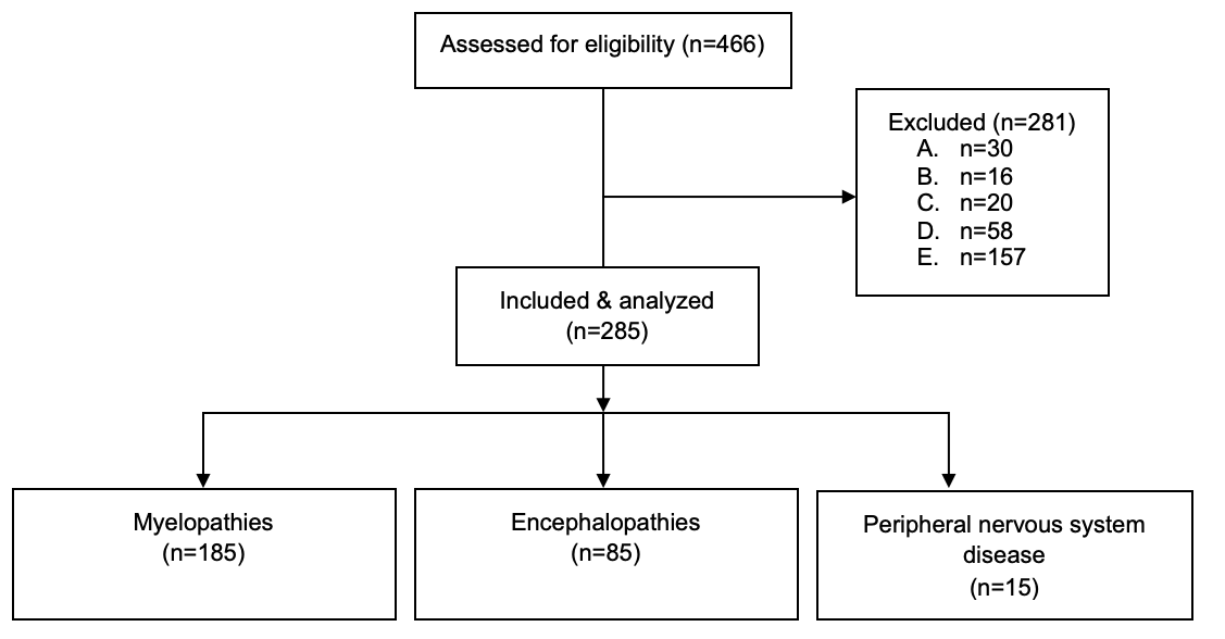
Figure 1: The Consolidated Standards of Reporting Trials (CONSORT) flow diagram of pug dogs referred to the Neurology & Neurosurgery department of Dick White Referrals (UK) from January 2013 to December 2020. A. Incomplete medical records; B. Diagnosis of a non-neurological condition; C. Final diagnosis not reached; D. Radiographic screening for vertebral malformations; E. Re-presenting for a previously diagnosed neurological condition.
Patient signalment including age, gender, neuter status and body weight was analysed for each diagnoses group separately (Table 1) and is described in the specific sections below.
| Myelopathies | Encephalopathies | PNS | P value | |
|---|---|---|---|---|
| (n = 185) | (n = 85) | (n = 15) | ||
| Sex (n) | 46 M; 75 MN; | 18 M; 31 MN; | 5 M; 8 MN; | 0.08 |
| 18 F; 46 FS | 17 F; 19 FS | 2 F; 0 FS | ||
| Age (months) | 70 (57-77)a | 43 (30-55)a | 74 (37-89) | 0.0004 |
| BW (kg) | 8.7 (± 2.1)a | 7.8 (± 2.3)a,b | 9.6 (± 2.5)b | 0.0015 |
| Presence of pain [n (%)] | 61 (33%)a,b | 3 (3.5%)a | 1 (6.7%)b | < 0.0001 |
Table 1: Signalment and presence of pain at neurological examination for each the three final diagnoses obtained in a cohort of p
Table 1: Signalment and presence of pain at neurological examination for each the three final diagnoses obtained in a cohort of pug dogs referred to the Neurology & Neurosurgery department a single referral centre in the UK. PNS: peripheral nervous system; M: male entire; MN: male neutered; F: female entire; FN: female neutered; BW: body weight; n = number of cases. P value refers to the comparison between the three groups. In the same row, same superscript letter refers to p < 0.05 between groups.
Ambulatory paraparesis was the most common presenting symptom in 79 cases (27.6%), followed by seizures (n = 58, 20.3%), ataxia (n = 47, 16.4%) and spinal pain (n = 29, 10.1%). The remaining cases were referred for investigation of vestibular signs (n = 16, 5.6%), non-ambulatory paraparesis (n = 12; 4.2%), ambulatory tetraparesis (n = 9; 3.15%), behavioural changes / involuntary movements (n = 9; 3.15%), monoparesis (n = 7; 2.5%), paraplegia (n = 4; 1.4%), non-ambulatory tetraparesis (n = 3; 1.05%), muscle atrophy (n = 3; 1.05%), head trauma (n = 3; 1.05%), facial nerve paralysis (n = 2; 0.7%), hemiparesis (n = 2; 0.7%), visual deficits (n = 1; 0.35%) and generalized weakness (n = 1; 0.35%).
Investigations were performed according to clinician’s recommendation and owner’s consent; a total of 278 cases (97.5%) underwent MRI scan. CSF was collected in 89 (31.2%) cases. Serology for Toxoplasma gondii and Neospora caninum was performed in 49 (17.5%) cases.
Myelopathies were the most common diagnosis, followed by encephalopathies, and PNS diseases were the least common (Table 1). Pugs diagnosed with encephalopathies were younger (p = 0.0003) than those diagnosed with myelopathies. Pugs with myelopathies were more likely to be painful than both those with encephalopathies [reciprocal OR 13.45 (4.23 – 41.94), p < 0.0001] or PNS disease [OR 6.89. (1.08 – 74.24), p = 0.04].
Myelopathies
Myelopathies were diagnosed in 185/285 (64.9%) cases. MRI was performed in 183/185 (98.9%). Two cases (0.7%) were diagnosed with a myelopathy, specifically vertebral malformation, in the light of radiographic diagnosis. In 24/185 (13%) cases, CSF was obtained. CSF was compatible with meningomyelitis of unknown aetiology in 8 cases (MUA), while the remaining 16 cases were diagnosed with IVDD, SAD or VM. Serology for Toxoplasma gondii and Neospora caninum was tested 8/185 (4.32%) cases and were all found negative.
| Myelopathies | IVDDs | SAD | VMs | Others | P value |
|---|---|---|---|---|---|
| (n = 185) | (n = 89) | (n = 41) | (n = 41) | (n = 14) | |
| Sex | 19M, 38MN, 6F, 26FS | 10M, 14MN | 12M, 18MN | 5M, 5MN | 0.82 |
| 7F, 10FS | 3F, 8FS | 2F, 2FS | |||
| Age (month) | 72 (63 – 89)a | 78 (34 – 84) | 54 (10 - 72)a | 70 (29 – 92) | 0.0074 |
| Body weight (kg) | 8.8 ± 1.8 | 8.6 ± 2.2 | 8.3 ± 2.4 | 9.5 ± 2.6 | 0.32 |
| Presence of pain [n (%)] | 46 (51.7%)a,b | 1 (2.4%)a,c,d,e | 7 (17.1%)b,c | 6 (42.8)d,e | <0.0001 |
Table 2: Signalment and presence of pain at neurological examination for the most prevalent myelopathies in a cohort of pug dogs
Table 2: Signalment and presence of pain at neurological examination for the most prevalent myelopathies in a cohort of pug dogs referred to the Neurology & Neurosurgery department a single referral centre in the UK. IVDDs: intervertebral disc diseases including intervertebral disc extrusions, intervertebral disc protrusions and acute non-compressive nucleus pulposus extrusions; SAD: spinal arachnoid diverticulum; VMs: vertebral malformations; M: male entire; MN: male neutered; F: female entire; FS: female spayed; BW: body weight; n: number of cases. P value refers to the comparison of the four groups. In the same row, same superscript letter refers to p < 0.05 between groups.
Myelopathies (Table 2) included IVDDs 89/185 (48%), SAD 41/185 (22.2%), VMs 41/185 (22.2%) and others 14/185 (7.6%). In the latter, MUA (9/14; 64.3%), syringomyelia (2/14; 14.3%), spinal cord gliosis (1/14; 7.13%), discospondylitis (1/14; 7.13%), and spinal cord atrophy (1/14; 7.13%) were included. Within the IVDDs, 53/89 (59.6%) pugs were affected by IVDE, 29/89 (32.6%) by IVDP, and 7/89 (7.8%) by ANNPE.
IVDDs were most commonly diagnosed in the thoracolumbar region (39/89; 43.8%), followed by cervical (25/89; 28.1%), lumbar (17/89; 19.1%), or multifocal (8/89; 9.0%) neuroanatomical localisations. IVDE was most commonly found at the thoracolumbar spinal cord segments (28/53; 52.8%), followed by cervical (18/53; 34%), and lumbosacral (7/53; 13.2%). The most common site of extrusion was C2-C3 in the cervical region (9/18; 50%), T13-L1 in the thoracolumbar (10/28; 35.7%) and L4-L5 or L7-S1 in the lumbosacral region (3/7; 42.8% for each space). 52.8% (28/53) of cases diagnosed with IVDE were painful. The most prevalent cervical, thoracolumbar, and lumbosacral locations for IVDP were C2-C3 (7/29; 24.1%), T12-T13 (8/29; 27.5%) and L7-S1 (7/29; 24.1%) respectively. Of the pugs diagnosed with IVDP, 14/29 (48.3%) had multiple intervertebral disc protrusions. 41.3% (12/39) of cases diagnosed with IVDP were painful.
ANNPE was diagnosed in the cervical vertebral column in three cases (one case each of C3-C4, C4-C5 and C5-C6), and in the thoracolumbar vertebral column in four cases (two cases, at L3-L4, and once case each at T11-T12 and T12-T13). 57% (4/7) cases were noted painful at the time of initial examination prior to referral.
SAD was most commonly diagnosed at the thoracolumbar (27/41; 65.9%), followed by cervical (14/41; 34.1%) region. No lumbar SAD was identified. At the thoracolumbar level, SAD was more frequently identified at T11-T12 and T12-T13 (4/27; 14.8% for each location). A C2-C3 SAD was identified in the majority of cases with cervical localisation (10/14; 71.4%).
The majority of VMs were found in the thoracolumbar region (31/41; 75.6%), followed by lumbar (5/41; 12.2%), cervical (3/41; 7.3%) and multifocal (2/41; 4.9%). The thoracic vertebrae T5-T9 (29/41; 70.7%) were the most affected, with T7 vertebra being the most prevalent (22/41; 53.7%).
Pugs diagnosed with IVDDs were statistically older than pugs with VMs (p = 0.0041). Those with IVDDs were more likely to be painful than dogs diagnosed with SAD [reciprocal OR 42.79 (7.43 – 444.6), p < 0.0001] or VMs [reciprocal OR 5.20 (2.06 – 13), p = 0.0002]. Furthermore, dogs with SAD were less likely to be painful than pugs with VMs [reciprocal OR 8.24 (1.32 – 94.74), p = 0.06] and pugs with other myelopathies [OR 30 (3.48 – 349.4), p = 0.0006] or other myelopathies were more likely to be painful than pugs with VMs [reciprocal OR 3.63 (1.03 – 12.15), p = 0.07] (Table 2). One single case diagnosed with SAD (at the thoracolumbar vertebral column) was painful.
Encephalopathies
Encephalopathies were diagnosed in 85/285 (29.8%) cases. MRI was performed in 82/85 (96%) cases. The diagnoses for the remaining 3 cases were: idiopathic epilepsy Tier I, hepatic encephalopathy secondary to portosystemic shunt and traumatic brain injury, respectively. CFS was obtained in 58/85 (68.2%) cases. Serology for Toxoplasma gondii and Neospora caninum was tested in 36/85 (42.3%) cases.
Encephalopathies included idiopathic epilepsy (38/85; 44.7%), MUA (29/85; 34.1%) and other encephalopathies (18/85; 21.2%) (Table 3). In the latter, the following conditions were included: meningitis-otitis interna (6/18; 33.3%), traumatic brain injury (3/18; 16.7%), cerebellar infarct (3/18; 16.7%), hydrocephalus (2/18; 11.1%), obsessive compulsive disorder (2/18; 11.1%), hepatic encephalopathy secondary to portosystemic shunt (1/18; 5.5%) and extra-axial neoplasia (1/18; 5.5%).
| Idiopathic epilepsy | MUA | Others | P value | |
|---|---|---|---|---|
| (n = 85) | (n = 38) | (n = 29) | P value | (n = 18) |
| Sex | 6M, 16MN | 7M, 9MN | 5M, 6MN | 0.92 |
| Sex | 5F, 11 FS | 7F, 4FS | 3F, 4FS | 0.92 |
| Age (month) | 45 (29 – 62) | 34 (16 – 69) | 41 (15 – 66) | 0.83 |
| Body weight (kg) | 8.6 ± 2.2a | 7.1 ± 2.1a | 7.2 ± 2.4 | 0.021 |
Table 3: Signalment and presence of pain at neurological examination for the most prevalent encephalopathies in a cohort of pug d
Table 3: Signalment and presence of pain at neurological examination for the most prevalent encephalopathies in a cohort of pug dogs referred to the Neurology & Neurosurgery department a single referral centre in the UK. MUA: meningoencephalitis of unknown aetiology; M: male entire; MN: male neutered; F: female entire; FS: female spayed; BW: body weight; n: number of cases; n/a: not available. P value refers to the comparison of the four groups. In the same row, same superscript letter refers to p < 0.05 between groups.
Among those diagnosed with MUA, 5/29 (17.2%) were more specifically diagnosed with necrotizing meningoencephalitis (necrotizing meningoencephalitis) based on MRI findings and/or post-mortem report. No difference in age and sex was found between pugs with idiopathic epilepsy, MUA or other encephalopathies.
Peripheral Nervous System Disease
A PNS disease was diagnosed in 15/285 cases (5.2%): otitis media/externa (9/15; 60%), masticatory muscle myositis (3/15; 20%), facial nerve paralysis (2/15; 13.3%) and myasthenia gravis (1/15; 6.7%). MRI was performed in 12/15 of cases. The remaining three cases which did not undergo MRI were diagnosed with masticatory muscle myositis and myasthenia gravis. CSF was obtained in 7/15 cases (46.7%). Serology for Toxoplasma gondii and Neospora caninum was tested in 5/15 (33.3%) cases with a PNS disorder; one case was found positive for N. caninum and diagnosed with masticatory muscle myositis on the basis of positive 2M antibody titre.
Discussion
This is the largest study reporting a wide range of neurological conditions in a large sample of pugs from a single referral centre in the UK to the authors’ knowledge. Myelopathies were overrepresented, with IVDE, IVDP, ANNPE, SAD and VMs comprising almost 60% of all diagnosed conditions. Conditions of the PNS were rarely reported in this population. In accordance with the previously reported high prevalence of gait abnormalities in pug dogs [9], the most frequent presenting complaints were: ambulatory paraparesis and ataxia, when referring to myelopathies and seizures, when referring to encephalopathies.
A recent study reporting the prevalence of neurological conditions in French bulldogs, found that IVDDs and SAD were the most frequent diagnoses [10], results similar to those of the present study, supporting possible predisposition of both breeds to develop myelopathies. Approximately one third of the total population of pugs in this study were affected by IVDDs. Whilst previous studies into IVDDs in dogs have included pugs, their prevalence was low compared to other breeds such as Dachshund, Pembroke Welsh Corgi and French Bulldog [11, 12]. The present study supports that IVDDs should be included amongst the main differentials for pugs presenting with myelopathies. According to our results, IVDDs are more likely to be painful than other myelopathies, and are more commonly localised at thoracolumbar level. In the present study, when a pug was comfortable on analgesia at the time of presentation, then the referring notes were used to record the presence or absence of pain. This may explain the high proportion of painful pugs diagnosed with ANNPE, since vocalisation and spinal hyperaesthesia are common at initial presentation [13].
SAD was diagnosed on 14.4% (41/285) of the total population of pugs included in this study. Mauler, et al. [6] found that pugs were the most commonly affected breed in a population of dogs presenting with SAD. SAD is generally described as a non-painful condition in dogs [6, 14], and the present study supports this statement having found only one case experiencing pain at neurological examination.
VMs, most commonly affecting the thoracic vertebrae, were the cause for clinical signs in 14.4% (41/285) of the total population of pugs included. In the present study, only findings suspected responsible for the clinical signs were reported, thus it is likely that the prevalence of VMs was higher and in many cases incidental. A recent study investigated the presence of CAPD and its association with clinical signs of myelopathy, and failed to confirm the latter [15]. Similarly, a study on thoracolumbar VMs in pugs failed to reach an association between the presence of VMs and relevant clinical signs, although the majority of VMs were adjacent to an area of focal spinal cord pathology, suggesting a possible clinical impact [16]. Previously, clinically relevant thoracic VMs, such as hemivertebrae, were described in 5% of all pugs [17, 18], although most dogs were free from clinical signs. In our study, pugs diagnosed with VMs were younger compared to those diagnosed with myelopathies, suggesting that this condition should be included in the differentials for young pugs with gait abnormalities usually non-painful. One case of spinal cord gliosis and one case of spinal cord atrophy were documented. Spinal cord gliosis and atrophy could be considered features indicative of constrictive myelopathy [19], which is a differential in pugs presenting with progressive gait abnormalities and urinary and/or faecal incontinence [19, 20]. Recent literature suggests that the development of constrictive myelopathy is common in pugs and often a result of several coexisting conditions, including CAPD, IVDP, and SAD, whilst CAPD alone is often incidental and not responsible for clinical signs [21].
Idiopathic epilepsy was the most represented encephalopathy. Pugs diagnosed with encephalopathies were significantly younger when compared to myelopathies or PNS diseases. Although pugs are not among the breeds with an identified genetic background for idiopathic epilepsy [22], a high prevalence of seizures was documented among pugs presenting to primary veterinary care in the UK [23]. Further studies are required to evaluate a possible genetic background for idiopathic epilepsy in pugs. MUA was the second most prevalent encephalopathy found but the diagnosis was mainly antemortem. Therefore, it is possible that the prevalence of MUA cases has been over or underestimated. Post-mortem examination, performed in 5/29 of the MUA cases, revealed the presence of necrotizing meningoencephalitis (NME), also known as pug dog encephalitis. NME was first reported in 1989 [24] and its imaging and histopathological features have been better characterised since. Two specific risk loci have been also associated to the development of NME in pugs [25]. A predisposition for young adult female pugs has been described [26], which is supported by the findings of our study. The small population of pugs diagnosed with NME in this present study could have also been affected by the referral population setting, considering that pugs with clinical signs compatible with NME may have been euthanised at the primary care veterinary centres without referral on the suspicion for poor prognosis.
Otitis was frequently associated with signs suggestive of peripheral vestibular disease, as previously described [27]. Masticatory muscle myositis has been reported in pugs [28], although it is not among the most commonly reported breeds, in agreement to the findings of the present study only a small number of patients was diagnosed with this condition. Future studies regarding the most common diseases of the PNS in brachycephalic breed dogs are warranted.
There are several limitations in the present study arising mainly from its retrospective nature. The caseload might be biased by the topographical differences in the prevalence of neurological disorders in the UK compared to other countries, and the fact that only a single and referral hospital was included. Cases were managed by different neurologists over a period of several years and the diagnostic steps for each specific disease were not standardized. The presence or absence of pain was documented based on referring notes and administration of analgesic at the time of admission, and pain scores were not available. Only findings suspected to be responsible for the presenting clinical signs were reported, thus other, coexisting conditions may be underrepresented if considered incidental (such as CAPD or other VM). Cases without a final diagnosis were excluded from the study, which may have biased the frequency of some conditions over others. For example, pugs presenting in refractory status epilepticus, may have been euthanised prior to investigations, cases with mild neurological signs may have been discharged without pursuing further investigations, or financial factors may have precluded allowing investigations, thus not reaching a definitive diagnosis. Finally, histopathological confirmation was not available in most cases of MUA, leading to a suspected rather than confirmed diagnosis for this disease, and not allowing more specific classification of the type of MUA for each case.
Conclusion
This is the first study reporting various neurological conditions in a large population of pugs. Myelopathies are the most common group of neurological diseases diagnosed in pugs, and they tend to be more frequently localised at the thoracolumbar level. The presence or absence of pain at neurological examination might be useful to differentiate among different myelopathies. Encephalopathies are the second most common category of neurological diseases diagnosed in pugs, and those dogs present significantly younger than those with myelopathies. PNS diseases are uncommon in this breed. The results of the present study can form the basis for future studies and be of interest for veterinarians, pug breeders and owners.
Ethical Approval
Ethical approval was obtained from the School’s Committee for Animal Research and Ethics (CARE); School of Veterinary Medicine University of Nottingham. Signed informed owner consent to use clinical information for retrospective studies was granted at the time of the animal’s admission.
References
-
Bedford E (2023) UK: Top 20 dog breeds by registered number 2022.
-
O’Neill D, Sahota J, Brodbelt D, Church D, Packer R, et al. (2022) Health of Pug dogs in the UK: disorder predispositions and protections. Canine Medicine and Genetics 9(1): 4.
-
Packer R, O’Neill D, Fletcher F, Farnworth M (2019) Great expectations, inconvenient truths, and the paradoxes of the dog-owner relationship for owners of brachycephalic dogs. PLoS One 14(7): e0219918.
-
Packer R, O’Neill D, Fletcher F, Farnworth M (2020) Come for the looks, stay for the personality? A mixed methods investigation of reacquisition and owner recommendation of Bulldogs, French Bulldogs and Pugs. PLoS One 15(8): e0237276.
-
De Risio L, Adams V, Dennis R, McConnell F (2009) Association of clinical and magnetic resonance imaging findings with outcome in dogs with presumptive acute non-compressive nucleus pulposus extrusion: 42 cases (2000-2007). J Am Vet Med Assoc 234(4): 495-504.
-
Mauler D, De Decker S, De Risio L, Volk H, Dennis R, et al. (2013) Signalment, Clinical Presentation, and Diagnostic Findings in 122 Dogs with Spinal Arachnoid Diverticula. J Vet Int Med 28(1): 175-181.
-
De Risio L, Bhatti S, Muñana K, Penderis J, Stein V, et al. (2015) International veterinary epilepsy task force consensus proposal: diagnostic approach to epilepsy in dogs. BMC Vet Res 11: 176.
-
Granger N, Smith P, Jeffery N (2010) Clinical findings and treatment of non-infectious meningoencephalomyelitis in dogs: A systematic review of 457 published cases from 1962 to 2008. The Veterinary Journal 184(3): 290-297.
-
Rohdin C, Jäderlund K, Ljungvall I, Lindblad-Toh K, Häggström J (2018) High prevalence of gait abnormalities in pugs. Vet Rec 182(6): 167.
-
Mayousse V, Desquilbet L, Jeandel A, Blot S (2017) Prevalence of neurological disorders in French bulldog: a retrospective study of 343 cases (2002–2016). BMC Vet Res 13(1).
-
Aikawa T, Fujita H, Kanazono S, Shibata M, Yoshigae Y (2012) Long-term neurologic outcome of hemilaminectomy and disk fenestration for treatment of dogs with thoracolumbar intervertebral disk herniation: 831 cases (2000–2007). J AM Vet med Assoc 241(12): 1617-1626.
-
Baumhardt R, Ripplinger A, Aiello G, Schwab M, Ferrarin D, et al. (2020) Clinical management of dogs with presumptive diagnosis of thoracolumbar intervertebral disc disease: 164 cases (2006-2017). Pesquisa Veterinária Brasileira 40(1): 55-60.
-
Fenn J, Drees R, Volk HA, De Decker S (2016) Comparison of clinical signs and outcomes between dogs with presumptive ischemic myelopathy and dogs with acute non-compressive nucleus pulposus extrusion. J AM Vet Med Assoc 249(7): 767-775.
-
Flegel T, Müller MK, Truar K, Löffler C, Oechtering G (2013) Thoracolumbar spinal arachnoid diverticula in 5 pug dogs. Can Vet J 54(10): 969.
-
Ban J, Park J, Kim H, Yoon K, Oh M, Lee Y, et al. (2023) Investigation of canine caudal articular process dysplasia of thoracic vertebrae using computed tomography: Prevalence and characteristics. Front Vet Sci 10: 1066420.
-
Rohdin C, Häggström J, Ljungvall I, Nyman Lee H, De Decker S, et al. (2018) Presence of thoracic and lumbar vertebral malformations in pugs with and without chronic neurological deficits. Vet J 241: 24-30.
-
Bertram S, Ter Haar G, Decker S (2019) Congenital malformations of the lumbosacral vertebral column are common in neurologically normal French Bulldogs, English Bulldogs, and Pugs, with breed‐specific differences. Vet Radiol Ultrasound 60(4): 400-408.
-
Ryan R, Gutierrez-Quintana R, Ter Haar G, De Decker S (2017) Prevalence of thoracic vertebral malformations in French bulldogs, Pugs and English bulldogs with and without associated neurological deficits. Vet J 221: 25- 29.
-
Lourinho F, Holdsworth A, McConnell J, Gonçalves R, Gutierrez‐Quintana R, et al. (2020) Clinical features and MRI characteristics of presumptive constrictive myelopathy in 27 pugs. Vet Radiol Ultrasound 61(5): 545-554.
-
Rohdin C, Ljungvall I, Häggström J, Leijon A, Lindblad‐ Toh K, et al. (2020) Thoracolumbar meningeal fibrosis in pugs. J Vet Int Med 34(2): 797-807.
-
Wachowiak IJ, Patterson JS, Winger KM, Smiler KL, Cole R, et al. (2023) Thoracolumbar myelopathies in Pug Dogs. J Vet Int Med 37(2): 618-625.
-
Hülsmeyer V, Fischer A, Mandigers P, DeRisio L, Berendt M, et al. (2015) International Veterinary Epilepsy Task Force’s current understanding of idiopathic epilepsy of genetic or suspected genetic origin in purebred dogs. BMC Vet Res 11: 175.
-
Erlen A, Potschka H, Volk H, Sauter-Louis C, O’Neill D (2018) Seizure occurrence in dogs under primary veterinary care in the UK: prevalence and risk factors. J Vet Int Med 32(5): 1665-1676.
-
Cordy D, Holliday T (1989) A Necrotizing Meningoencephalitis of Pug Dogs. Vet Pathol 26(3): 191- 194.
-
Barber R, Schatzberg S, Corneveaux J, Allen A, Porter B, et al. (2011) Identification of Risk Loci for Necrotizing Meningoencephalitis in Pug Dogs. J Hered 102(1): S40-S46.
-
Levine J, Fosgate G, Porter B, Schatzberg S, Greer K (2008) Epidemiology of Necrotizing Meningoencephalitis in Pug Dogs. J Vet Int Med 22(4): 961-968.
-
Garosi L, Dennis R, Penderis J, Lamb C, Targett M, et al. (2001) Results of magnetic resonance imaging in dogs with vestibular disorders: 85 cases (1996-1999). J AM Vet Med Assoc 218(3): 385-391.
-
Evans J, Levesque D, Shelton G (2004) Canine Inflammatory Myopathies: A Clinicopathologic Review of 200 Cases. J Vet Int Med 18(5): 679-691.
- The Digital Stethoscope: Harnessing AI in Veterinary Medicine Without Losing Our Healing Touch
- Meningoencephalomyelitis of Unknown Etiology: Short-Term Effect of Two Treatment Protocols on Cerebrospinal Fluid
- Safety and Efficacy of the HomeoPet Cough in Domestic Pets –A Clinical and Correction Analysis Based Upon User Response Survey
- Non Human Animals Responses to Social Loss
- Owner Reported Clinical Outcomes of a Homeopathic Proprietary Preparation for the Treatment of Upper Respiratory and Nasal Disorders in Companion Animals
- Effects and Diagnostic Approach of Ultrasound in Veterinary Practice: A Systematic Review