Bovine Tuberculosis: Ethiopian Perspective
Bovine tuberculosis is a significant zoonotic disease affecting livestock and humans worldwide, causing substantial economic losses and posing serious public health risks. This review aims to provide an overview of bovine tuberculosis in Ethiopia, highlighting its prevalence, risk factors, transmission routes, clinical manifestations, and diagnostic methods. Mycobacterium bovis primarily causes bovine tuberculosis, which is characterized by granulomatous lesions in various organs. Transmission occurs through inhalation, ingestion of contaminated materials, and direct contact with infected animals. The age, breed, herd size, and management practices are some of the risk factors for bovine tuberculosis. In Ethiopia, the disease is prevalent but poorly controlled, lacking a national control program. Effective diagnosis relies on methods such as the tuberculin skin test, culture, and PCR. Control measures include test-and-slaughter programs, movement restrictions, biosecurity protocols, and public health education. This review underscores the urgent need for a comprehensive national control strategy, enhanced surveillance, public awareness campaigns, and investment in research to mitigate the impact of bovine tuberculosis in Ethiopia, thereby safeguarding both animal and human health.
Introduction
There are a number of bacterial zoonoses, such as anthrax, brucellosis, campylobacteriasis, glanders, colibacillosis, leptospirosis, listeriosis, melioidosis, necrobacillosis, salmonellosis, tuberculosis, yersiniosis, etc., that cause morbidity as well as mortality in humans and animals [1, 2]. Among these, bovine tuberculosis is a serious zoonotic disease that affects both domestic animals and humans globally, leading to substantial financial losses in the animal production industry, especially in intensive farming systems [3, 4, 5]. A group of related bacteria, including Mycobacterium tuberculosis, Mycobacterium bovis, Mycobacterium africanum, Mycobacterium canettii, Mycobacterium caprae, Mycobacterium pinnipedii, and Mycobacterium microti, cause this chronic granulomatous disease [6, 7]. Mycobacterium bovis is the main cause of bovine tuberculosis in animals, and its transmission to humans poses a significant public health risk [7, 8]. The disease is characterized by the development of granulomas in various tissues and organs, such as the kidneys, lungs, lymph nodes, and intestines [9]. WHO has mentioned that around 5% of cattle are affected by tuberculosis, making it a major disease of economic and public health importance [2].
The bacilli exit the host through the respiratory discharges, milk, feces, urine, semen, and genital discharges. These body excretions can contaminate grazing pastures, drinking water, feed troughs, or fomites, potentially acting as sources of infection for other animals. Common routes of infection include inhalation of Mycobacterium bovis bacilli and ingestion of contaminated milk, pasture, and water [10].
Direct contact with infected animals or consumption of unpasteurized milk and dairy products are two additional ways that may spread Mycobacterium bovis to humans [11]. Because it reduces meat and milk outputs and causes the disposal of corpses or parts judged unfit for human use, bovine tuberculosis poses a serious threat to communities that depend on cattle. Also, it makes it more difficult for goods made from animals to be traded internationally. As a source of infection for both humans and animals, the disease has the potential to disrupt conservation efforts in wildlife populations [12, 13].
In Ethiopia, there are currently no national policies or strategies in place for controlling bovine tuberculosis, despite it being one of the top three diseases affecting dairy production in urban and peri-urban areas in terms of prevalence and impact on households [14]. The prevalence of bovine tuberculosis among livestock in the country remains poorly understood, with most research concentrated in the central regions, particularly around Addis Ababa. It is important to assess the disease status across all regions of Ethiopia where such studies have not been conducted to inform the development of a national control program for bovine tuberculosis in the future. Therefore, the objective of this review is to highlight the current information and status of bovine tuberculosis in Ethiopia.
Literature Review
Definition
Bovine tuberculosis is a chronic infectious disease in cattle, marked by the development of granulomatous tubercles in various organs. This condition is caused by bacteria from the genus Mycobacterium [9].
Etiology
The microorganism responsible for bovine tuberculosis is Mycobacterium bovis, a member of the genus Mycobacterium. This bacterium is acid-fast, filamentous, curved, rod-shaped, aerobic, non-motile, non-spore-forming, and lacks a capsule [2, 7, 15, 16]. It is cultured on an egg yolk- based medium called Lowenstein-Jensen medium, which is supplemented with malachite green or crystal violet to inhibit unwanted growth. Mycobacterium bovis does not grow on blood agar plates and has a slow growth rate, requiring 6–8 weeks of incubation on Lowenstein-Jensen medium to produce visible colonies. Acid-fast staining reveals acid-fast-positive rod-shaped organisms in sputum smears [17, 18]. While the bacterium is as susceptible to heat and light as other vegetative organisms, it is highly resistant to chemical agents, which aids in obtaining pure cultures from contaminated samples [19].
Epidemiology
Geographical Distribution and Occurrences Bovine tuberculosis is present globally but has been significantly reduced or eliminated in many developed countries [20]. The disease does not respect geographical boundaries and affects a wide range of hosts, including economically important farm animals, wildlife, and humans [21].
Bovine tuberculosis (TB) can persist within livestock and wildlife populations, with human infections primarily resulting from animal-to-human transmission and rarely through human-to-human spread [22, 23]. Research on bovine TB in Ethiopia has been predominantly conducted in the Addis Ababa, Amhara, Oromia, and Southern regions, with a notable absence of published studies from the Benishangul-Gumuz, Harari, and Dire Dawa regions. Limited studies have also been carried out in the Afar, Gambella, Somali, and Tigray regions. The prevalence of bovine TB varies widely across different districts, ranging from 0.8% to 54.6%, with higher rates observed in intensive farming operations near cities and lower rates in rural grazing areas (Figure 1) [24].
![Figure 1: Epidemiology of bovine tuberculosis in Ethiopia. Source: Sibhat et al. [24]](/fulltextimages/13100/fig_1.png)
Source of Infection Infected cattle are the primary source of infection for other cattle. The bacteria are present in exhaled air, sputum, feces, milk, urine, and vaginal and uterine discharges. Even before visible lesions appear, cattle in the early stages of the disease can shed viable mycobacteria in nasal and tracheal mucus. In experimentally infected cattle, bacterial excretion typically begins around 90 days after infection. The bacteria are expelled through respiratory secretions, feces, milk, urine, semen, and genital discharges. These excretions can contaminate grazing pastures, drinking water, feed troughs, and fomites, potentially serving as sources of infection for other animals [10].
Wildlife reservoirs are crucial in the spread of bovine tuberculosis, especially in certain regions where some wildlife species act as significant maintenance hosts. These animals can transmit the disease to domestic cattle by contaminating pastures with their urine, accessing farm buildings and cattle troughs where they defecate and urinate directly onto the feed and through direct contact at pasture- bush margins [22, 25].
Transmission In Ethiopia, the close living conditions between humans and animals, combined with the practice of using cow dung to paint floors and walls, contribute to the transmission of bovine tuberculosis [26]. This environment increases the likelihood of tuberculosis spreading to humans. The primary route of infection for cattle is through inhalation of Mycobacterium bovis, which is facilitated by prolonged and close contact between infected and healthy animals. In some regions, ingestion of Mycobacterium bovis from contaminated pasture, water, or utensils is also a common route of infection. Although congenital and vertical transmissions, including genital transmission from infected reproductive organs, have been documented, these routes are now rarely observed in most areas [27].
Risk Factors Risk factors for bovine tuberculosis are divided into animal-level and herd-level risk factors.
Animal-Level Risk Factors: Age, breed, body condition, immune status, genetic resistance and susceptibility to bovine tuberculosis, vertical and pseudo-vertical transmissions, and auto-contamination are considered risk factors [28]. Older animals are more susceptible due to the chronic nature of bovine tuberculosis, stress, malnutrition, and immunosuppression, which all increase with age. Exotic and cross-bred cattle are more susceptible to bovine tuberculosis compared to the indigenous zebu breed [29, 30].
Herd-Level Risk Factors: A history of bovine tuberculosis outbreaks, instances of tuberculosis in household members, herd size, type of cattle enterprise, management practices, lack of diagnostic testing, reduced detection opportunities, introduction of purchased cattle into the herd, animal movements, presence of other domestic species, contact between animals and wildlife, and climate influences [31]. The spread of Mycobacterium bovis from one herd to another and across different areas is typically due to the movement of an infected animal from an infected herd into a non-infected, susceptible herd. The risk of infection within a herd increases with herd size, likely due to overcrowding, which raises the likelihood of contact between animals [30].
Pathogenesis
In animals, tuberculosis typically has a slow onset, with clinical signs often taking several months or longer to manifest. Infections can also remain latent for years and may reactivate later. In humans, systemic signs of infection can develop months to years after exposure, or the infection may remain latent until waning immunity permits reactivation of the organisms. Tuberculosis chancre, a type of skin lesion, usually appears 2-4 weeks after cutaneous exposure. The development and production of lesions also depend on the number of mycobacteria in the inoculum, their subsequent multiplication, and the type of host. A visible primary focus develops within 8 days of being affected by the bacteria. Calcification of the lesions commences about 2 weeks later [9].
Clinical Findings
In cattle, many cases of tuberculosis are asymptomatic, which complicates control efforts. When clinical signs do appear, they vary: respiratory forms can cause coughing and dyspnea, while digestive system involvement can lead to diarrhea and constipation [2, 32]. Tuberculosis is typically a chronic, debilitating disease, though it can sometimes be acute and rapidly progressive. Early infections often show no clinical signs. However, in the late stages, common symptoms include progressive emaciation, fluctuating fever, weakness, and loss of appetite. Animals with pulmonary involvement usually exhibit a moist cough, which is worse in the morning, during cold weather, or with exercise, and may also have dyspnea or tachypnea. In the terminal stage, animals can become extremely emaciated and develop severe respiratory distress [33].
Diagnosis
A presumptive diagnosis of tuberculosis in cattle is often based on the animal’s history, clinical signs, tuberculin skin tests, and/or necropsy findings [34, 35].
Tuberculin Skin Test The tuberculin skin test is the definitive screening method currently in use. It is widely employed in practice and is also prescribed by the OIE for international trade. Tuberculin is a solution containing protein material extracted from the cell wall of Mycobacteria. When this protein solution is injected into the skin of an infected animal, it triggers a localized inflammatory response in sensitized individuals, leading to the characteristic signs of a positive test. The preferred site for administering this test is the neck area [27].
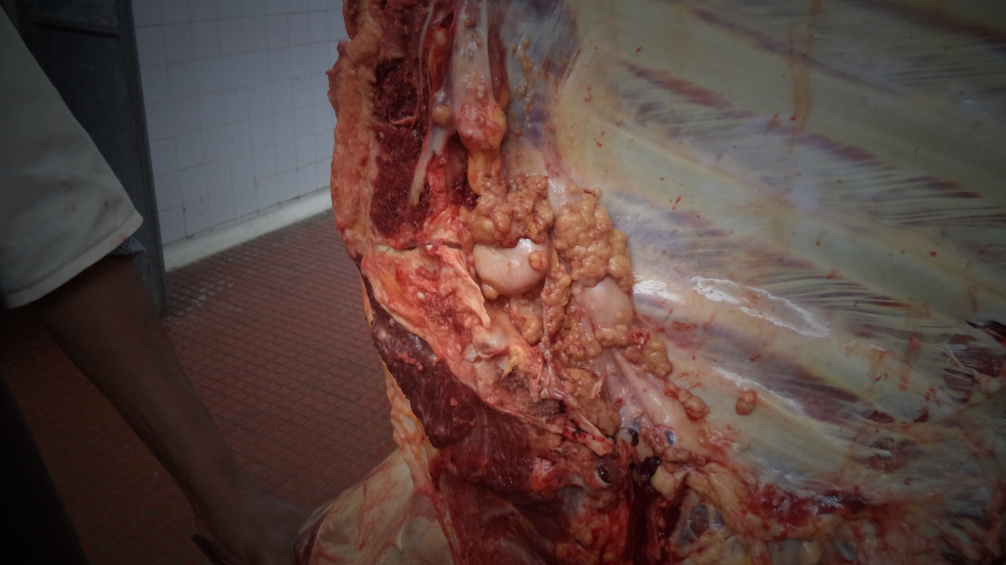
Postmortem Examination Bovine tuberculosis is marked by the formation of granulomas, where the bacteria are localized. These granulomas are typically yellowish and can be caseous, caseo-calcareous, or calcified, often with an encapsulated appearance. Some tubercles may be small and not visible to the naked eye unless the tissue is sectioned. In cattle, tubercles are commonly found in the lymph nodes, especially those of the head and thorax, and are also frequently present in the lungs, spleen, liver, and body cavity surfaces [9, 26, 36]. The tuberculous lesions were found in the sternal area of the thoracic cavity of adult cattle (Figure 2).
Laboratory Diagnosis Direct detection of Mycobacterium bovis in clinical samples taken from living animals or by laboratory culture is difficult. Sensitivity is often lacking in methods like polymerase chain reaction or culture that are intended to identify the organism or it’s DNA. Isolating and identifying the bacteria from specimens acquired from suspicious people and cultivated in Lowenstein-Jensen media is necessary for the final diagnosis of tuberculosis [2, 37].
Treatment
Treatment of tuberculosis in animals is generally not favored or considered cost-effective in countries focused on eradicating the disease. In humans, long-term treatment is necessary, and improper adherence to the regimen can lead to the development of multidrug-resistant, extremely drug- resistant, and even drug-resistant bacterial strains. Commonly used anti-tuberculous drugs for human tuberculosis, as recommended by the WHO, include isoniazid, streptomycin, and para-aminosalicylic acid, often administered through the Directly Observed Treatment, Short-course (DOTS) strategy [4].
Control and Prevention
Disease can be controlled through test-and-slaughter or test-and-segregation methods [38]. In developed countries, bovine tuberculosis has either been nearly eradicated or significantly reduced in farm animals to low levels through effective control and eradication programs [39]. In Ethiopia, however, animal tuberculosis remains endemic due to economic constraints that prevent the implementation of skin testing and slaughter control strategies, which have proven effective in more developed regions [40].
In many developing countries, including Ethiopia, the fundamental strategies needed for the control and elimination of bovine tuberculosis are either not implemented or applied inadequately due to several reasons including financial constraints, a scarcity of trained professional staff, lack of political will, and poor recognition of zoonotic importance of disease [7].
In Ethiopia, BCG vaccination is considered a primary control measure for bovine tuberculosis, complemented by test-and-slaughter strategies with partial or full government compensation. BCG vaccination involves administering the vaccine to calves at 6 weeks of age. Since calves are immune-competent at birth and are naturally exposed to environmental mycobacteria early on, they typically mount a strong immunological response to these antigens by 6 weeks. Vaccination at this age helps induce a high level of immunity [41]. Control measures in traditional extensive production systems face significant challenges due to the large number of livestock, the mobility of animals, and various social and economic factors [39].
Status of Bovine Tuberculosis in Ethiopia
Bovine tuberculosis is an endemic disease of cattle, widely distributed across major livestock-producing regions in Ethiopia, leading to high morbidity and mortality [35]. The prevalence of bovine tuberculosis varies with breed and management system, with higher rates observed in intensive or semi-intensive production systems compared to extensive systems [42]. Recent studies in Ethiopia show a wide range of prevalence rates: from 2.38% [43] to 39% [3] at the individual level.
In Ethiopia, the prevalence of bovine tuberculosis varies depending on the geographical areas, breeds, and husbandry practices. The pastoral production system is related to the epidemiological factors that favor the transmission of bovine tuberculosis, which include large herd sizes, communal grazing and watering of diverse species of animals, including camels, cattle, goats, and sheep, and extensive seasonal mobility within and outside the districts, which creates favorable conditions for a wide range of interspecies contact [44].
In studies conducted in extensive production systems in the Highlands, bovine tuberculosis prevalence rates have been reported between 2.388% and 5.5% [43]. In various towns across Ethiopia, studies have shown that the prevalence of bovine tuberculosis in dairy farms ranges from 5.16% [45] to 39% [3] at the individual level, and from 22.41% to 60.20% [46] at the herd level. Exotic breeds are found to be more susceptible to Mycobacterium bovis compared to cross-bred and local breeds, exhibiting higher incidence and prevalence rates.
Economic Importance and Public Health Significance
Bovine tuberculosis is highly prevalent in dairy commercial and urban/peri-urban production systems, particularly where exotic, graded, or crossbred animals are kept. The disease results in considerable economic losses, primarily due to reduced and foregone production rather than mortality. Its impacts include decreased productivity, movement restrictions, screening costs, culling of affected animals, and trade restrictions, all contributing to significant economic losses related to animal health [47].
Bovine tuberculosis can reduce the productive efficiency of affected animals by 10–25%, with direct losses including a 10–18% decrease in milk production and a 15% reduction in meat production) [12]. It is a major bacterial zoonosis that is transmitted to humans through aerosols and the consumption of raw milk. The disease remains prevalent in developing countries, including Ethiopia, where it is endemic. However, the epidemiology and zoonotic significance of bovine tuberculosis is not well understood due to a lack of comprehensive national investigations [48].
Conclusion and Recommendation
Bovine tuberculosis presents a significant challenge in Ethiopia, where it remains endemic and poses substantial economic and public health risks. The disease, primarily caused by Mycobacterium bovis, affects livestock across various production systems, with high prevalence rates reported in both intensive and extensive farming systems. Despite the implementation of some control measures, such as BCG vaccination and test-and-slaughter methods, the lack of a comprehensive national strategy, coupled with financial constraints and limited resources, impedes effective management and eradication efforts. The economic losses attributed to bovine tuberculosis primarily from reduced productivity and trade restrictions highlight the urgent need for a more robust and coordinated approach to control the disease [49].
Based on the above conclusion, the following recommendations were forwarded:
- Increase the scope and frequency of surveillance to provide accurate and up-to-date data on bovine tuberculosis prevalence and distribution.
- Invest in improving diagnostic infrastructure and technologies to enhance the sensitivity and accuracy of bovine tuberculosis detection.
- Support research into bovine tuberculosis epidemiology, zoonotic transmission, and effective control measures. Facilitate the sharing of knowledge and best practices among stakeholders, including government agencies, veterinary services, and farmers.
References
-
Pal M (2005) Importance of zoonoses in public health. Indian Journal of Animal Sciences 75(5): 586-591.
-
Pal M (2007) Zoonoses 2nd (Edn.). Satyam Publishers.
-
Tulu B, Zewede A, Belay M, Zeleke M, Girma M, et al. (2021) Epidemiology of bovine tuberculosis and its zoonotic implication in Addis Ababa milkshed, Central Ethiopia. Front Vet Sci 8: 595511.
-
Pal M, Tolawak D, Bikila U (2022) Zoonotic importance of bovine tuberculosis in Ethiopia: An overview. Research in Veterinary Science and Medicine 2(7): 1-5.
-
Tewodros SS (2023) Review on bovine tuberculosis and its status in Ethiopia. Int J Adv Res Biol Sci 10(9): 119- 127.
-
Forrellad MA, Klepp LI, Gioffré A, Sabio G, Morbidoni HR, et al. (2013) Virulence factors of the Mycobacterium tuberculosis complex. Virulence 4(1): 3-66.
-
Pal M (2015) Bovine tuberculosis and its zoonotic implications. Addis Ababa University.
-
OIE (2009) Manual of Diagnostic Tests and Vaccines for Terrestrial Animals. World Organization for Animal Health.
-
Radostits OM, Gay C, Hinchcliff KW, Constable PD (2007) A textbook of the diseases of cattle, sheep, goats, pigs and horses. Veterinary Medicine 14: 1576-1580.
-
Russel DG (2003) Highlighting the parallels between human and animal tuberculosis. JVME 30: 140-142.
-
Sa’idu AS, Okolocha EC, Dzikwi AA, Gamawa AA, Ibrahim S, et al. (2015) Public health implications and risk factors assessment of Mycobacterium bovis infections among abattoir personnel in Bauchi state, Nigeria. J Vet Med (1): 718193.
-
Muller B, Dürr S, Alonso S, Hattendorf J, Laisse CJ, et al. (2013) Zoonotic Mycobacterium bovis induced tuberculosis in humans. Emerg Infect Dis 19(6): 899- 908.
-
OIE (2013) Tool for the Evaluation of Performance of Veterinary Services. Paris.
-
LMP (2015) Livestock Health Priorities in the Ethiopian Livestock Master Plan. Ethiopia LMP Brief.
-
Carslake D, Grant W, Green LE, Cave J, Greaves J, et al. (2011) Endemic cattle diseases: comparative epidemiology and governance. Philos Trans R Soc Lond Biol Sci 366(1573): 1975-1986.
-
Higgins J, Camp P, Farrell D, Bravo D, Pate M, et al. (2011) Identification of Mycobacterium spp. of veterinary importance using rpoB gene sequencing. BMC Veterinary Research 7: 1-14.
-
Caulfield AJ, Wengenack NL (2016) Diagnosis of active tuberculosis disease: From microscopy to molecular techniques. J Clin Tuberc and Other Mycobact Dis 4: 33- 43.
-
Forbes BA, Hall GS, Miller MB, Novak SM, Rowlinson MC, et al. (2018) Practical guidance for clinical microbiology laboratories: mycobacteria. Clin Microbiol Rev 31(2): e00038-17
-
Parkale DD, Kulkarni A (2011) Bovine Tuberculosis and its control. Livestock Development Officer WRDDL 7: 1-9.
-
Kemal J, Sibhat B, Abraham A, Terefe Y, Tulu KT et al. (2019) Bovine tuberculosis in eastern Ethiopia: prevalence, risk factors and its public health importance. BMC Infectious Diseases 19: 1-9.
-
Smith NH, Gordon SV, Ricardo RD, Hadley RS, Hewinson RG (2006) Bottlenecks and broomsticks: the molecular evolution of Mycobacterium bovis. Nature Reviews Microbiology 4(9): 670-681.
-
Wichatitsky GM, Caron A, Kock R, Tschopp R, Munyeme M, et al. (2013) A review of bovine tuberculosis at the wildlife–livestock–human interface in sub-Saharan Africa. Epidemiol Infect 141(7): 1342-1356.
-
Caron A, Wichatitsky GM, Roger F (2014) Bovine tuberculosis: a double-edged issue at the human/ livestock/wildlife interface in Africa. Empres-animal health 44(2): 10-13.
-
Sibhat B, Asmare K, Demissie K, Ayelet G, Mamo G, et al. (2017) Bovine tuberculosis in Ethiopia: a systematic review and meta-analysis. Prev Vet Med 147: 149-157.
-
Dejene SW (2017) Eco-epidemiology of Bovine Tuberculosis (bTB) in an African savanna: The conflict between traditional pastoralist adaptations and disease transmission in the modern era. Wageningen University. 137: 36-42.
-
Pal M, Zenebe N, Amare T, Woldemariam T (2017) An abattoir based study on bovine tuberculosis in Debre Zeit, Ethiopia. World’s Veterinary Journal (3): 101-107.
-
OIE (2018) Bovine tuberculosis. In: Manual of Diagnostic Tests and Vaccines for Terrestrial Animals. World Organization for Animal Health, pp: 1061-1062.
-
Humblet MF, Boschiroli ML, Saegerman C (2009) Classification of worldwide bovine tuberculosis risk factors in cattle: a stratified approach. Veterinary Research 40(5).
-
Vordermeier M, Ameni G, Berg S, Bishop R, Robertson BD, et al. (2012) The influence of cattle breed on susceptibility to bovine tuberculosis in Ethiopia. Comp Immunol Microbiol Infect Dis 35(3): 227-232.
-
Broughan JM, Judge J, Ely E, Delahay RJ, Wilson G, et al. (2016) A review of risk factors for bovine tuberculosis infection in cattle in the UK and Ireland. Epidemiol Infect 144(14): 2899-2926.
-
Gutema KP, Beyene DM, Disasa D (2021) A Review on: Epidemiology of bovine tuberculosis in Ethiopia. Academic Journal of Animal Diseases 10(1): 1-9.
-
Dametto LL, Santos ED, Santos LR, Dickel EL (2020) Bovine tuberculosis: diagnosis in dairy cattle through the association of analyzes. Pesquisa Veterinaria Brasileira 40(1): 12-16.
-
Van RI, Godfroid J, Michel A, Rutten V (2008) Bovine tuberculosis as a model for human tuberculosis: advantages over small animal models. Microbes and Infection 10 (7): 711-715.
-
Birhanu T, Mezgebu E, Ejeta E, Gizachew A, Nekemte E (2015) Review on diagnostic techniques of bovine tuberculosis in Ethiopia. Report and Opinion 7(1): 7-14.
-
Woldemariam T, Pal M, Zewude A, Mamo G, Ameni G (2021) Study on the prevalence of tuberculosis in cattle at selected abattoirs in Ethiopia. Journal of Research in Microbiology 2(2): 9-13.
-
Ninios T, Lundén J, Korkeala H, Fredriksson A (2014) Meat inspection and control in the slaughterhouse. John Wiley and Sons.
-
GLI (2014) Global laboratory initiative Advancing TB diagnosis. World Health Organization.
-
Anaelom NJ, Ikechukwu OJ, Sunday EW, Nnaemeka UC (2010) Zoonotic tuberculosis: A review of epidemiology, clinical presentation, prevention and control. JPHE 2(6): 118-124.
-
Ayele WY, Neill SD, Zinsstag J, Weiss MG, Pavlik I (2004) Bovine tuberculosis: an old disease but a new threat to Africa. Int J Tuberc Lung Dis 8(8): 924-937.
-
Lafourcade BM, Inwald J, Ostyn A, Durand B, Hughes S, et al. (2001) Molecular typing of Mycobacterium bovis isolates from Cameroon. J Clin Microbiol 39(1): 222-227.
-
Gutema FD, Agga GE, Makita K, Smith RL, Mourits M, et al. (2020) Evaluation of the control options of bovine tuberculosis in Ethiopia using a multi-criteria decision analysis. Front Vet Sci 7: 586056.
-
Shitaye JE, Tsegaye W, Pavlik I (2007) Bovine tuberculosis infection in animal and human populations in Ethiopia: a review. Vet Med 52(8): 317-332.
-
Gumi B, Schelling E, Firdessa R, Erenso G, Biffa D, et al. (2012) Low prevalence of bovine tuberculosis in Somali pastoral livestock, southeast Ethiopia. Tropical Animal Health and Production 44: 1445-1450.
-
Mamo G, Abebe F, Worku Y, Hussein N, Legesse M (2013) Bovine tuberculosis and its associated risk factors in pastoral and agro-pastoral cattle herds of Afar Region, Northeast Ethiopia. Journal of Veterinary Medicine and Animal Health 5(6): 171-179.
-
Mekonnen GA, Conlan AJ, Berg S, Ayele BT, Alemu A, et al. (2019) Prevalence of bovine tuberculosis and its associated risk factors in the emerging dairy belts of regional cities in Ethiopia. Prev Vet Med 168: 81-89.
-
Almaw G, Mekonnen GA, Mihret A, Aseffa A, Taye H, et al. (2021) Population structure and transmission of Mycobacterium bovis in Ethiopia. Microbial genomics 7(5): 000539.
-
FAO (2019) Livestock health, livelihoods and the environment in Ethiopia. An integrated analysis.
-
Akalu B (2017) Review on epidemiology of bovine tuberculosis in Ethiopia. Academic Journal of Animal Diseases 6(3): 57-66.
-
Ameni G, Aseffa A, Engers H, Young D, Hewinson G, et al. (2006) Cattle husbandry in Ethiopia is a predominant factor affecting the pathology of bovine tuberculosis and gamma interferon responses to mycobacterial antigens. Clin Vaccine Immunol 13(9): 1030-1036.
- The Digital Stethoscope: Harnessing AI in Veterinary Medicine Without Losing Our Healing Touch
- Meningoencephalomyelitis of Unknown Etiology: Short-Term Effect of Two Treatment Protocols on Cerebrospinal Fluid
- Safety and Efficacy of the HomeoPet Cough in Domestic Pets –A Clinical and Correction Analysis Based Upon User Response Survey
- Non Human Animals Responses to Social Loss
- Owner Reported Clinical Outcomes of a Homeopathic Proprietary Preparation for the Treatment of Upper Respiratory and Nasal Disorders in Companion Animals
- Effects and Diagnostic Approach of Ultrasound in Veterinary Practice: A Systematic Review